. 2022 Mar 8;119(10):e2119866119.
doi: 10.1073/pnas.2119866119. Epub 2022 Mar 3.

Dual-function Spot 42 RNA encodes a 15-amino acid protein that regulates the CRP transcription factor

Affiliations

Dual-function Spot 42 RNA encodes a 15-amino acid protein that regulates the CRP transcription factor

Jordan J Aoyama et al. Proc Natl Acad Sci U S A. .

Abstract

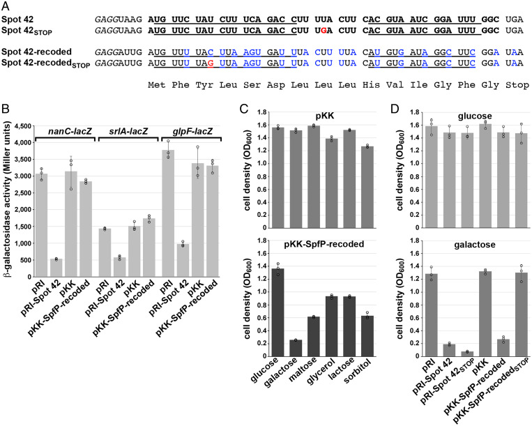
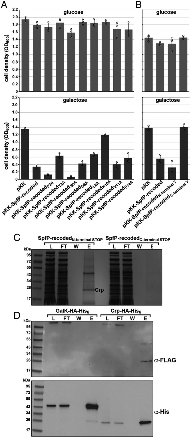
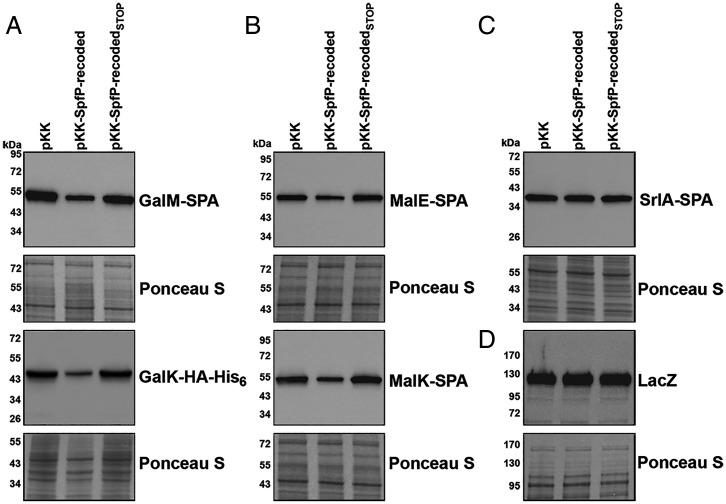
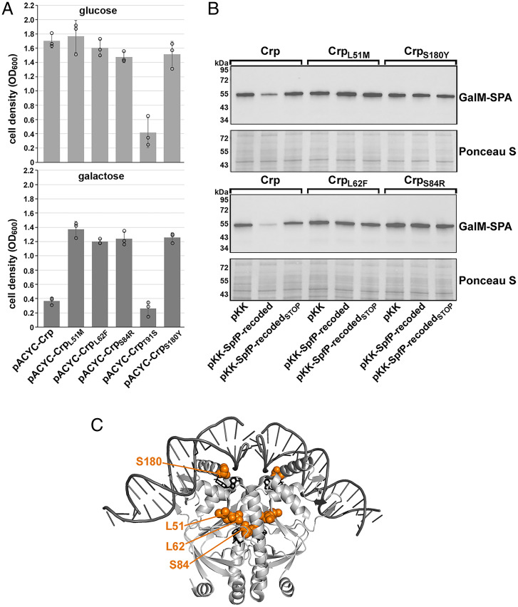
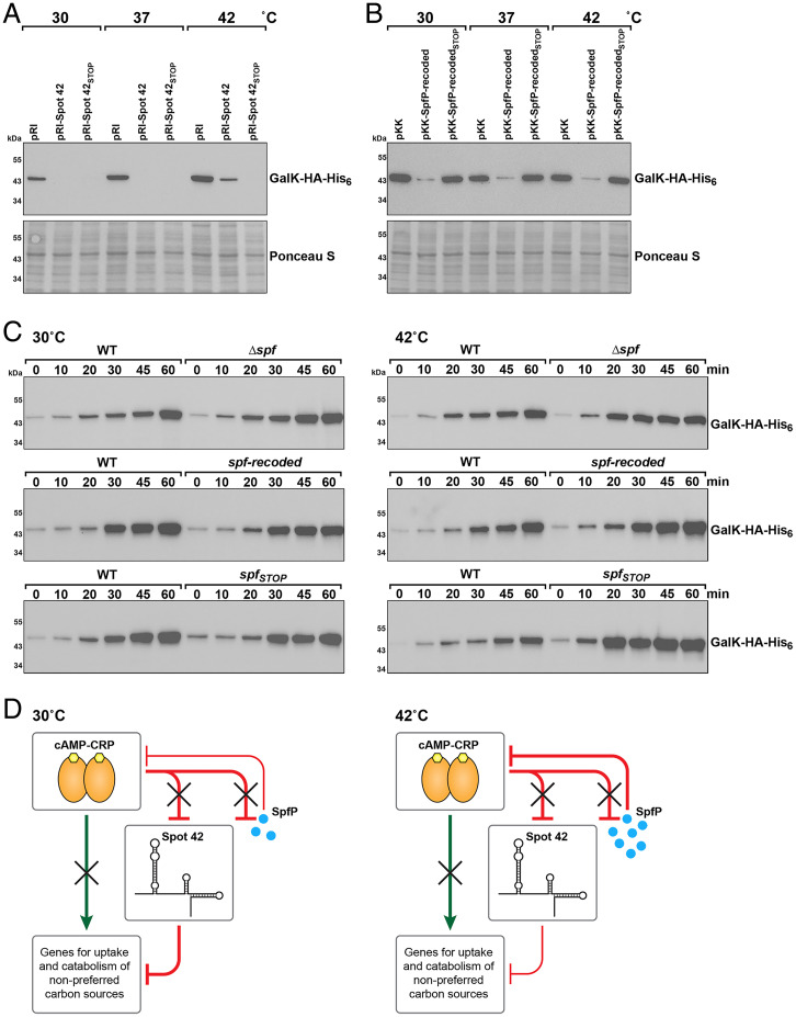
SignificanceDual-function RNAs base pair with target messenger RNAs as small regulatory RNAs and encode small protein regulators. However, only a limited number of these dual-function regulators have been identified. In this study, we show that a well-characterized base-pairing small RNA surprisingly also encodes a 15-amino acid protein. The very small protein binds the cyclic adenosine monophosphate receptor protein transcription factor to block activation of some promoters, raising the question of how many other transcription factors are modulated by unidentified small proteins.

Keywords: SpfP; carbon catabolite repression; dual-function RNA; sRNA; small protein.


Conflict of interest statement

The authors declare no competing interest.

Figures

Fig. 1.
Fig. 1.
Spot 42 is a dual-function sRNA. (A) Diagram of the Spot 42 RNA and sequence of the spf promoter and coding region. Boxes and text in light blue denote SpfP coding sequence, and the yellow box and highlighted text denote the region of Spot 42 base pairing with target mRNAs. The Spot 42 transcript is denoted by the gray box and is indicated in bold, with the +1 site of transcription (position 1988001 of the E. coli K-12 genome) in green font and the 3′-end of the transcript in red font. The SpfP ORF ribosome-binding site, start codon (ATG), and stop codon (TAA) are indicated by black boxes, the potential σ70 −10 sequence is underlined, and a predicted CRP-binding site (36) is highlighted in light gray on the sequence. (B) Diagram of Spot 42 secondary structure (taken from ref. 37) with the Shine–Dalgarno sequence, start codon, and stop codon boxed. Single-stranded regions involved in base pairing with mRNAs are highlighted in yellow. The Hfq-binding regions are indicated in blue, and double-stranded regions are indicated in red. (C) Expression of SpfP protein and Spot 42 RNA. Cells expressing SpfP–SPA (GSO1119) were grown to an OD600 of ∼0.4 at 30 °C in LB and then transferred to 30 °C, 42 °C, or 45 °C. Immunoblotting analysis was carried out on samples collected before transfer and 5 and 10 min after transfer. Anti-FLAG antibody was used to detect the SPA tag, and the Ponceau S stain documents approximately equal loading of the samples. The theoretical molecular weight of the 84–amino acid SPA-tagged SpfP protein is 9.8 kDa. For total RNA isolated for WT cells grown under the same conditions, the Spot 42 and 5S RNAs were detected with primers specific to each of these transcripts.
Fig. 2.
Fig. 2.
SpfP expression impacts growth on galactose. (A) Sequence of region encoding 15–amino acid SpfP. Nucleotides changed in the recoded derivative are indicated in blue, and nucleotides changed in the STOP mutants are indicated in red. (B) β-galactosidase assay of Δspf cells with nanC-lacZ (GSO440), srlA-lacZ (GSO441), or glpF-lacZ (GSO519) transformed with pRI, pRI-Spot 42, pKK, or pKK–SpfP-recoded. The cells were grown to an OD600 of ∼1.0 in LB supplemented with 0.2% arabinose. (C) Growth assays of Δspf::kan cells (GSO433) transformed with pKK or pKK–SpfP-recoded. Strains were grown in M63 minimal medium supplemented with the indicated carbon sources all at 0.2% except for glycerol, which was at 0.4%. (D) Growth assays of Δspf::kan cells (GSO433) transformed with pRI, pRI-Spot42, pRI-Spot42STOP, pKK, pKK–SpfP-recoded, or pKK–SpfP-recoded STOP. Strains were grown in M63 minimal medium supplemented with either glucose (Top) or galactose (Bottom). For C and D, cells grown overnight in LB with ampicillin were diluted to an OD600 of ∼0.05 in M63 minimal medium with the indicated carbon sources and grown for 16 h, at which point the OD600 was measured. For B, C, and D, bars correspond to the average of three biological replicates with circles corresponding to individual data points and error bars representing 1 SD.
Fig. 3.
Fig. 3.
SpfP copurifies with CRP. (A) Growth assays of Δspf::kan cells (GSO433) expressing alanine substitution derivatives of pKK–SpfP-recoded. Strains were grown in M63 minimal medium supplemented with either glucose (Top) or galactose (Bottom). (B) Growth assays of Δspf::kan cells (GSO433) transformed with pKK–SpfP-recoded derivatives with either an additional N-terminal Y or C-terminal Y. Strains were grown in M63 minimal medium supplemented with either glucose (Top) or galactose (Bottom). For A and B, cells were assayed as in Fig. 2, and bars correspond to the average of three biological replicates with circles corresponding to individual data points and error bars representing 1 SD. (C) CRP copurifies with biotin-tagged SpfP. Δspf::kan cells (GSO433) with the SpfP-recodedN-terminal STOP or SpfP-recodedC-terminal STOP plasmids and the orthogonal tRNA and aminoacyl tRNA pair were grown in M63 glucose medium with 1 mM p-AzF to an OD600 of ∼0.5 and induced with 0.2% arabinose for 3 h. Cell lysates were treated with biotin–PEG-alkyne to biotinylate SpfP and passed over streptavidin beads. Fractions from the lysate (L), flow-through (FT), wash (W), and eluants (E) for each sample were subjected to SDS-PAGE followed by Coomassie blue staining. The unique ∼25-kDa band enriched in the eluant from the SpfP-expressing cells was excised from the gel and identified by mass spectrometry. (D) N-terminally FLAG-tagged SpfP copurifies with CRP–HA-His6 but not GalK–HA-His6. Δspf cells expressing CRP–HA-His6 or GalK–HA-His6 from the chromosome (GSO1061 and GSO1060, respectively) were transformed with pKK–SpfP-recodedN-FLAG and grown in LB until an OD600 of ∼0.5 was reached. The cell lysate was incubated overnight with 50 µL of Pierce anti-HA magnetic beads. The beads were collected using a magnet, and proteins were eluted with Laemmeli buffer. Immunoblots of fractions from the lysate (L), flow-through (FT), wash (W), and eluants (E) separated by SDS-PAGE were probed with anti-FLAG (Top) and anti-His (Bottom) antibodies. A cross-reacting band of high molecular weight is detected with anti-FLAG antibodies.
Fig. 4.
Fig. 4.
SpfP overexpression leads to down-regulation of CRP-activated genes. (A) Immunoblotting analysis of GalM–SPA and GalK–HA-His6 levels in Δspf strains with chromosomal galM-SPA::kan or galK-HA-His6::kan (GSO1066 and GSO106, respectively) transformed with pKK, pKK–SpfP-recoded, or pKK–SpfP-recodedSTOP. Cells were grown in M63 galactose to an OD600 of ∼0.5. Anti-FLAG (Top) or anti-His antibody (Bottom) were used to detect GalM–SPA or GalK–HA-His6, respectively. (B) Immunoblotting analysis of MalE–SPA (Top) and MalK–SPA (Bottom) levels in Δspf strains with chromosomal malE-SPA::kan or malK-SPA::kan (GSO1067 and GSO1068, respectively) transformed with pKK, pKK–SpfP-recoded, or pKK–SpfP-recodedSTOP. Cells were grown in M63 maltose to an OD600 of ∼0.5. Anti-FLAG antibodies were used to detect both proteins. (C) Immunoblotting analysis of SrlA–SPA levels in Δspf strains with chromosomal srlA-SPA::kan (GSO449) transformed with pKK, pKK–SpfP-recoded, or pKK–SpfP-recodedSTOP. Cells were grown in M63 sorbitol to an OD600 of ∼0.5. Anti-FLAG antibody was used to detect the protein. (D) Immunoblotting analysis of LacZ levels in the Δspf strain (GSO1059) transformed with pKK, pKK–SpfP-recoded, or pKK–SpfP-recodedSTOP. Cells were grown in M63 lactose to an OD600 of ∼0.5. Anti-β-galactosidase antibody was used to detect the protein. For A, B, C, and D, the Ponceau S–stained membrane documents approximately equal loading of the samples.
Fig. 5.
Fig. 5.
Mutations that relieve the SpfP effect on CRP map to AR3. (A) Growth assays of Δspf Δcrp cells (GSO1133) expressing pKK–SpfP-recoded and pACYC-CRP derivatives with L51M, L62F, S84R, T91S, or S180Y mutations in M63 minimal medium supplemented with either glucose (Top) or galactose (Bottom). Bars correspond to the average of three biological replicates with circles corresponding to individual data points and error bars representing 1 SD. (B) Immunoblotting analysis of GalM–SPA levels in Δspf Δcrp strains with chromosomal galM-SPA::kan (GSO1134) transformed with pKK, pKK–SpfP-recoded, or pKK–SpfP-recodedSTOP and pACYC-crpL51M, pACYC-crpL62F, pACYC-crpS84R, or pACYC-crpS180Y grown in M63 galactose to an OD600 of ∼0.5. Anti-FLAG antibody was used to detect GalM–SPA, and the Ponceau S–stained membrane documents approximately equal loading of the samples. (C) Structure of CRP bound to DNA (38) (Protein Data Bank [PDB] 1ZRF) depicted with PyMol with the L51, L62, S84, and S180 side chains highlighted in orange (numbering is shifted by one amino acid in PDB 1ZRF).
Fig. 6.
Fig. 6.
Spot 42 and SpfP have different effects at different temperatures. (A) Immunoblotting analysis of GalK–HA-His6 levels in a Δspf galK-HA-His6::kan strain (GSO1060) transformed with pRI, pRI-Spot 42, or pRI-Spot 42STOP and grown in LB at 30 °C, 37 °C, and 42 °C. (B) Immunoblotting analysis of GalK–HA-His6 levels in a Δspf galK-HA-His6::kan strain (GSO1060) transformed with pKK, pKK–SpfP-recoded, or pKK–SpfP-recodedSTOP and grown in LB at 30 °C, 37 °C, and 42 °C. For A and B, samples were collected at an OD600 of ∼0.5. Anti-His antibody was used to detect the HA-His6 tag. The Ponceau S–stained membrane documents approximately equal loading of the samples. (C) Immunoblotting analysis of GalK–HA-His6 levels in galK-HA-His6::kan (GSO1057), Δspf galK-HA-His6::kan (GSO1060), spf-recoded galK-HA-His6::kan (GSO1077), or spfSTOP::kan galK-HA-His6 (GSO1075) grown in M63 glucose to an OD600 of ∼0.4 at 30 °C (Left) or 42 °C (Right). Cells were collected and resuspended in M63 galactose at 30 °C or 42 °C, respectively, with samples collected at the indicated times. (D) The small protein SpfP reinforces the multioutput feedforward loop between CRP and Spot 42. For cells grown in the absence of glucose, CRP directly increases transcription of targets and represses Spot 42. When glucose is available (shown here), the Spot 42 RNA represses CRP-activated targets through base pairing, particularly at lower temperatures (Left), and the small protein SpfP blocks CRP-dependent activation, particularly at higher temperature (Right) (adapted from ref. 16).

Similar articles

Cited by

References

    1. Brückner R., Titgemeyer F., Carbon catabolite repression in bacteria: Choice of the carbon source and autoregulatory limitation of sugar utilization. FEMS Microbiol. Lett. 209, 141–148 (2002). - PubMed
    1. Görke B., Stülke J., Carbon catabolite repression in bacteria: Many ways to make the most out of nutrients. Nat. Rev. Microbiol. 6, 613–624 (2008). - PubMed
    1. Kolb A., Busby S., Buc H., Garges S., Adhya S., Transcriptional regulation by cAMP and its receptor protein. Annu. Rev. Biochem. 62, 749–795 (1993). - PubMed
    1. Zheng D., Constantinidou C., Hobman J. L., Minchin S. D., Identification of the CRP regulon using in vitro and in vivo transcriptional profiling. Nucleic Acids Res. 32, 5874–5893 (2004). - PMC - PubMed
    1. Busby S., Ebright R. H., Transcription activation by catabolite activator protein (CAP). J. Mol. Biol. 293, 199–213 (1999). - PubMed

LinkOut - more resources