Review
. 2022 Jan-Dec;14(1):2041342.
doi: 10.1080/19490976.2022.2041342.

Mucus, commensals, and the immune system

Affiliations
Review

Mucus, commensals, and the immune system

Qing Zhao et al. Gut Microbes. 2022 Jan-Dec.

Abstract

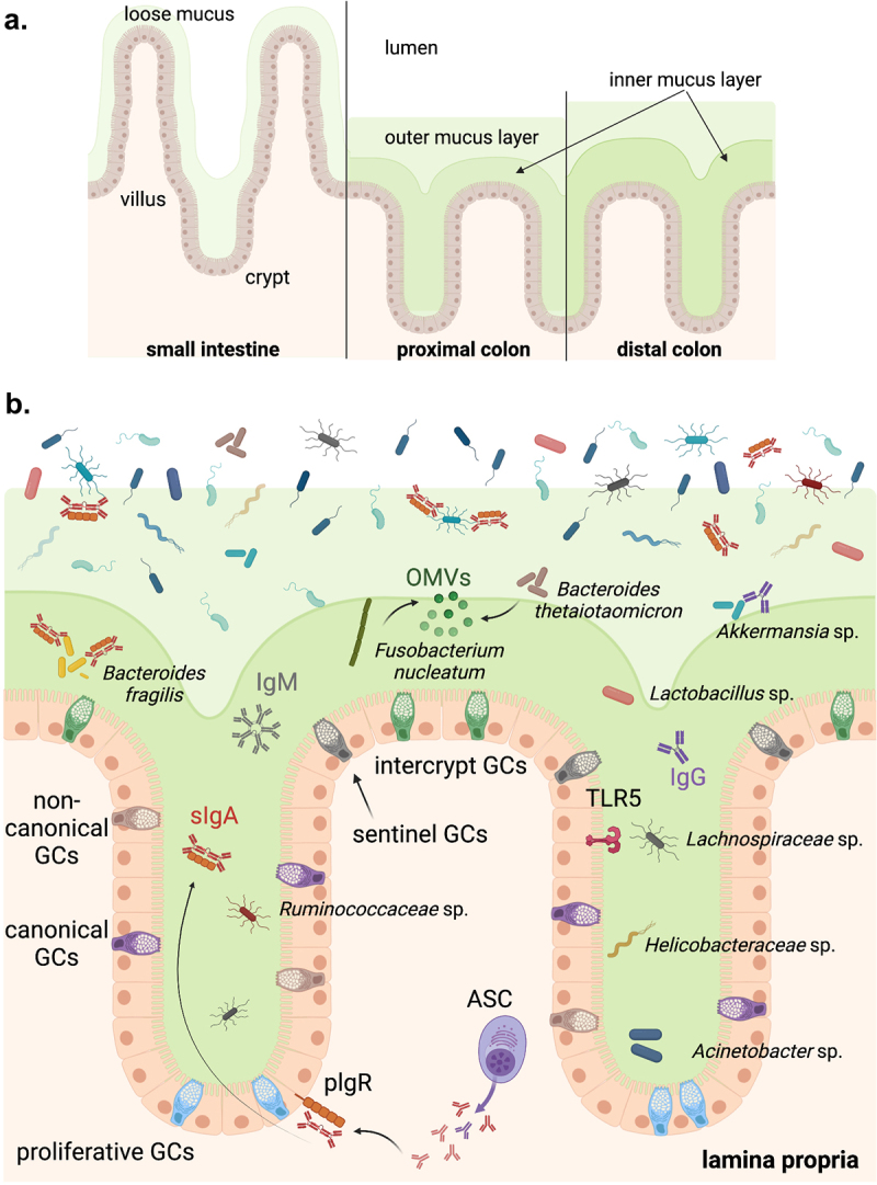
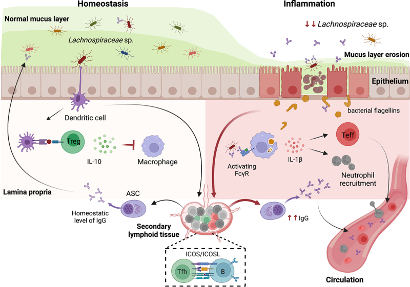
The immune system in the large intestine is separated from commensal microbes and comparatively rare enteric pathogens by a monolayer of diverse epithelial cells overlaid with a compact and adherent inner mucus layer and a looser outer mucus layer. Microorganisms, collectively referred to as the mucus-associated (MA) microbiota, physically inhabit this mucus barrier, resulting in a dynamic and incessant dialog to maintain both spatial segregation and immune tolerance. Recent major findings reveal novel features of the crosstalk between the immune system and mucus-associated bacteria in health and disease, as well as disease-related peripheral immune signatures indicative of host responses to these organisms. In this brief review, we integrate these novel observations into our overall understanding of host-microbiota mutualism at the colonic mucosal border and speculate on the significance of this emerging knowledge for our understanding of the prevention, development, and progression of chronic intestinal inflammation.

Keywords: Colon mucus layer; T-dependent; T-independent; anti-commensal IgA; anti-commensal IgG; flagellin; lachnospiraceae; mucus-associated bacteria.


Conflict of interest statement

No potential conflict of interest was reported by the author(s).

Figures

Figure 1.
Figure 1.
Host-microbiota spatial dynamics at the gut epithelial border. (A) The small intestinal epithelium is covered with a loose mucus layer, whereas a dense inner mucus layer and a looser outer mucus layer overlay the colonic epithelium. Both the density and thickness of the inner mucus layer increases from the proximal to the distal colon. (B) The colonic epithelial layer includes multiple subtypes of mucus-secreting goblet cells (GC). Crypt GCs include canonical and non-canonical GCs, as well as proliferative GCs that locate at the base of the crypts, whereas intercrypt GCs are found on the surface epithelium. TLR5, the receptor for bacterial flagellin, is constitutively and uniquely expressed on the luminal surface of colonic epithelial cells in the proximal colon. Microbial density is significantly higher in the outer compared to the inner mucus layer and the crypts. Bacteria including Bacteroides thetaiotaomicron and Fusobacterium nucleatum can bind mucin and release outer membrane vesicles (OMVs) which can lead to immune activation. Flagellated bacteria belonging to the Lachnospiraceae, Ruminococcaceae, and Helicobacteraceae families can colonize the crypts of the proximal colon. In addition to the physical barriers of the epithelial and mucus layers, lamina propria plasma cells produce anti-commensal immunoglobulins (Ig) including IgM, IgA, and IgG which help to regulate bacterial activation of the immune system. The most widely studied, IgA is transported into the lumen via the polymeric Ig receptor (PIGR) and helps to sequester bacteria and bacterial antigens away from the gut epithelium, but also facilitates colonization by bacteria such as Bacteroides fragilis.
Figure 2.
Figure 2.
Examples of the crosstalk between mucus-associated bacteria and the intestinal immune system in health and disease. Under homeostatic conditions, the peaceful existence in the mucus layer of bacteria, including members of the Lachnospiraceae and Ruminococcaceae families, is aided by coordinated antigen collection by lamina propria myeloid cells including dendritic cells and macrophages. Intestinal dendritic cells promote the differentiation and/or maintenance of IL-10-producing Treg cells. IL-10 inhibits commensal-dependent activation of lamina propria macrophages, maintain the macrophages in a quiescent state. In addition, the presentation of commensal-derived antigens by B cells in the gut-draining lymph nodes induces differentiation into IgG+ antibody-secreting cells (ASCs) with the aid of ICOS-ICOSL-dependent T-follicular helper (Tfh) cells. This T-dependent ASC-derived IgG crosses the epithelium into the mucus layer and helps regulate the resident microbial communities. In contrast, the inflammatory state is characterized by an erosion of the mucus layer resulting in increased availability and opsonization of commensal antigens. This enables dysregulated differentiation and expansion of pro-inflammatory/effector CD4 T cells as well as hyperproduction of IgG antibodies to antigens including Lachnospiraceae flagellins. Excessive activation of intestinal macrophages via the interaction of IgG immune complexes with activating Fc gamma receptors (FcγR) induces a pro-inflammatory phenotype including secretion of cytokines such as IL-1β which can promote neutrophil recruitment. In CD, the chronic inflammatory state is also characterized by an overall reduction in the abundance of Lachnospiraceae bacteria and a breach in the so-called ‘mucosal firewall’ resulting in effector CD4 T cells and anti-commensal IgG antibodies entering the systemic circulation.

References

    1. Bunker JJ, Flynn T, Koval J, Shaw D, Meisel M, McDonald B, Ishizuka I, Dent A, Wilson P, Jabri B, et al. Innate and adaptive humoral responses coat distinct commensal bacteria with immunoglobulin A. Immunity. 2015;43:541–19. doi: 10.1016/j.immuni.2015.08.007. - DOI - PMC - PubMed
    1. Palm NW, De zoete M, Cullen T, Barry N, Stefanowski J, Hao L, Degnan P, Hu J, Peter I, Zhang W, et al. Immunoglobulin A coating identifies colitogenic bacteria in inflammatory bowel disease. Cell. 2014;158:1000–1010. doi: 10.1016/j.cell.2014.08.006. - DOI - PMC - PubMed
    1. Miranda RL, Conway T, Leatham MP, Chang DE, Norris WE, Allen JH, Stevenson SJ, Laux DC, Cohen PS.. Glycolytic and gluconeogenic growth of Escherichia coli O157:H7 (EDL933) and E. coli K-12 (MG1655) in the mouse intestine. Infect Immun. 2004;72:1666–1676. doi: 10.1128/IAI.72.3.1666-1676.2004. - DOI - PMC - PubMed
    1. Ravcheev DA, Thiele I. Comparative genomic analysis of the human gut microbiome reveals a broad distribution of metabolic pathways for the degradation of host-synthetized mucin glycans and utilization of mucin-derived monosaccharides. Front Genet. 2017;8:111. doi: 10.3389/fgene.2017.00111. - DOI - PMC - PubMed
    1. Riva A, Kuzyk O, Forsberg E, Siuzdak G, Pfann C, Herbold C, Daims H, Loy A, Warth B, Berry D, et al. A fiber-deprived diet disturbs the fine-scale spatial architecture of the murine colon microbiome. Nat Commun. 2019;10:4366. doi: 10.1038/s41467-019-12413-0. - DOI - PMC - PubMed

Publication types