Case Reports
. 2022 Mar 3;16(3):e0010226.
doi: 10.1371/journal.pntd.0010226. eCollection 2022 Mar.

In vivo reflectance confocal microscopy, dermoscopy, high-frequency ultrasonography, and histopathology features in a case of chromoblastomycosis

Affiliations
Case Reports

In vivo reflectance confocal microscopy, dermoscopy, high-frequency ultrasonography, and histopathology features in a case of chromoblastomycosis

Gabriela A Giraldelli et al. PLoS Negl Trop Dis. .
No abstract available


Conflict of interest statement

The authors have declared that no competing interests exist.

Figures

Fig 1
Fig 1. A well-defined moderate severity verrucous violaceous plaque, raised edges, measuring 4.5 × 3.0 cm with central ulceration and black dots in the periphery.
Fig 2
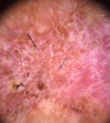
Fig 2. Dermoscopy shows black dots (black arrow), shiny white blotches and strands (white arrows), and yellowish-orange area (blue arrow).
DermLite IV DL4; 3 Gen, polarized mode, ×10 magnification.
Fig 3
Fig 3. Reflectance confocal microscopy (basic image 0.5 × 0.5 mm) at the level of the stratum corneum and epidermis evidencing 2 small round hyperreflective bodies. VivaScope 1500.
Fig 4
Fig 4. High-frequency ultrasound with Doppler shows epidermal thickening, hyperechogenic round structures (arrow), dermal fibrosis, and increased vascularization.
My Lab Touch, 22 MHz linear transducer.
Fig 5
Fig 5. Histopathology of a skin biopsy reveals grouped fungal structures encircled by a granulomatous infiltrate in the dermis.
HE, ×400.

References

    1. Queiroz-Telles F, de Hoog S, Santos DW, Salgado CG, Vicente VA, Bonifaz A, et al.. Chromoblastomycosis. Clin Microbiol Rev. 2017;30:233–76. doi: 10.1128/CMR.00032-16 . - DOI - PMC - PubMed
    1. Santos DWCL, de Azevedo CMPES, Vicente VA, Queiroz-Telles F, Rodrigues AM, de Hoog GS, et al.. The global burden of chromoblastomycosis. PLoS Negl Trop Dis. 2021;12(15):e0009611. doi: 10.1371/journal.pntd.0009611 . - DOI - PMC - PubMed
    1. Okuda C, Ito M, Oka K. Relationship between tissue reactions and morphological changes of the fungi in chromoblastomycosis: morphometry and electron microscopy. Arch Dermatol Res. 1992;284:95–9. doi: 10.1007/BF00373377 - DOI - PubMed
    1. Subhadarshani S, Yadav D. Dermoscopy of chromoblastomycosis. Dermatol Pract Concept. 2017;7:23–4. doi: 10.5826/dpc.0704a06 . - DOI - PMC - PubMed
    1. Chauhan P, Jindal R, Shirazi N. Dermoscopy of chromoblastomycosis. Indian Dermatol Online J. 2019;106:759–60. doi: 10.4103/idoj.IDOJ_213_18 . - DOI - PMC - PubMed

Publication types