. 2022 May 15;434(9):167516.
doi: 10.1016/j.jmb.2022.167516. Epub 2022 Feb 28.

SARS-CoV-2 Nucleocapsid Protein Targets a Conserved Surface Groove of the NTF2-like Domain of G3BP1

Affiliations

SARS-CoV-2 Nucleocapsid Protein Targets a Conserved Surface Groove of the NTF2-like Domain of G3BP1

Mahamaya Biswal et al. J Mol Biol. .

Abstract

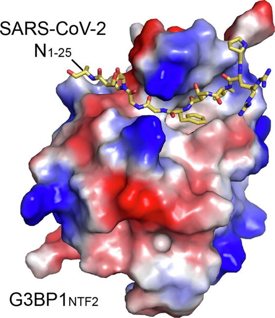
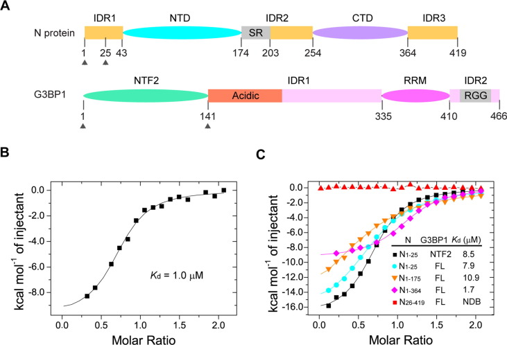
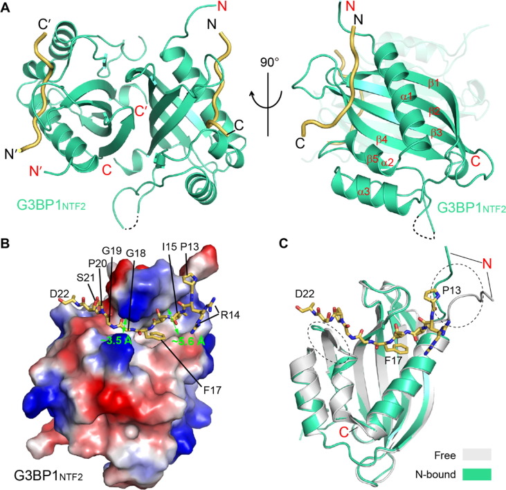
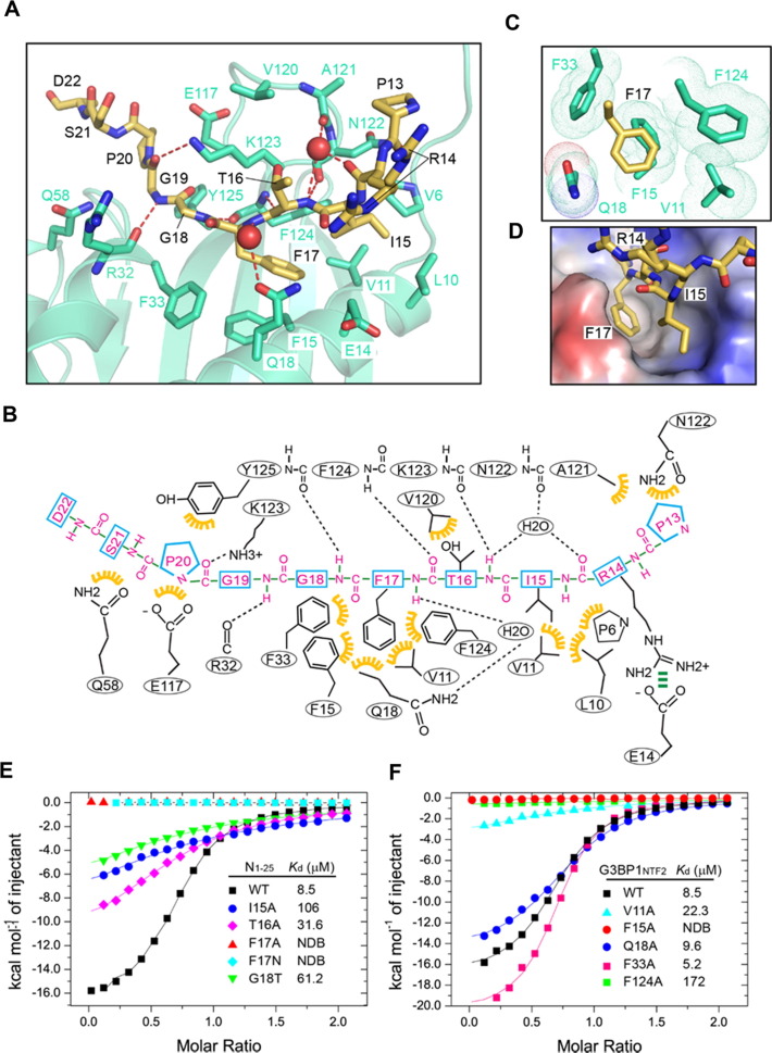
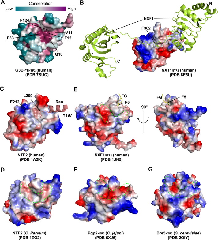
Stress granule (SG) formation mediated by Ras GTPase-activating protein-binding protein 1 (G3BP1) constitutes a key obstacle for viral replication, which makes G3BP1 a frequent target for viruses. For instance, the SARS-CoV-2 nucleocapsid (N) protein interacts with G3BP1 directly to suppress SG assembly and promote viral production. However, the molecular basis for the SARS-CoV-2 N - G3BP1 interaction remains elusive. Here we report biochemical and structural analyses of the SARS-CoV-2 N - G3BP1 interaction, revealing differential contributions of various regions of SARS-CoV-2 N to G3BP1 binding. The crystal structure of the NTF2-like domain of G3BP1 (G3BP1NTF2) in complex with a peptide derived from SARS-CoV-2 N (residues 1-25, N1-25) reveals that SARS-CoV-2 N1-25 occupies a conserved surface groove of G3BP1NTF2 via surface complementarity. We show that a φ-x-F (φ, hydrophobic residue) motif constitutes the primary determinant for G3BP1NTF2-targeting proteins, while the flanking sequence underpins diverse secondary interactions. We demonstrate that mutation of key interaction residues of the SARS-CoV-2 N1-25 - G3BP1NTF2 complex leads to disruption of the SARS-CoV-2 N - G3BP1 interaction in vitro. Together, these results provide a molecular basis of the strain-specific interaction between SARS-CoV-2 N and G3BP1, which has important implications for the development of novel therapeutic strategies against SARS-CoV-2 infection.

Keywords: NTF2-like domain; SARS-CoV-2 nucleocapsid protein; pathogen-host interaction; ras GTPase-activating protein-binding protein 1; stress granule.


Conflict of interest statement

Conflict of Interest The authors declare no competing interest.

Figures

None
Graphical abstract
Figure 1
Figure 1
Biochemical characterization of the interaction between SARS-CoV-2 N and G3BP1. (A) Domain architecture of SARS-CoV-2 N and G3BP1, with individual domains color coded. The SR-rich region within the IDR2 of SRS-CoV-2 N, the acidic region within the IDR1 of G3BP1 and the RG-rich region (RGG) within the IDR2 of G3BP1 are labeled. The protein fragments (residues 1–25 of SARS-CoV-2 N protein and 1–139 of G3BP1) used for crystallographic study are delimited by arrows. (B) ITC binding assays for full-length SARS-CoV-2 N and G3BP1. (C) ITC binding assays for the truncated fragments of SARS-CoV-2 N and G3BP1. FL, full length; NDB, no detectable binding.
Figure 2
Figure 2
Structural overview of the SARS-CoV-2 N125 − G3BP1NTF2 complex. (A) Orthogonal views of the SARS-CoV-2 N1–25 − G3BP1NTF2 complex, with SARS-CoV-2 N1–25 colored in yellow orange and G3BP1NTF2 colored in green. The traceable N- and C-termini of SARS-CoV-2 N1–25 or G3BP1NTF2 are labeled with “N” and “C”, respectively. The region (residue S47) with untraceable electron density is shown in dashed line. (B) Electrostatic surface view of the G3BP1NTF2 domain bound to the SARS-CoV-2 N1–25 peptide (stick representation). The widths of two distinct groove regions of G3BP1NTF2 are marked. For clarity, only one monomer of the G3BP1NTF2 homodimer is shown. (C) Structural overlay of the G3BP1NTF2 domain, free (grey) and in complex with SARS-CoV-2 N1–25 (green). The two structurally divergent regions are circled with dotted lines.
Figure 3
Figure 3
Structural and biochemical characterizations of the SARS-CoV-2 N125 − G3BP1NTF2 interaction. (A) Close-up view of the SARS-CoV-2 N1–25 − G3BP1NTF2 interaction. The interacting residues are shown in stick representation. The hydrogen bonds are shown as dashed lines. The water molecules are shown as red spheres. (B) Schematic view of the SARS-CoV-2 N1–25 − G3BP1NTF2 interaction. Hydrogen bonds and electrostatic interactions are indicated by black and green dashed lines, respectively, and van der Waals contacts are indicated by yellow gears. (C) Close-up view of the hydrophobic pocket harboring residue F17 of SARS-CoV-2 N1–25. The van der Waals radii of the G3BP1NTF2 residues are shown in dots. (D) Close-up view of the electrostatic surface of G3BP1NTF2 harboring the side chains of I15 and F17 of SARS-CoV-2 N1–25. (E) ITC binding assays for wild-type G3BP1NTF2 titrated with wild-type or mutant SARS-CoV-2 N1–25 peptide. (F) ITC binding assays for wild-type or mutant G3BP1NTF2 titrated with the wild-type SARS-CoV-2 N1–25 peptide. NDB, no detectable binding.
Figure 4
Figure 4
Structural comparison of SARS-CoV-2 N125 − G3BP1NTF2 with other G3BP1NTF2 complexes. (A) Structural overlay between the SARS-CoV-2 N1–25 (yellow orange) − G3BP1NTF2 (green) and the SFV nsP3449–471 (magenta) − G3BP1NTF2 (wheat) complex. The phenylalanine residues inserting into the hydrophobic pocket of G3BP1NTF2 are labeled and shown in stick representation. (B) Structural overlay between the SARS-CoV-2 N1–25 (yellow orange) − G3BP1NTF2 (green) and the Caprin1363–382 (slate) − G3BP1NTF2 (yellow) complex. The phenylalanine residues inserting into the hydrophobic pocket of G3BP1NTF2 are labeled and shown in stick representation. (C) Sequence alignment of the G3BP1NTF2-interacting peptides, with the P-2, P1 and P2 sites colored in magenta, red and green, respectively. (D) Structural alignment of the G3BP1NTF2-interacting peptides, with the P–2 and P1 sites, as well as the N- and C-termini, labeled.
Figure 5
Figure 5
Structural evolution of the NTF2 domains. (A) Color-coded sequence conservation of the G3BP1NTF2 domain, analyzed using the ConSurf server (https://consurf.tau.ac.il). (B) Electrostatic surface of the NXT1NTF2 domain bound to the LRR (NXF1LRR)-NTF2 (NXF1NTF2) domain linker of the NXF1 protein (limon), with the side chain of NXF1 F362 shown in stick representation. (C) Electrostatic surface of human NTF2 domain bound to residues 197–212 of one Ran molecule (light pink), with the side chain of Ran L209 shown in stick representation. For clarity, the rest of Ran structure is not shown. (D-G) Electrostatic surface of C. parvum NTF2 (D), human NXF1NTF2 (E), C. jejuni Pgp2NTF2 (F) and S. cerevisiae Bre5NTF2 (G), with the individual surface grooves circled by dotting lines. Note that human NXF1NTF2 binds to the FG peptide through a hydrophobic pocket positioned separately from the groove.

References

    1. Wu A., Peng Y., Huang B., Ding X., Wang X., Niu P., Meng J., Zhu Z., et al. Genome Composition and Divergence of the Novel Coronavirus (2019-nCoV) Originating in China. Cell Host Microbe. 2020;27:325–328. - PMC - PubMed
    1. Chang C.K., Hou M.H., Chang C.F., Hsiao C.D., Huang T.H. The SARS coronavirus nucleocapsid protein–forms and functions. Antiviral Res. 2014;103:39–50. - PMC - PubMed
    1. Hsieh P.K., Chang S.C., Huang C.C., Lee T.T., Hsiao C.W., Kou Y.H., Chen I.Y., Chang C.K., et al. Assembly of severe acute respiratory syndrome coronavirus RNA packaging signal into virus-like particles is nucleocapsid dependent. J. Virol. 2005;79:13848–13855. - PMC - PubMed
    1. McBride R., van Zyl M., Fielding B.C. The coronavirus nucleocapsid is a multifunctional protein. Viruses. 2014;6:2991–3018. - PMC - PubMed
    1. Liu X., Verma A., Garcia G., Jr., Ramage H., Lucas A., Myers R.L., Michaelson J.J., Coryell W., et al. Targeting the coronavirus nucleocapsid protein through GSK-3 inhibition. Proc. Natl. Acad. Sci. U. S. A. 2021;118 - PMC - PubMed

Publication types

MeSH terms