

Pulmonary lymphangitis sarcomatosis: a rare cause of severe progressive dyspnoea
- PMID: 35241446
- PMCID: PMC8895897
- DOI: 10.1136/bcr-2021-246128
Pulmonary lymphangitis sarcomatosis: a rare cause of severe progressive dyspnoea
Abstract
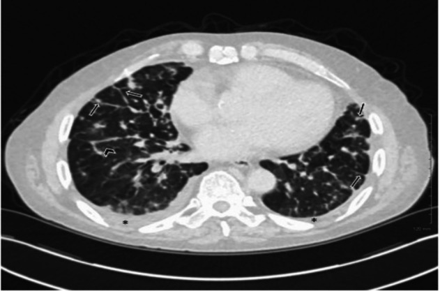
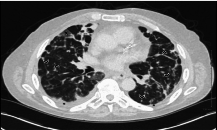
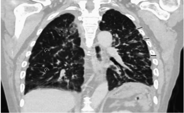
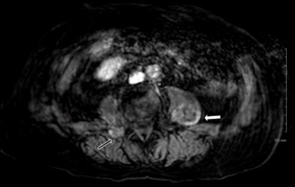
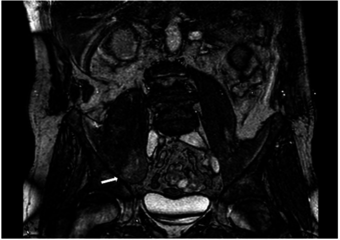
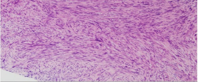
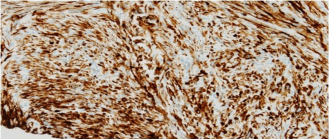
Pulmonary lymphangitis carcinomatosis is a complication of malignancy with a poor prognosis. We describe an unusual case in which it caused ventilatory failure and unfortunately death in a previously well male in his 70s. Abnormal chest imaging led to a wide differential diagnosis with Bronchoscopy confirming malignant cells. MRI of his pelvis and biopsy was done diagnosis of metastatic leiomyosarcoma, a particularly aggressive malignancy. Sarcoma-related lymphangitis carcinomatosis is rarely described in the literature and this is the first case to our knowledge of its association with leiomyosarcoma.
Keywords: lung cancer (oncology); radiology; respiratory system.
© BMJ Publishing Group Limited 2022. No commercial re-use. See rights and permissions. Published by BMJ.
Conflict of interest statement
Competing interests: None declared.
Figures
Similar articles
-
Pulmonary lymphangitis carcinomatosis: systematic review and meta-analysis of case reports, 1970-2018.Postgrad Med. 2019 Jun;131(5):309-318. doi: 10.1080/00325481.2019.1595982. Epub 2019 Apr 5. Postgrad Med. 2019. PMID: 30900501
-
Pulmonary lymphangitic carcinomatosis as a primary manifestation of gastric carcinoma in a young adult: a case report and review of the literature.BMC Res Notes. 2012 Nov 16;5:638. doi: 10.1186/1756-0500-5-638. BMC Res Notes. 2012. PMID: 23158653 Free PMC article. Review.
-
Pulmonary tumour embolism and lymphangitis carcinomatosa: a case report and review of the literature.J Cardiothorac Surg. 2022 May 7;17(1):105. doi: 10.1186/s13019-022-01832-8. J Cardiothorac Surg. 2022. PMID: 35525999 Free PMC article. Review.
-
[Clinical features and diagnosis of pulmonary lymphangitic carcinomatosis].Ai Zheng. 2006 Sep;25(9):1127-30. Ai Zheng. 2006. PMID: 16965655 Chinese.
-
Asymptomatic lymphangitis carcinomatosis due to squamous cell lung carcinoma.Indian J Chest Dis Allied Sci. 2005 Apr-Jun;47(2):121-3. Indian J Chest Dis Allied Sci. 2005. PMID: 15832957
Cited by
-
Pulmonary Lymphangitis Poses a Major Challenge for Radiologists in an Oncological Setting during the COVID-19 Pandemic.J Pers Med. 2022 Apr 12;12(4):624. doi: 10.3390/jpm12040624. J Pers Med. 2022. PMID: 35455740 Free PMC article.
-
Radiation Recall Pneumonitis: The Open Challenge in Differential Diagnosis of Pneumonia Induced by Oncological Treatments.J Clin Med. 2023 Feb 10;12(4):1442. doi: 10.3390/jcm12041442. J Clin Med. 2023. PMID: 36835977 Free PMC article. Review.
References
-
- Zielińska-Leś I, Kamiński J, Kozielski J. Trudności diagnostyczne zmian rozsianych w płucach [Diagnostic difficulties in diffuse pulmonary infiltrations]. Wiad Lek 2006;59:724–6. - PubMed
Publication types
MeSH terms
LinkOut - more resources
Full Text Sources
Medical