. 2022 Mar-Apr;36(2):657-666.
doi: 10.21873/invivo.12750.

In Vivo Imaging of Ischemia/Reperfusion-mediated Aminopeptidase N Expression in Surgical Rat Model Using 68Ga-NOTA-c(NGR)

Affiliations

In Vivo Imaging of Ischemia/Reperfusion-mediated Aminopeptidase N Expression in Surgical Rat Model Using 68Ga-NOTA-c(NGR)

Gergely Farkasinszky et al. In Vivo. 2022 Mar-Apr.

Abstract

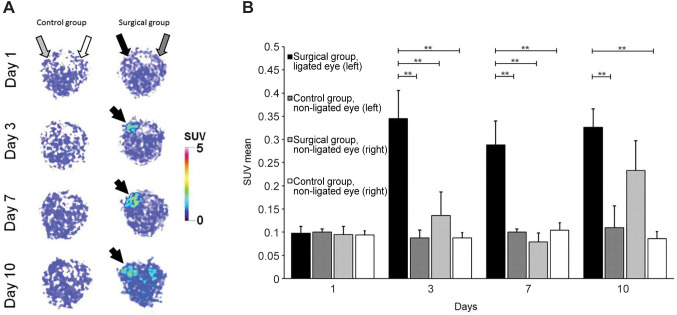
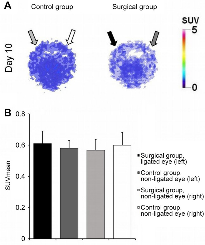
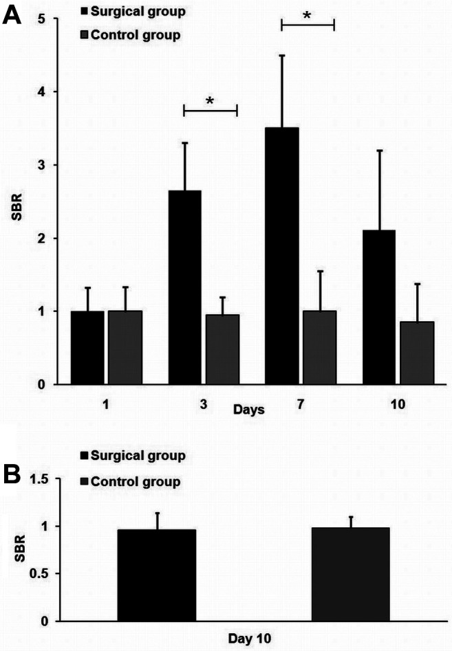
Background/aim: Previous studies have already shown that 68Gallium(68Ga)-labeled NGR-based radiopharmaceuticals specifically bind to the neoangiogenic molecule Aminopeptidase N (APN/CD13). The aim of this study was to evaluate the applicability of 68Ga-NOTA-c(NGR) in the in vivo detection of the temporal changes of APN/CD13 expression in the diabetic retinopathy rat model using positron emission tomography (PET).

Materials and methods: Ischemia/reperfusion injury was initiated by surgical ligation of the left bulbus oculi of rats. In vivo PET imaging studies were performed after the surgery using 68Ga-NOTA-c(NGR).

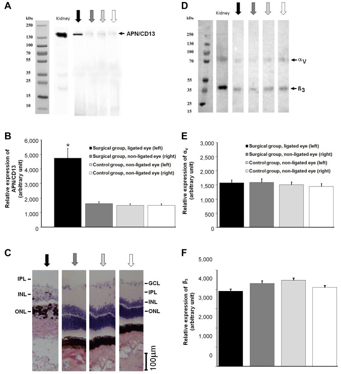
Results: Significantly higher 68Ga-NOTA-c(NGR) uptake was observed in the surgically-ligated left bulbus, compared to the bulbus of the non-surgical group at each investigated time point. The western blot and histological analysis confirmed the increased expression of the neo-angiogenic marker APN/CD13.

Conclusion: 68Ga-NOTA-c(NGR) is a suitable radiotracer for the detection of the temporal changes of the ischemia/reperfusion-mediated expression of APN/CD13 in the surgically induced diabetic retinopathy rat model.

Keywords: 68Ga-NOTA-c(NGR); Aminopeptidase N; Angiogenesis; NGR peptide; ischemia/reperfusion; positron emission tomography.


Conflict of interest statement

The Authors declare no conflicts of interest.

Figures

Figure 1
Figure 1. PET imaging following NGR administration. (A) Representative transaxial decay-corrected PET images of the control (A panel, left) and surgical group (A panel, right) 1, 3, 7 and 10 days after ocular I/R surgery and 90 min after intravenous injection of 68Ga-NOTA-c(NGR). Black arrow points to the ligated eye (I/R) of the surgical group, dark grey arrow points to the non-ligated eye (non-I/R) of the surgical group, grey arrow points to the non-ligated eye (non-I/R) of the (non-surgical)control group, white arrow points to the non-ligated eye (non-I/R) of the (non-surgical) control group. (B) Quantitative analysis of the PET images 1, 3, 7 and 10 days after ocular I/R surgery and 90 min after intravenous injection of 68Ga-NOTA-c(NGR). PET: Position emmission tomography; SUV: standard uptake value; I/R: ischemia/reperfusion. Significance levels: p≤0.05 (*) and p≤0.01 (**).
Figure 2
Figure 2. PET imaging following RGD administration. (A) Representative transaxial decay-corrected PET images of the control (A panel, left) and surgical group (A panel, right) 10 days after ocular I/R surgery and 90 min after the intravenous injection of 68GaNODAGA-[c(RGD)]2. Black arrow points to the ligated eye (I/R) of the surgical group, dark grey arrow points to the non-ligated eye (non-I/R) of the surgical group, grey arrow points to the non-ligated eye (nonI/R) of the (non-surgical) control group, white arrow points to nonligated eye (non-I/R) of the (non-surgical) control group. (B) Quantitative analysis of the PET images 10 days after ocular I/R surgery and 90 min after intravenous injection of 68Ga-NODAGA-[c(RGD)]2. PET: Position emmission tomography; SUV: standard uptake value; I/R: ischemia/reperfusion.
Figure 3
Figure 3. Quantitative assessment of SBR. Assessment 1, 3, 7 and 10 days after ocular I/R using 68Ga-NOTA-c(NGR) (A), and 10 days after ocular I/R using 68Ga-NODAGA-[c(RGD)]2 (B) PET imaging. SBR: Signal-to-background ratio; SUV: standard uptake value; I/R: ischemia/reperfusion. Significance level: p≤0.05 (*).
Figure 4
Figure 4. Western blot and histopathological analysis of neo-angiogenic marker expression of the ligated and non-ligated eyes of the experimental groups 10 days after the surgery. (A) APN/CD13 expression. (B) Schematic diagram of arbitrary unit values of APN/CD13 expression (quantitative western blot analysis). (C) Representative hematoxylin-eosin staining of the ligated and non-ligated eyes of the experimental groups, showing severe retinal damage in the ganglion cell layer, inner plexiform layer, and in the nuclear layers. (D) AlphaVβ3 integrin receptor expression. (E) and (F) Schematic diagram of arbitrary unit values of αV and β3 integrin receptor expression, respectively (quantitative western blot analysis). Black arrow: ligated eye (I/R) of the surgical group, dark grey arrow: non-ligated eye (non-I/R) of the surgical group, grey arrow: non-ligated eye (non-I/R) of the (non-surgical) control group, white arrow: non-ligated eye (non-I/R) of the (non-surgical) control group. I/R: Ischemia/reperfusion. Significance level: p≤0.05 (*). GCL: Ganglion cell layer; IPL: inner plexiform layer; INL: inner nuclear layer; ONL: outer nuclear layer.

Similar articles

Cited by

References

    1. Blair M. Diabetes mellitus review. Urol Nurs. 2016;36(1):27–36. - PubMed
    1. DeFronzo RA, Ferrannini E, Groop L, Henry RR, Herman WH, Holst JJ, Hu FB, Kahn CR, Raz I, Shulman GI, Simonson DC, Testa MA, Weiss R. Type 2 diabetes mellitus. Nat Rev Dis Primers. 2015;1:15019. doi: 10.1038/nrdp.2015.19. - DOI - PubMed
    1. Kitabchi AE, Umpierrez GE, Miles JM, Fisher JN. Hyperglycemic crises in adult patients with diabetes. Diabetes Care. 2009;32(7):1335–1343. doi: 10.2337/dc09-9032. - DOI - PMC - PubMed
    1. Ribeiro L, Bandello F, Tejerina AN, Vujosevic S, Varano M, Egan C, Sivaprasad S, Menon G, Massin P, Verbraak FD, Lund-Andersen H, Martinez JP, Jürgens I, Smets E, Coriat C, Wiedemann P, Ágoas V, Querques G, Holz FG, Nunes S, Neves C, Cunha-Vaz J, Evicr Net Study Group Characterization of retinal disease progression in a 1-year longitudinal study of eyes with mild nonproliferative retinopathy in diabetes type 2. Invest Ophthalmol Vis Sci. 2015;56(9):5698–5705. doi: 10.1167/iovs.15-16708. - DOI - PubMed
    1. IDF Diabetes Atlas Group Update of mortality attributable to diabetes for the IDF Diabetes Atlas: Estimates for the year 2013. Diabetes Res Clin Pract. 2015;109(3):461–465. doi: 10.1016/j.diabres.2015.05.037. - DOI - PubMed

Substances

LinkOut - more resources