. 2023 Feb;72(2):275-294.
doi: 10.1136/gutjnl-2021-325520. Epub 2022 Mar 3.

Epithelial RAC1-dependent cytoskeleton dynamics controls cell mechanics, cell shedding and barrier integrity in intestinal inflammation

Affiliations

Epithelial RAC1-dependent cytoskeleton dynamics controls cell mechanics, cell shedding and barrier integrity in intestinal inflammation

Luz Del Carmen Martínez-Sánchez et al. Gut. 2023 Feb.

Abstract

Objective: Increased apoptotic shedding has been linked to intestinal barrier dysfunction and development of inflammatory bowel diseases (IBD). In contrast, physiological cell shedding allows the renewal of the epithelial monolayer without compromising the barrier function. Here, we investigated the role of live cell extrusion in epithelial barrier alterations in IBD.

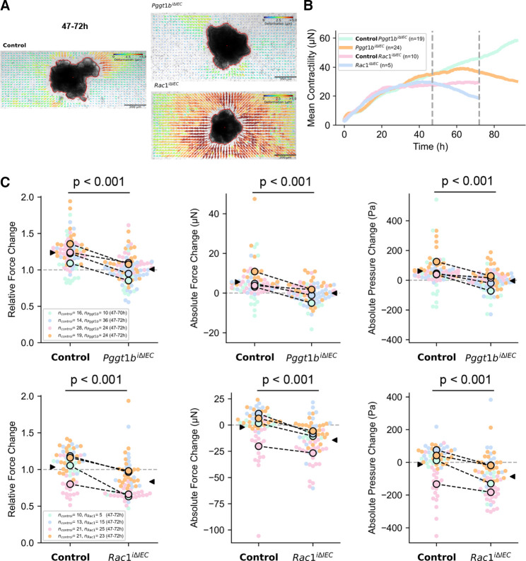
Design: Taking advantage of conditional GGTase and RAC1 knockout mice in intestinal epithelial cells (Pggt1b iΔIEC and Rac1 iΔIEC mice), intravital microscopy, immunostaining, mechanobiology, organoid techniques and RNA sequencing, we analysed cell shedding alterations within the intestinal epithelium. Moreover, we examined human gut tissue and intestinal organoids from patients with IBD for cell shedding alterations and RAC1 function.

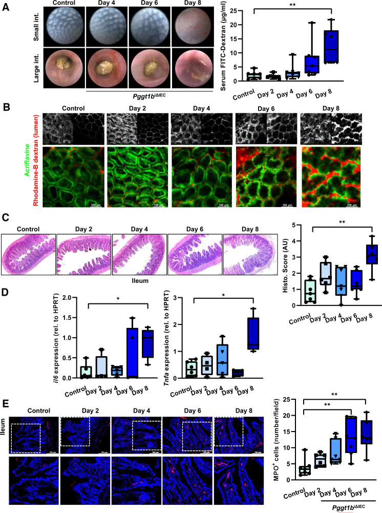
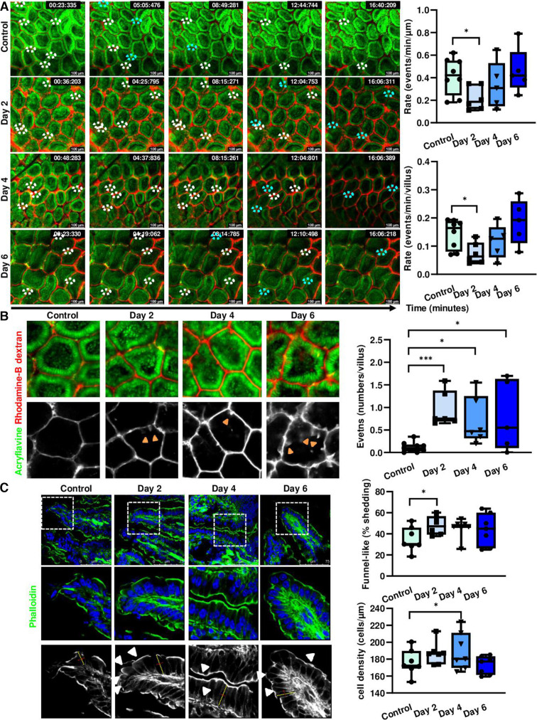
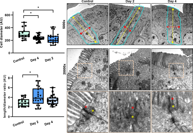
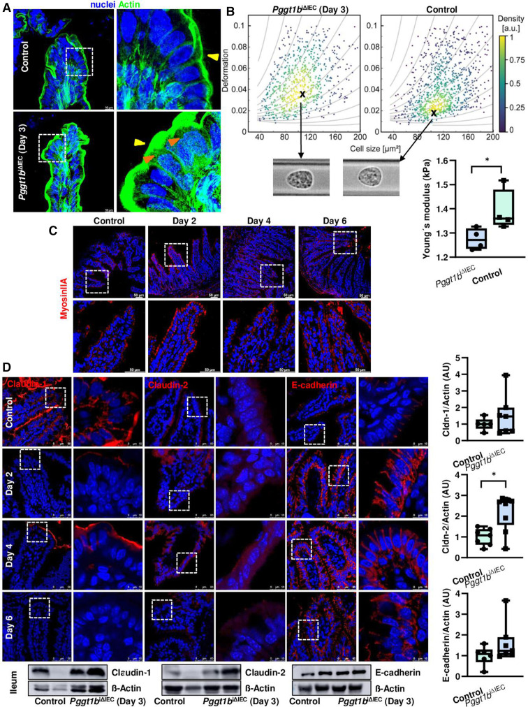
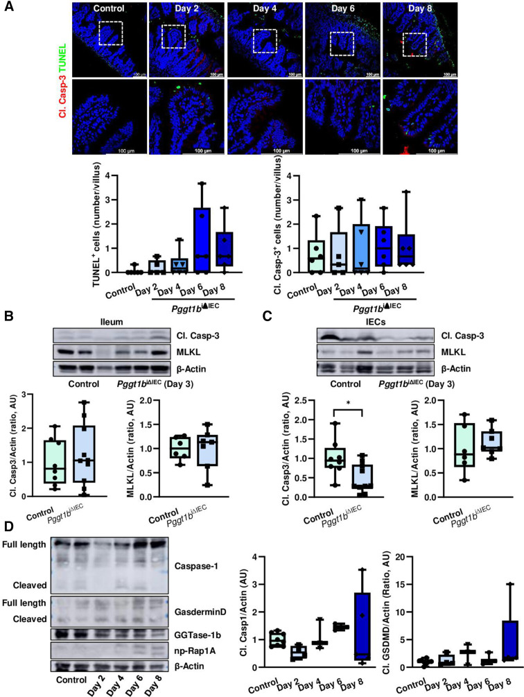
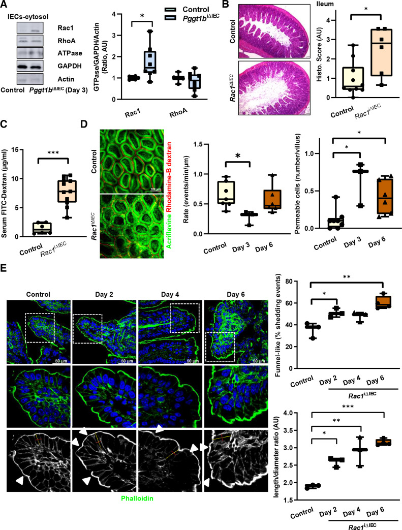
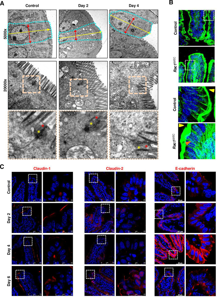
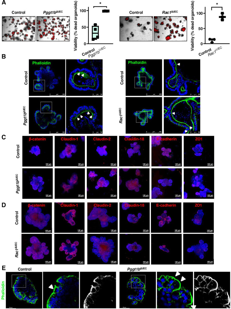
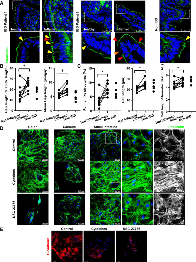
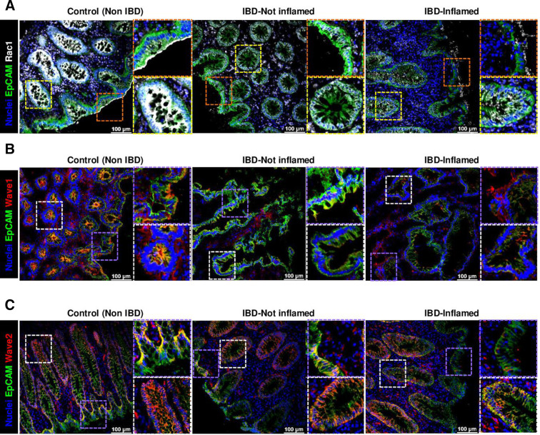
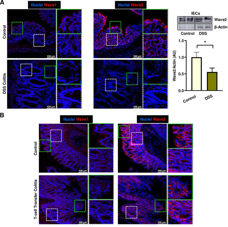
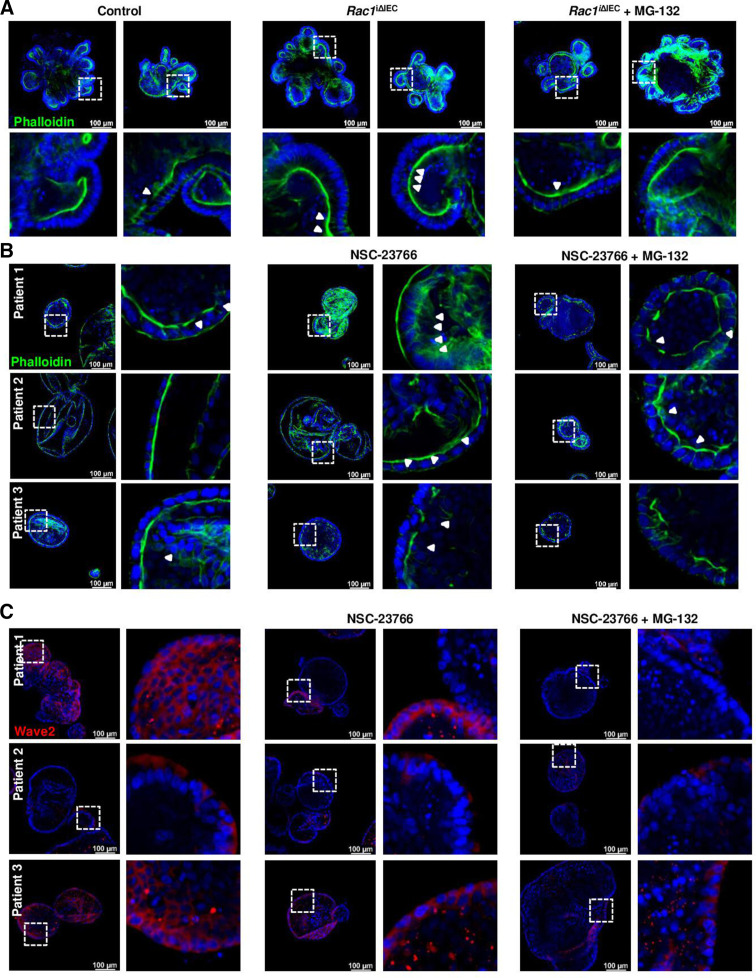
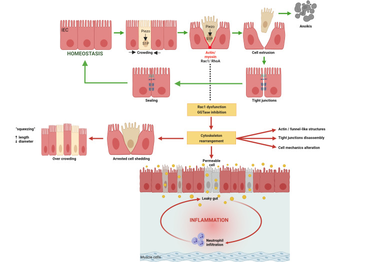
Results: Epithelial Pggt1b deletion led to cytoskeleton rearrangement and tight junction redistribution, causing cell overcrowding due to arresting of cell shedding that finally resulted in epithelial leakage and spontaneous mucosal inflammation in the small and to a lesser extent in the large intestine. Both in vivo and in vitro studies (knockout mice, organoids) identified RAC1 as a GGTase target critically involved in prenylation-dependent cytoskeleton dynamics, cell mechanics and epithelial cell shedding. Moreover, inflamed areas of gut tissue from patients with IBD exhibited funnel-like structures, signs of arrested cell shedding and impaired RAC1 function. RAC1 inhibition in human intestinal organoids caused actin alterations compatible with arresting of cell shedding.

Conclusion: Impaired epithelial RAC1 function causes cell overcrowding and epithelial leakage thus inducing chronic intestinal inflammation. Epithelial RAC1 emerges as key regulator of cytoskeletal dynamics, cell mechanics and intestinal cell shedding. Modulation of RAC1 might be exploited for restoration of epithelial integrity in the gut of patients with IBD.

Keywords: IBD; actin cytoskeleton; epithelial barrier; intestinal epithelium; tight junction.


Conflict of interest statement

Competing interests: None declared.

Figures

Figure 1
Figure 1
Intestinal disease due to inhibition of prenylation within IECs over time. (A) Assessment of tissue integrity in vivo/ ex vivo and intestinal permeability in vivo. Endoscopy pictures of colon and small intestine tissue (left); transmucosal passage of orally administered FITC-Dextran (4 KDa); serum concentration (µg/mL) (right). (B) Representative pictures of intravital microscopy analysis of barrier function of small intestine using luminal acriflavine (green) and rhodamineB-dextran (red). (Control, n=8; day 2, n=6; day 4, n=5; day 6, n=5; day 8, n=2). (C) Histology analysis of ileum tissue using H&E staining. Representative pictures (left), and corresponding score (right). (D) Gene expression of IL-6 and TNF-α in ileum tissue (RT-qPCR; six independent experiments). (E) MPO immunofluorescence staining in cross-sections from ileum. Representative pictures (left), and corresponding quantification (right). Data are expressed as box-plots (Min to Max); seven independent experiments, except where indicated. One-way ANOVA, Dunnett’s multiple comparisons test. *P≤0.050; ** P≤0.001. ANOVA, analysis of variance.
Figure 2
Figure 2
Cell shedding alterations and overcrowding in GGTase-deficient small intestine (time course study). (A, B) Intravital microscopy analysis of cell shedding using luminal acriflavine (green) and rhodamineB-dextran (red). (Control, n=8; day 2, n=6; day 4, n=5; day 6, n=5). Multiple unpaired t-test (A) Representative pictures of time sequences; white ellipses indicate cell shedding events in progress; turquoise ellipses indicate completed cell shedding events, cell is shown to be in the lumen, out of the epithelial layer. In some cases, the Z-position has been corrected based on changes in the focus plane during image acquisition due to tissue contraction or peristalsis. Quantification of cell shedding rate considering exclusively events which are completed during the duration of the image acquisition; (number of cell shedding events/time/length) (top), and in a single villus (events/minute) (bottom), at a determined focus plane near the lumen (five villi/video; two videos/mouse). (B) Representative pictures of single villus from intravital microscopy experiment (left); orange arrows indicate permeable cells. Quantification of permeable cells (dextran is detected inside the cell) (events/villus; 10 villi/picture; two pictures/mouse). (C) F-actin fibre staining using AlexaFluor488-phalloidin in ileum tissue (green). Seven independent experiments. Mixed effect analysis. Representative pictures (left); quantification of funnel-like structures, indicated by white arrows (top right); quantification of cell density (number of cells/µm of basement membrane length) (bottom right). Data are expressed as box-plots (Min to Max). *P≤0.050; ***P≤0.0001.
Figure 3
Figure 3
TEM analysis of ileum tissue from control and Pggt1b iΔIEC mice (days 2 and 4). Quantification of cell diameter and cell length/diameter ratio (left); representative pictures (right). Cell shape is indicated by dotted turquoise lines; red arrows indicate cell diameter (between two lateral membranes) and yellow arrows indicate cell length (between basal and apical membrane). Within the AJC, tight junction zone is indicated by red asterisks, while yellow asterisks indicate Adherens junction zone. One sample/group. Data are expressed as box-plots (Min to Max). *P≤0.050. AJC, apical junction complex; TEM, transmission electron microscopy.
Figure 4
Figure 4
Cytoskeleton rearrangement and altered cell mechanics in small intestine on deletion of Pggt1b. (A) F-actin fibre staining using AlexaFluor488-phalloidin in ileum tissue (green). High resolution confocal microscopy (Leica Stellaris). Representative pictures of a maximum projection (system optimised z-stack). Yellow arrows indicate apical actin network; orange arrows indicate redistribution of actin fibres. Three independent experiments. (B) Representative pictures of Myosin IIA staining (red) in ileum tissue (five independent experiments). Confocal microscopy (Leica SP8). (C) RT-FDC analysis from small intestine IECs (n=4/group). (D) Expression and redistribution of selected candidate AJC proteins in ileum tissue (claudin-1, claudin-2 and E-cadherin). Immunostaining (top, red signal) and western blot (bottom). Band densitometry quantification (right). Minimum five independent experiments. Data are expressed as box-plots (Min to Max). One-way ANOVA, Dunnett’s multiple comparisons test. *P≤0.050. AJC, apical junction complex; ANOVA, analysis of variance; RT-FDC, real time fluorescence deformability cytometry.
Figure 5
Figure 5
Cell death activation on inhibition of prenylation within small intestinal epithelium. (A) TUNEL (green) and cleaved caspase-3 (red) staining in ileum cross-sections from control and Pggt1b iΔIEC mice at different time points. Representative pictures (top) and quantification (bottom) of TUNEL+ (left) and Cl. Casp-3+/villus (right). Six independent experiments. (B, C) Western blot analysis of cleaved caspase-3 and MLKL. Representative blots (top) and band densitometry quantification (bottom). (B) Ileum tissue (Cl. Casp-3; n=8, control; n=10, Pggt1b iΔIEC) (MLKL; n=6, control; n=7, Pggt1b iΔIEC). (C) Small intestine isolated IECs (Cl. Casp-3; n=8, control; n=10, Pggt1b iΔIEC) (MLKL; n=6, control; n=7, Pggt1b iΔIEC). (D) Detection of Caspase-1 and Gasdermin-D cleavage in small intestine IECs (western blot analysis). Representative blots (left) and band densitometry quantification (right). Four independent experiments. Data are expressed as box-plots (Min to Max). One-way ANOVA, Dunnett’s multiple comparisons test (A, D). Unpaired t test (B, C). *P≤0.050.
Figure 6
Figure 6
RAC1 function within small intestine IECs is crucial for the maintenance of intestinal homeostasis. (A) Subcellular localisation of RAC1 and RHOA within small intestine IECs from control and Pggt1b iΔIEC mice at day 3 on tamoxifen treatment. Cytosolic proteins are separated via centrifugation gradient and small GTPases are detected via western blotting. Representative blots (left) and band densitometry quantification (right). Three experiments (n=6, control; n=8, Pggt1b iΔIEC). (B–E). Phenotype of Rac1 iΔIEC mice. (B–D) Intestinal pathology and cell shedding alterations in Rac1 iΔIEC mice. (B) Histology analysis of ileum tissue using H&E staining (day 7). Representative pictures (left) and histology score (right) (n=10, control; n=6, Rac1 iΔIEC). (C) Assessment of intestinal permeability in vivo. Transmucosal passage of orally administered FITC-Dextran; serum concentration (µg/mL) (n=6, Control; n=11, Rac1 iΔIEC). (D) Intravital microscopy analysis of cell shedding using luminal acriflavine (green) and rhodamineB-dextran (red) in small intestine. Representative pictures (left); and quantification of cell shedding rate (number of cell shedding events occurring over time in a single villus at a determined focus plane (events/minute/µm), and permeable cells (dextran is detected inside the cell) (events/villus). (n=8, Control; n=3, day 3; n=6, day 6). (E) Time course study. Three independent experiments. F-actin fibre staining using AlexaFluor488-phalloidin (green) in ileum tissue. Representative pictures (left); and quantification of funnel-like structures, indicated by white arrows (% of total cell shedding events) (top right); quantification of cell length/diameter ratio (bottom right). One-way ANOVA, Dunnett’s multiple comparisons test or unpaired t test. *P≤0.050; **P≤0.001; ***P≤0.0001. ANOVA, analysis of variance.
Figure 7
Figure 7
Cytoskeleton rearrangement in RAC1-deficient intestinal epithelium. (A) Electron microscopy analysis of ileum tissue. Representative pictures. Cell shape is indicated by dotted turquoise lines; red arrows indicate cell diameter (between two lateral membranes) and yellow arrows indicate cell length (between basal and apical membrane). Within the AJC, tight junction zone is indicated by red asterisks; while yellow asterisks indicate Adherens junction zone. One sample/group. (B) F-actin fibre staining using AlexaFluor488-phalloidin (green). High resolution confocal microscopy (Leica Stellaris). Maximum projection (system optimised z-stack). Yellow arrows indicate apical actin network; orange arrows indicate actin fibres. (C) Detection of selected candidate AJC proteins in ileum tissue (claudin-1, claudin-2, E-cadherin). Immunostaining (red signal). Three independent experiments. AJC, apical junction complex.
Figure 8
Figure 8
Epithelial intrinsic mechanisms in small intestine organoids. GGTase-deficient and RAC1-deficient organoids generated from small intestinal crypts. (A) Cell viability measured by PI incorporation (red). Representative pictures (left), and corresponding quantification (% of dead organoids) (right). (Pggt1b, four experiments; Rac1, 3 experiments). (B) F-actin fibre staining using AlexaFluor488-phalloidin (green) (Pggt1b, five experiments; Rac1, four experiments). White arrows indicate funnel-like structures or arrested cell shedding events. (C, D). Analysis of candidate AJC proteins by immunostaining. Maximum projection from z-stacks (system optimised). (C) GGTase-deficient organoids. Four experiments; except for β-catenin and Claudin-1, three experiments. (D) RAC1-deficient organoids. Three experiments; except for claudin-2, five experiments. (E) Apical-out GGTase-deficient organoids, representative pictures of Phalloidin staining. One experiment. Data are expressed as box-plots (Min to Max). Paired t-test. *P≤0.050. AJC, apical junction complex.
Figure 9
Figure 9
3D traction-force microscopy in control, GGTase and RAC1-deficient small intestine organoids. (A) Organoid-generated matrix deformations between 47 hours and 72 hours of time-lapse imaging (48–73 hours after completion of collagen polymerisation). Representative control organoid shows inward-directed matrix deformation, indicative of force increase, whereas representative Pggt1b iΔIEC and Rac1 iΔIEC organoids show outward-directed deformations, indicative of force relaxation. (B) Mean contractility over time for control, Pggt1b iΔIEC and Rac1 iΔIEC organoids, each pair from the same replicate experiment. Grey lines indicate the 47 hours and 72 hours time point for which force development is reported in the figures below. The measurement of the Pggt1b iΔIEC and corresponding control organoids was carried out over 90 hours to demonstrate that the force trends continue. (C) Relative and absolute changes in contractile force and absolute changes in contractile pressure between 47 hours and 72 hours. Each point represents the data from an individual organoid, colours represent four biological replicates. Black circles represent the mean value for each biological replicate, and black arrows represent the mean value of all organoids. P values are calculated from a two-sided Student’s t-test assuming unequal variances. Paired t-test. *P≤0.050.
Figure 10
Figure 10
Analysis of cell shedding and overcrowding in small intestine sections from human patients with IBD. (A–C) F-actin fibre staining using AlexaFluor488-phalloidin (green) (n=13, total; n=5, Control; n=8, CD). (A) Representative pictures (top); yellow arrows indicate epithelial gaps; orange arrow indicates microerosions. (B, C) Quantification. (B) Gap length (% of epithelial length), and mean gap length (µm/gap). (C) Funnel-like structures (red triangles) (% of total cells) and calculated length/diameter ratio (AU). (D, E) 2D/monolayer organoids generated from human intestinal crypts (three patients), and treated with NSC-23766 (100 µM) or a cytokine cocktail (IL-1β 10 ng/mL, IL-6 10 ng/mL and TNF-α; 20 ng/mL). (D) Phalloidin staining. (E) E-cadherin staining. Paired t-test. *P≤0.050. IBD, inflammatory bowel disease.
Figure 11
Figure 11
Analysis of RAC1 pathway in human IBD. (A) RAC1 immunostaining (white), counterstained with EpCAM (green) and Hoechst (blue). Representative pictures, showing expression and subcellular localisation at the epithelial surface (top) and crypts (bottom) (n=20, total; n=9, Control; n=11, IBD). Wave1 (B) and Wave2 (C) immunostaining (red) (Wave1, n=13, total; n=5, Control; n=8, IBD); (Wave2, n=17, total; n=6, Control; n=11, IBD).
Figure 12
Figure 12
Analysis of RAC1 pathway in experimental colitis. WAVE1 and WAVE2 expression in mouse experimental colitis models. (A) DSS-induced colitis. Representative pictures from immunostaining in colon samples (left), and WB from isolated IECs (right). Immunostaining (n=10, total; n=5, Control; n=5); WB (n=13, total; n=6, Control; n=7, DSS). (B) Adoptive lymphocyte TC. Representative pictures from immunostaining in colon samples (n=14, total; n=6, Control; n=8). Data are expressed as mean±SEM. Unpaired t-test. *P≤0.050. TC, transfer colitis.
Figure 13
Figure 13
Interfering with RAC1 pathway in organoids. (A) RAC1-deficient small intestine organoids treated with the proteasome inhibitor MG132 (1 µM). Phalloidin staining. Three experiments. (B, C) Human organoids treated with the RAC1 inhibitor NSC-23766 (100 µM) with or without the proteasome inhibitor MG132 (n=3). (B) Phalloidin staining. (C) Wave2 staining.
Figure 14
Figure 14
Mechanism behind intestinal inflammation induced by inhibition of GGTase1 or RAC1 within intestinal epithelial cells. Figure has been created with BioRender.com.

Comment in

References

    1. Turner JR. Intestinal mucosal barrier function in health and disease. Nat Rev Immunol 2009;9:799–809. 10.1038/nri2653 - DOI - PubMed
    1. Maloy KJ, Powrie F. Intestinal homeostasis and its breakdown in inflammatory bowel disease. Nature 2011;474:298–306. 10.1038/nature10208 - DOI - PubMed
    1. Kiesslich R, Goetz M, Angus EM, et al. . Identification of epithelial gaps in human small and large intestine by confocal endomicroscopy. Gastroenterology 2007;133:1769–78. 10.1053/j.gastro.2007.09.011 - DOI - PubMed
    1. Lim LG, Neumann J, Hansen T, et al. . Confocal endomicroscopy identifies loss of local barrier function in the duodenum of patients with Crohn's disease and ulcerative colitis. Inflamm Bowel Dis 2014;20:892–900. 10.1097/MIB.0000000000000027 - DOI - PubMed
    1. Vivinus-Nébot M, Frin-Mathy G, Bzioueche H, et al. . Functional bowel symptoms in quiescent inflammatory bowel diseases: role of epithelial barrier disruption and low-grade inflammation. Gut 2014;63:744–52. 10.1136/gutjnl-2012-304066 - DOI - PubMed

Publication types