. 2022 Mar 3;5(1):202.
doi: 10.1038/s42003-022-03121-5.

Cytoskeletal dynamics regulates stromal invasion behavior of distinct liver cancer subtypes

Affiliations

Cytoskeletal dynamics regulates stromal invasion behavior of distinct liver cancer subtypes

Ryan Y Nguyen et al. Commun Biol. .

Abstract

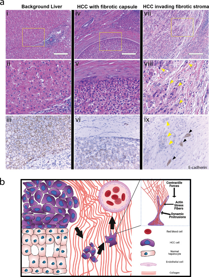
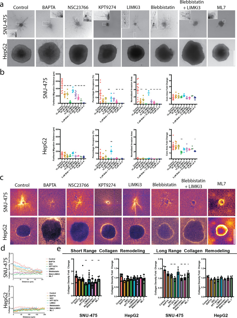
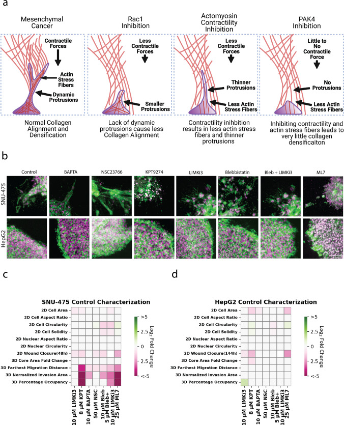
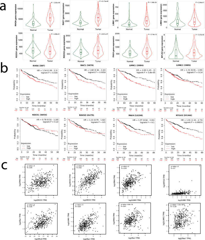
Drug treatment against liver cancer has limited efficacy due to heterogeneous response among liver cancer subtypes. In addition, the functional biophysical phenotypes which arise from this heterogeneity and contribute to aggressive invasive behavior remain poorly understood. This study interrogated how heterogeneity in liver cancer subtypes contributes to differences in invasive phenotypes and drug response. Utilizing histological analysis, quantitative 2D invasion metrics, reconstituted 3D hydrogels, and bioinformatics, our study linked cytoskeletal dynamics to differential invasion profiles and drug resistance in liver cancer subtypes. We investigated cytoskeletal regulation in 2D and 3D culture environments using two liver cancer cell lines, SNU-475 and HepG2, chosen for their distinct cytoskeletal features and invasion profiles. For SNU-475 cells, a model for aggressive liver cancer, many cytoskeletal inhibitors abrogated 2D migration but only some suppressed 3D migration. For HepG2 cells, cytoskeletal inhibition did not significantly affect 3D migration but did affect proliferative capabilities and spheroid core growth. This study highlights cytoskeleton driven phenotypic variation, their consequences and coexistence within the same tumor, as well as efficacy of targeting biophysical phenotypes that may be masked in traditional screens against tumor growth.


Conflict of interest statement

The authors declare the following competing interests: B.E.E. is a cofounder of Osmol Therapeutics, a company that is targeting NCS1 for therapeutic purposes. The remaining authors declare no competing interests.

Figures

Fig. 1
Fig. 1. Distinct morphological differences and invasive modes of HCC.
a Background liver shows well ordered liver lobule and portal tract with mild chronic inflammation, portal and periportal fibrosis (i–iii), HCC encapsulated by a fibrotic capsule (iv–vi), and HCC tumor cells invading into the fibrotic stroma (vii–ix). Samples are hematoxylin and eosin stained. Lower panels show IHC staining of E-cadherin in approximately corresponding regions. Scale bars: 200 μm. Yellow arrows indicate stromal invasion of HCC in collagen while black arrows indicate bile ductules. b Our working model illustrates invasion modes of HCC. While HCC tumor cells can proliferate against the fibrotic capsule, HCC tumor cells can also invade into the fibrotic stroma. HCC tumor cells can send out dynamic protrusions and exert contractile forces on the collagen to allow for alignment and densification which will facilitate further invasion. Made with Biorender.com.
Fig. 2
Fig. 2. Molecular and physical differences between SNU-475 and HepG2 cells.
a Representative immunofluorescent images of SNU-475 cells and HepG2 cells. Green is F-actin, magenta is DAPI, and blue is Vimentin. Scale bar is 25 μm. b Immunoblotting of SNU-475 and HepG2 cells for E-cadherin, p-cofilin, and p-MLC. Black vertical separation lines for p-cofilin and p-MLC blot indicate that measurements are separated lanes of the same blot. c Scratch assay analyses and d corresponding representative images of scratch assays for SNU-475 and HepG2 wounds over 48 h. Yellow dotted lines indicate wound area. Single dotted line indicates that the wound is closed. N = 11 replicates for each cell line. e SNU-475 (top panel) and HepG2 spheroids (bottom panel) embedded in collagen matrices and imaged over 5 days. Images depict brightfield and reflectance microscopy of each spheroid. Scale bar: 500 μm. f After 5 days of culture in collagen gel, the percentage occupancy of the disseminated cells at each of the distances from the spheroid periphery is calculated (i) and the percentage occupancy at 100 μm away from the spheroid periphery are compared between the two cell lines (ii). The total areas of the disseminated cells after normalization (iii) and the farthest cell migration distances (iv) are compared between the two cell lines. The collagen densification as a function of distance from the spheroid (v) and core area fold change are compared across cell lines (vi). Plots show mean ± SEM. N = 14 spheroids for each cell line.Unpaired t-test with Welch’s correction is performed. *P ≤ 0.05, **P ≤ 0.01.
Fig. 3
Fig. 3. 2D Molecular and migratory phenotype profiling of SNU-475 and HepG2 cells.
a Pathway diagram illustrating the effect of each drug used on cytoskeletal machinery. b Scratch assay bar plots for SNU-475 and HepG2 for all drugs tested. Scratch assay bar plots at 48 h and 144 h for all drugs tested for SNU-475 and HepG2 cells, respectively. N ≥ 2 replicates. Plots show mean ± SEM with conditions where N > 2. c Representative immunofluorescence images of SNU-475 spheroids (first row) and HepG2 spheroids (second row) treated with BAPTA (Ca2+ chelator), NSC23766 (Rac1 inhibitor), KPT9274 (PAK4 inhibitor), LIMKi3 (LIMK inhibitor), blebbistatin (myosin inhibitor inhibitor), both blebbistatin and LIMKi3, aor ML-7 (MLCK inhibitor), respectively. Green represents F-actin and magenta is DAPI. Green represents F-actin and magenta is DAPI. Scale bar: 30 μm. d SNU-475 (i) and HepG2 (ii) cellular area and solidity. n > 30 cells for each condition from N = 2 independent experiments. e p-MLC western blots for drug conditions for SNU-475 and HepG2 cells. Plots show mean ± SD. One-way ANOVA with Tukey post-hoc testing was performed. Significant difference (p < 0.05) was detected between any two of the conditions. *P < 0.05, **P < 0.01.
Fig. 4
Fig. 4. Characterization of F-actin and lamellipodial dynamics under drug treatments for SNU-475 and HepG2.
Coherency measurement on F-actin as an indicator for cytoskeleton organization and cell morphology under drug treatments for a SNU-475 and b HepG2. Stress fiber-rich HCC cell line SNU-475 contains more organized actin and well-defined morphology and is more sensitive to the cytoskeleton drugs, compared to HepG2. Significance is compared between control and each drug condition. (From left to right) Coherency of actin stress fiber (SF) intensity and proportion of SF of total F-actin. n > 12 cells for each condition from N = 2 independent experiments. a Rac1 inhibitor (NSC23766) does not affect stress fiber formation but reduces lamellipodia. BAPTA seems to induce stress fiber building up and induce thin protrusions on the cell periphery. b The selected inhibitors do not significantly reduce total F-actin intensity for HepG2 cells. c Representative images denoting traced lamellipodia for SNU-475 cells. Yellow outline indicates lamellipodia tracing. d Metrics denoting SNU-475 number of cells with lamellipodia, lamellipodia area percentage of cell area, and lamellipodia area after no treatment, 8 μm KPT treatment, and 50 μm NSC treatment. e Representative images denoting traced lamellipodia for HepG2 cells. Yellow outline indicates lamellipodia tracing. f Metrics denoting HepG2 number of cells with lamellipodia, lamellipodia area percentage of cell area, and lamellipodia area after no treatment, 8 μm KPT treatment, and 50 μm NSC treatment. Plots show mean ± SEM. n > 12 cells for each condition from N = 2 independent experiments. One-way ANOVA with Tukey post-hoc testing was performed. Significant difference (p < 0.05) was detected between any two of the conditions. *P < 0.05, **P < 0.01, #P < 0.0001.
Fig. 5
Fig. 5. Characterization of SNU-475 and HepG2 3D spheroid invasion under drug treatments.
a Representative images of SNU-475 (top row) or HepG2 (bottom row) spheroids 5 days after seeding in collagen gels. Arrows indicate circular morphology of invading SNU-475 cells. Scale bar, 500 μm. b We use fractional occupancy, normalized invasion area, and farthest migration distance to quantify the spheroid invasiveness, and calculate the core area fold change on day 5 normalized by that of day 0 to quantify the spheroid size growth for SNU-475 and HepG2 spheroids. n > 4 spheroids for each condition from N = 2 independent experiments. c Representative images of reflectance microscopy to indicate collagen densification and alignment for NSC23766, KPT9274, Blebbistatin, and Blebbistatin + LIMKi3 treatment for both cell lines. Scale bar, 500 μm. d Average normalized collagen density profiles for SNU-475 SNU-475 (top row) or HepG2 (bottom row) spheroids 5 days after seeding in collagen gels. Measurements are normalized to the last 30 data points of each plot profile. Plots show mean collagen density. e Quantification of short-range and long-range collagen remodeling. The short-range collagen remodeling metric is determined as the collagen density fold change immediately outside the periphery of the spheroid (30 μm). Long-range collagen density is determined as the collagen density fold change 80 μm away from the spheroid boundary. Plots show mean ± SD. n > 4 spheroids for each condition from N = 2 independent experiments. One-way ANOVA with Tukey post-hoc testing was performed. Significant difference (p < 0.05) was detected between any two of the conditions. *P < 0.05, **P < 0.01.
Fig. 6
Fig. 6. HCC invasion schematic and heatmap summary.
a Schematic illustrating how altering cytoskeletal dynamics will affect collagen densification and subsequent migration. b Representative immunofluorescence images of SNU-475 spheroids (first row) and HepG2 spheroids (second row) treated with BAPTA (Ca2+ chelator), NSC23766 (Rac1 inhibitor), KPT9274 (PAK4 inhibitor), LIMKi3 (LIMK inhibitor), blebbistatin (myosin inhibitor inhibitor), both blebbistatin and LIMKi3, or ML-7 (MLCK inhibitor), respectively. Green represents F-actin and magenta is DAPI. Scale bar: 50 μm. Heatmap illustrating 2D and 3D metrics characterized in this study for c SNU-475 and d HepG2 cells. Heatmap values are colored if the difference between two conditions is considered significantly different (p < 0.05). White indicates no significant difference between conditions. Color intensity is determined by log2 fold change of the group on the x-axis over control. Green signifies upregulation while pink signifies downregulation. One-way ANOVA with Tukey post-hoc testing was performed to test for significance.
Fig. 7
Fig. 7. Rho family of small GTPases, PAK4, LIM kinase, and myosin light chain kinase 2 (MYLK2) are correlated with more invasive liver cancer.
a Gene expression levels of RhoA, Rac1, LIMK1, LIMK2, ROCK1, ROCK2, PAK4, and MYLK2 are upregulated in HCC compared to normal liver tissue. b Kaplan–Meier analysis (n = 364 patients) reveals that RhoA, Rac1, LIMK1, LIMK2, ROCK1, ROCK2, PAK4, and MYLK2 are linked to poor prognosis of HCC. c Correlation analysis shows expressions of Rac1, RhoA, and LIMK1 are positively correlated, suggesting the signaling axes Rac1-LIMK and RhoA-LIMK may be active in HCC. For each correlation plot, p < 10−16.

References

    1. Losic B, et al. Intratumoral heterogeneity and clonal evolution in liver cancer. Nat. Commun. 2020;11:1–15. - PMC - PubMed
    1. Vyas M, Zhang X. Hepatocellular carcinoma: role of pathology in the era of precision medicine. Clin. Liver Dis. 2020;24:591–610. - PubMed
    1. Dagogo-Jack I, Shaw AT. Tumour heterogeneity and resistance to cancer therapies. Nat. Rev. Clin. Oncol. 2018;15:81–94. - PubMed
    1. Xin Wei Wang SST. The biological and clinical challenge of liver cancer heterogeneity. Hepatic Oncol. 2014;1:349. - PMC - PubMed
    1. Northcott JM, Dean IS, Mouw JK, Weaver VM. Feeling stress: the mechanics of cancer progression and aggression. Front. Cell Dev. Biol. 2018;6:17. - PMC - PubMed

Publication types