. 2022 Aug;40(8):1270-1275.
doi: 10.1038/s41587-022-01232-2. Epub 2022 Mar 3.

Efficient discovery of SARS-CoV-2-neutralizing antibodies via B cell receptor sequencing and ligand blocking

Affiliations

Efficient discovery of SARS-CoV-2-neutralizing antibodies via B cell receptor sequencing and ligand blocking

Andrea R Shiakolas et al. Nat Biotechnol. 2022 Aug.

Abstract

Although several monoclonal antibodies (mAbs) targeting severe acute respiratory syndrome coronavirus 2 (SARS-CoV-2) have been approved for coronavirus disease 2019 (COVID-19) therapy, development was generally inefficient, with lead generation often requiring the production and testing of numerous antibody candidates. Here, we report that the integration of target-ligand blocking with a previously described B cell receptor-sequencing approach (linking B cell receptor to antigen specificity through sequencing (LIBRA-seq)) enables the rapid and efficient identification of multiple neutralizing mAbs that prevent the binding of SARS-CoV-2 spike (S) protein to angiotensin-converting enzyme 2 (ACE2). The combination of target-ligand blocking and high-throughput antibody sequencing promises to increase the throughput of programs aimed at discovering new neutralizing antibodies.


Conflict of interest statement

A.R.S. and I.S.G. are cofounders of AbSeek Bio. I.S.G., A.R.S. and K.J.K. are listed as inventors on antibodies described herein. I.S.G., A.R.S. and I.S. are listed as inventors on patent applications for the LIBRA-seq technology. J.E.C. has served as a consultant for Luna Biologics, is a member of the Scientific Advisory Board Meissa Vaccines and is Founder of IDBiologics. The Crowe laboratory has received funding support in sponsored research agreements from AstraZeneca, IDBiologics and Takeda. The Georgiev laboratory at VUMC has received unrelated funding from Takeda Pharmaceuticals. The remaining authors declare no competing interests.

Figures

Fig. 1
Fig. 1. Antibody discovery using LIBRA-seq with ligand blocking.
a, Experimental setup of three LIBRA-seq experiments: experiment 1, LIBRA-seq with ligand blocking; experiment 2, LIBRA-seq with a SARS-CoV-2 S (SARS2 S) titration; experiment 3, LIBRA-seq with a SARS-CoV-2 S titration and ligand blocking. For experiments 2 and 3, six different aliquots of S protein were added in a titration series (1–6); HIV ZM197 envelope and influenza hemagglutinin H1 NC99 were used as controls. b–d, Left, after next-generation sequencing, hundreds of B cells (dots) were recovered that had paired heavy/light chain sequencing information and antigen reactivity information for the three experiments. For experiments 1 (b), 2 (c) and 3 (d), select LIBRA-seq scores for all cells per experiment are shown as open circles (n = 828, 829 and 957, respectively). Antibodies selected for expression and validation are highlighted and numbered in light blue. Right, LIBRA-seq scores for the selected antibodies for all antigens from each experiment are shown as a heat map from –2 (tan) to 2 (purple); scores outside of this range are shown as the minimum and maximum values. For experiments 1 and 3, antibodies with negative scores for ACE2 are shown above the dotted line, while antibodies with positive scores for ACE2 are shown below the dotted line and are controls. For experiment 2, all SARS-CoV-2-reactive antibodies are shown above the dotted line, whereas influenza-specific antibody 53181-3 is shown as a control below the dotted line.
Fig. 2
Fig. 2. Validation and characterization of selected antibodies.
a, ELISA area under the curve (AUC) values for binding to SARS-CoV-2 recombinant antigen proteins and a negative control influenza hemagglutinin protein are shown for antibodies (rows) in each experiment and were calculated from data in Extended Data Fig. 2b. b, Kd (M) values of antibodies for SARS-CoV-2 RBD or NTD (based on epitope shown in a) were determined by biolayer interferometry; ND, not done. c, Percent reduction in ACE2 binding, as measured by ELISA, is shown as a heat map from 0 to 100% (white to blue) reduction in binding compared to SARS-CoV-2 binding only. d, Vesicular stomatitis virus (VSV)–SARS-CoV-2 neutralization half-maximum inhibitory concentration (IC50) values are shown as a heat map from high potency (red) to low potency (green). Non-neutralizing antibodies are shown as white.
Fig. 3
Fig. 3. Assessment of LIBRA-seq with ligand blocking.
a, Predicted neutralizing antibodies were defined as the subset of selected antibodies with negative ACE2 LIBRA-seq scores from experiments 1 (n = 7 antibodies) and 3 (n = 6 antibodies) and all antibodies with high LIBRA-seq scores (>1) for SARS-CoV-2 S from experiment 2 (n = 7 antibodies). The percentage of neutralizing antibodies from the set of predicted neutralizers is shown for each experiment. b, The IC50 values (µg ml–1) for SARS-CoV-2 neutralization by real-time cell analysis (RTCA) with VSV–SARS-CoV-2 (IC50 values for each antibody are shown as single dots) are plotted for the set of predicted neutralizers. The horizontal lines represent the geometric mean for each experiment. Non-neutralizing antibodies are shown as >10 µg ml–1. c, Spearman correlation of ACE2 LIBRA-seq score (x axis) and percent reduction in ACE2 binding to SARS-CoV-2 (y axis) for antibodies from experiments 1 and 3; Spearman r = –0.54, P = 0.017 (two tailed, 95% confidence interval).
Fig. 4
Fig. 4. Antibody neutralization of SARS-CoV-2 variants.
Authentic SARS-CoV-2 neutralization for a panel of antibodies is shown against USA-WA1 and variants (Alpha, Beta, Gamma and Delta). Data represent the percent neutralization as mean ± s.d. The IC50 values calculated in GraphPad Prism software by four-parameter best-fit analysis are shown on the right.
Fig. 5
Fig. 5. Structural characterization of antibodies 5317-4 and 5317-10.
a, A 9-Å-resolution cryo-EM structure of the Fab–S complex for 5317-4 Fab (orange) and 5317-10 Fab (pink). S protomers are shown in green, blue and red. b, Fab–S complex structure modeled with ACE2 (purple); ECD, extracellular domain.
Fig. 6
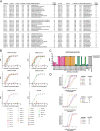
Fig. 6. Discovery of cross-reactive ACE2-blocking coronavirus antibodies using LIBRA-seq with ligand blocking.
a, Schematic of LIBRA-seq with ligand blocking applied to cross-reactive antibody discovery. b, For identification of cross-reactive coronavirus antibodies with ligand-blocking capabilities, all IgGs recovered from the LIBRA-seq experiment (n = 2,569) are shown, with LIBRA-seq scores for SARS-CoV (x axis) and SARS-CoV-2 (y axis). Each dot represents a cell, and the color of the dots shows the ACE2 LIBRA-seq score, with a color heat map shown on the right. c, Cells selected for expression and validation are shown in blue (ACE2 score < –1) or gray (ACE2 score ≥ –1). Of these selected cells, eight had high LIBRA-seq scores (>1) for SARS-CoV-2 and SARS-CoV and low scores (<–1) for ACE2. Additional candidates with a variety of scores for SARS-CoV-2, SARS-CoV and ACE2 were also selected for expression and validation as controls. d, The eight IgGs with high LIBRA-seq scores for SARS-CoV-2 and SARS-CoV and low scores for ACE2 are shown above the dotted line. Control antibodies with other LIBRA-seq score patterns are shown below the dotted line. For each antibody, complementarity-determining region (CDR) sequences and lengths are shown at the amino acid level, and V-gene and J-gene identities are shown at the nucleotide level. LIBRA-seq scores for antigens included in the screening library (SARS-CoV-2 S, SARS-CoV S, ACE2, HIV ZM197 envelope and influenza hemagglutinin H1 NC99) are shown as a heat map; the heat map is colorized from negative (tan) to zero (white) to positive (purple). Scores outside of this range are shown as the minimum and maximum values. e,f, ELISA AUC values from binding to coronavirus S proteins, influenza hemagglutinin H1 NC99 (negative control) (e) and recombinant antigen domains (f) are shown as heat maps from minimum (white) to maximum (purple) binding. g, Percent reduction in ACE2 binding by ELISA is shown for SARS-CoV-2 and SARS-CoV S proteins and displayed as a heat map from 0% (white) to 100% (blue). h, For the eight IgGs with high LIBRA-seq scores for SARS-CoV-2 and SARS-CoV and low scores for ACE2, the percent reduction in ACE2 binding due to antibody blocking by ELISA is shown for SARS-CoV (x axis) and SARS-CoV-2 (y axis).
Extended Data Fig. 1
Extended Data Fig. 1. Schematic representation of LIBRA-seq experiments.
a. An antigen screening library of oligonucleotide-labeled antigens was generated. This library consisted of SARS-CoV-2 spike antigens and negative controls. Additionally, oligo-labeled ACE2 (the SARS-CoV-2 spike host cell receptor) was included. The antigen screening library was mixed with donor PBMCs. This approach allowed for assessment of B cell ligand blocking functionality from the sequencing experiment. b. An antigen screening library containing an antigen titration was generated, with a goal of identifying high affinity antibodies from LIBRA-seq. In this experiment, six different amounts of oligo-labeled SARS-CoV-2 S protein, each labeled with a different barcode, were included in a screening library. C. Schematic of LIBRA-seq with S titrations and ACE2 included for ligand blocking.
Extended Data Fig. 2
Extended Data Fig. 2. Characterization of LIBRA-seq-identified antibodies.
a. Genetic characteristics for monoclonal antibodies prioritized for expression and validation. VH, JH, VL, JL inferred gene segment identity is shown at the nucleotide level. CDRH3 and CDRL3 amino acid sequence and length are also shown. b. ELISA binding of antibodies to SARS-CoV-2 spike, SARS-CoV-2 S1, SARS-CoV-2 RBD, SARS-CoV-2 NTD, SARS-CoV-2 S2 and influenza hemagglutinin H1 NC99. Data are represented as mean ± SEM of technical duplicates and represent one of at least two independent experiments (n = 2). c. ACE2 blocking ELISA. Antibodies were added to spike, and recombinant ACE2 was added and detected. Antibodies that block ACE2 binding show a reduction in absorbance compared to ACE2 binding without competitor (dotted line). ELISAs were performed at one antibody concentration, and data are represented as mean ± SEM of technical triplicates and represent one of at least two independent experiments (n = 2). d. Antibodies were tested in a VSV SARS-CoV-2 real time cell analysis (RTCA) neutralization assay. Neutralization curves and IC50 values are shown. Data are represented as mean ± S.D. of technical triplicates, and represent one of two independent experiments (n = 2).
Extended Data Fig. 3
Extended Data Fig. 3. Characterization of selected cross-reactive antibodies.
a. For the IgGs that showed high LIBRA-seq scores (>1) for both SARS-CoV-2 and SARS-CoV, the percent of cells with low ACE2 scores (<-1) is shown. b. ELISA binding of antibodies to SARS-CoV-2 spike, SARS-CoV spike, influenza hemagglutinin H1 NC99, SARS-CoV-2 S1, SARS-CoV-2 RBD, and SARS-CoV-2 S2. Data are represented as mean ± SEM of technical duplicates and represent one of at least two independent experiments (n = 2). c. ACE2 blocking ELISA. ACE2 binding without competitor is shown as a dotted line. ELISAs were performed at one antibody concentration, and data are represented as mean ± SEM of technical triplicates and represent one of at least two independent experiments (n = 2).

Update of

References

    1. Jiang S, Hillyer C, Du L. Neutralizing antibodies against SARS-CoV-2 and other human coronaviruses. Trends Immunol. 2020;41:355–359. doi: 10.1016/j.it.2020.03.007. - DOI - PMC - PubMed
    1. Zost SJ, et al. Potently neutralizing and protective human antibodies against SARS-CoV-2. Nature. 2020;584:443–449. doi: 10.1038/s41586-020-2548-6. - DOI - PMC - PubMed
    1. Chi X, et al. A neutralizing human antibody binds to the N-terminal domain of the spike protein of SARS-CoV-2. Science. 2020;369:650–655. doi: 10.1126/science.abc6952. - DOI - PMC - PubMed
    1. Brouwer PJM, et al. Potent neutralizing antibodies from COVID-19 patients define multiple targets of vulnerability. Science. 2020;369:643–650. doi: 10.1126/science.abc5902. - DOI - PMC - PubMed
    1. Rogers TF, et al. Isolation of potent SARS-CoV-2 neutralizing antibodies and protection from disease in a small animal model. Science. 2020;369:956–963. doi: 10.1126/science.abc7520. - DOI - PMC - PubMed

Publication types