Case Reports
. 2022 Feb 17;17(4):1288-1292.
doi: 10.1016/j.radcr.2022.01.060. eCollection 2022 Apr.

Computed tomography findings of Crouzon syndrome: A case report

Affiliations
Case Reports

Computed tomography findings of Crouzon syndrome: A case report

Juan Guillermo Arámbula Neira et al. Radiol Case Rep. .

Abstract

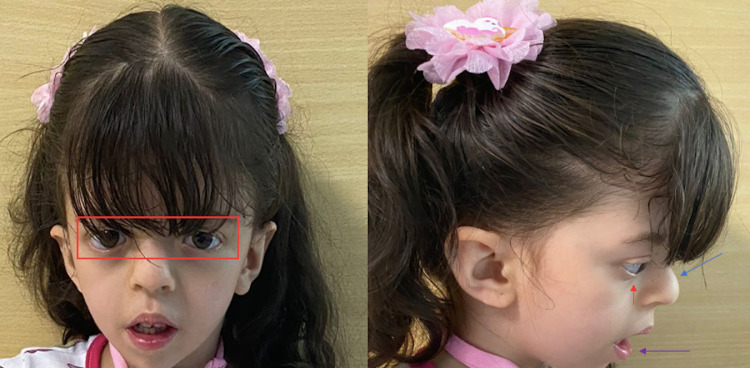
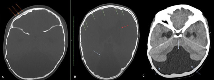
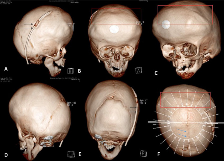
Crouzon syndrome is a genetic condition characterized by a premature fusion of skull sutures resulting in head and facial deformities. Crouzon syndrome is usually suspected at birth through physical examination or in the antenatal period via ultrasonographic assessment. Once Crouzon syndrome is suspected, advanced imaging methods such as three-dimensional computed tomography must be requested, showing early signs of cranial sutures fusion. In this paper, we present a case of a six-year-old girl who was taken to a pediatrician control appointment due to abnormal facies. During the physical examination, a suspicion of Crouzon syndrome was raised. Therefore, a head computed tomography was requested, showing asymmetrical calvarium thickening, diffuse indentation of the inner table of the skull, and moderate hydrocephalus with a big cyst in the posterior fossa. Due to these findings, the patient was remitted to maxillofacial surgery for further evaluation; however, the medical appointment could not be achieved as a consequence of the poor medical insurance of the girl. This paper aims to describe and discuss the computed tomography findings of Crouzon syndrome.

Keywords: Brain diseases; Child; Craniofacial dysostosis; Craniosynostoses; Crouzon syndrome; Exophthalmos; Tomography X-ray computed.


Figures

Fig 1
Fig. 1
Physical examination showing hypertelorism (red box), proptosis (red arrow), beaked nose (blue arrow), micrognathia, and retrognathia (purple arrow).
Fig 2
Fig. 2
Non-contrast axial head CT in bone window shows asymmetrical calvarium thickening in the right frontal bone (orange arrows) (A). Moderate hydrocephalus is seen (red arrow) (B). A big cyst located in the posterior fossa (blue arrows), and diffuse indentation of the inner table of the skull are evident (green arrows) (B, C).
Fig 3
Fig. 3
Three-dimensional CT skull reconstruction. An increased diameter of the head is evident (A–F). A correctly placed ventriculoperitoneal valve is shown (black arrow) (A, E). The total fusion of both coronal sutures is appreciated (red box) (B, C, F). Partial fusion of the posterior region of the sagittal suture is seen (blue arrow) (F). Lambdoid sutures are normal.

Similar articles

Cited by

References

    1. Mathijssen IMJ. Guideline for care of patients with the diagnoses of craniosynostosis: working group on craniosynostosis. J Craniofac Surg. 2015;26(6):1735–1807. doi: 10.1097/SCS.0000000000002016. - DOI - PMC - PubMed
    1. Al-Namnam NM, Hariri F, Thong MK, Rahman ZA. Crouzon syndrome: genetic and intervention review. J oral Biol craniofacial Res. 2019;9(1):37–39. doi: 10.1016/j.jobcr.2018.08.007. - DOI - PMC - PubMed
    1. Balyen L, Deniz Balyen LS, Pasa S. Clinical characteristics of Crouzon syndrome. Oman J Ophthalmol. 2017;10(2):120–122. doi: 10.4103/0974-620X.209111. - DOI - PMC - PubMed
    1. Rannan-Eliya S V, Taylor IB, De Heer IM, Van Den Ouweland AMW, Wall SA, et al. Paternal origin of FGFR3 mutations in Muenke-type craniosynostosis. Hum Genet. 2004;115(3):200–207. doi: 10.1007/s00439-004-1151-5. - DOI - PubMed
    1. Kaur H, Singh Waraich H, Sharma CM. Crouzon syndrome: a case report and review of literature. Indian J Otolaryngol Head Neck Surg. 2006;58(4):381–382. doi: 10.1007/BF03049602. - DOI - PMC - PubMed

Publication types

LinkOut - more resources