Review
. 2022 Feb 10:33:e00710.
doi: 10.1016/j.btre.2022.e00710. eCollection 2022 Mar.

Heterologous production of chondroitin

Affiliations
Review

Heterologous production of chondroitin

Márcia R Couto et al. Biotechnol Rep (Amst). .

Abstract

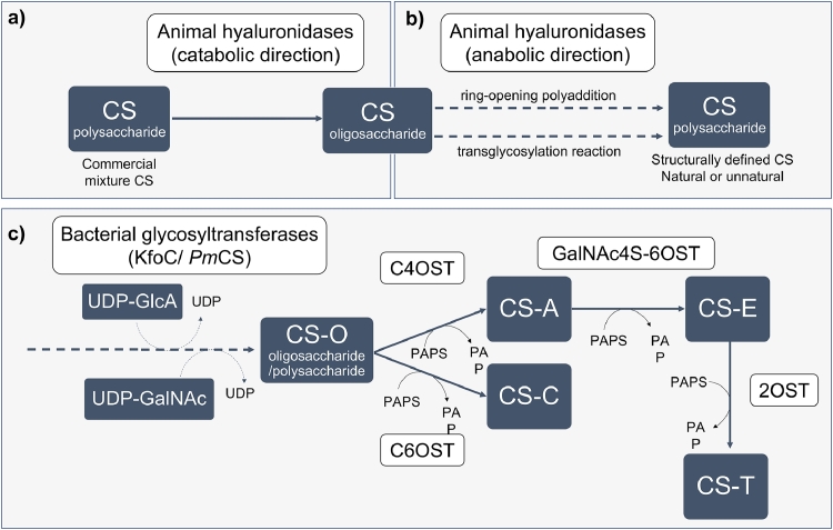
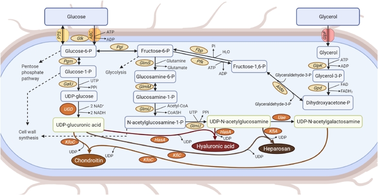
Chondroitin sulfate (CS) is a glycosaminoglycan with a broad range of applications being a popular dietary supplement for osteoarthritis. Usually, CS is extracted from animal sources. However, the known risks of animal products use have been driving the search for alternative methods and sources to obtain this compound. Several pathogenic bacteria naturally produce chondroitin-like polysaccharides through well-known pathways and, therefore, have been the basis for numerous studies that aim to produce chondroitin using non-pathogenic hosts. However, the yields obtained are not enough to meet the high demand for this glycosaminoglycan. Metabolic engineering strategies have been used to construct improved heterologous hosts. The identification of metabolic bottlenecks and regulation points, and the screening for efficient enzymes are key points for constructing microbial cell factories with improved chondroitin yields to achieve industrial CS production. The recent advances on enzymatic and microbial strategies to produce non-animal chondroitin are herein reviewed. Challenges and prospects for future research are also discussed.

Keywords: Biosynthetic pathway; Chondroitin; Glycosaminoglycans; Heterologous production; Metabolic engineering; Microbial fermentation.


Conflict of interest statement

The authors declare that they have no known competing financial interests or personal relationships that could have appeared to influence the work reported in this paper.

Figures

Fig. 1
Fig. 1
Structures of the main glycosaminoglycans (GAGs) a) hyaluronic acid, b) keratan sulfate, c) chondroitin and chondroitin sulfate, d) dermatan sulfate, and e) heparosan, heparan sulfate and heparin. Monomers of the disaccharide building blocks are abbreviated as GlcA - d-glucuronic acid, GlcNAc – N-acetyl-d-glucosamine, Gal – d-galactose, GalNAc – N-acetyl-d-galactosamine, IdoA – l-iduronic acid, GlcN, d-glucosamine. Hyaluronic acid (a) does not go under post-polymerization modifications. Keratan sulfate (b) has di-sulfated, mono-sulfated and non-sulfated disaccharide units (each R6 = H or SO3H) due to O-sulfotransferases action. Chondroitin (c) is the simple non-sulfated backbone (R2, R3, R4 and R6 = H) which can be modified by different tissue-specific O-sulfotransferases to form chondroitin sulfate (each R2, R3, R4 and R6 = H or SO3H). Dermatan sulfate (d) is formed from chondroitin through epimerization of GlcA into IdoA by tissue-specific epimerases followed by O-sulfotransferases (each R2, R4 and R6 = H or SO3H). Heparosan (e) has non-modified sugar moieties, that can be further modified through actions of tissue-specific N-sulfotransferases, C5 epimerases and O-sulfotransferases to generate the sulfated forms heparan sulfate and heparin (R2 from uronic acid = H or SO3H; when the hexosamine unit is GlcN, R2 in that unit = SO3H, while R2 = Ac when the unit is GlcNAc; other groups R3, R6 = H or SO3H). Heparin has more sulfate groups and IdoA content than heparan sulfate. Depending on the GAG type and source the molecular size can generally vary between 4 and 200 mer (n = 4 – 200). Exceptionally, the highest size can be found for hyaluronic acid that can achieve 20,000 repeating units.
Fig. 2
Fig. 2
Enzymatic synthesis of chondroitin sulfate (CS). a) Depolymerizing enzymes, such as animal hyaluronidases, can be used to obtain CS oligosaccharides from CS polysaccharides; b) the same type of enzymes is able to, under different conditions, polymerize the CS oligosaccharides through chemoenzymatic approaches. c) Bacterial glycosyltransferases (such as chondroitin synthase from Escherichia coli K4, KfoC, or from Pasteurella multocida type F, PmCS) act by transferring alternate residues of glucuronic acid (GlcA) and acetylgalactosamine (GalNAc), using uridine diphosphate (UDP)-GlcA and UDP-GalNAc as donors, to the nonreducing end of a chondroitin chain acceptor to elongate the chondroitin oligosaccharide/ polysaccharide backbones. Sulfotransferases such as 4-O-sulfotransferase (C4OST), 6-O-sulfotransferase (C6OST), N-acetylgalactosamine 4-sulfate 6-O-sulfotransferase (GalNAc4S-6OST), and 2-O-sulfotransferase (2OST) that require the presence of 3′-phosphoadenosine-5′-phosphosulfate (PAPS) as sulfate donor, convert the unsulfated backbone (CS-O) in CS with different sulfation patterns such as CS-A, CS-C, CS-E and CS-T. Only CSs with a homogenous defined sulfation pattern are shown although a CS chain may have different CS units if a combination of sulfotransferases is used. Dashed arrows represent polymerization steps.
Fig. 3
Fig. 3
Production of glycosaminoglycans in microbes and its possible use in the biosynthesis of microbial chondroitin, hyaluronic acid or heparosan. Depending on the microbial host, the heterologous expression of the enzymes shown in orange boxes might be required for glycosaminoglycans production. Enzyme abbreviations: ABC, adenosine triphosphate (ATP)-binding cassette transporters; Aldo, fructose-6-phosphate aldolase; Fbp, fructose-1, 6-bisphosphatase; GalU, uridine triphosphate:glucose-1-phosphate uridylyltransferase; Glk, glucokinase; GlmM, phosphoglucosamine mutase; GlmS, glucosamine-6-phosphate synthase; GlmU, glucosamine-1-phosphate N-acetyltransferase/N-acetylglucosamine-1-phosphate uridyltransferase; GlpF, Glycerol uptake facilitator protein; GlpK, glycerol kinase; Gpd, glyceraldehyde-3-phosphate dehydrogenase; HasA, hyaluronan synthase; KfiA, β−1, 3-glucuronyltransferase; KfiC, α−1, 4-N-acetylglucosaminyltransferase; KfoC, chondroitin synthase; Pfk, 6-phosphofructokinase; Pgi, glucose-6-phosphate isomerase; Pgm, phosphoglucomutase; PTS, phosphotransferase system; Uae, UDP-N-acetylglucosamine 4-epimerase; UGD, uridine diphosphate (UDP)-glucose 6-dehydrogenase.

References

    1. Kang Z., Zhou Z., Wang Y., Huang H., Du G., Chen J. Bio-based strategies for producing glycosaminoglycans and their oligosaccharides. Trends Biotechnol. 2018;36:806–818. doi: 10.1016/j.tibtech.2018.03.010. - DOI - PubMed
    1. Mende M., Bednarek C., Wawryszyn M., Sauter P., Biskup M.B., Schepers U., Bräse S. Chemical synthesis of glycosaminoglycans. Chem. Rev. 2016;116:8193–8255. doi: 10.1021/acs.chemrev.6b00010. - DOI - PubMed
    1. Casale J., Crane J.S. StatPearls Publishing; Treasure Island (FL): 2021. Biochemistry, Glycosaminoglycans.http://www.ncbi.nlm.nih.gov/pubmed/31335015 (accessed July 4, 2021) - PubMed
    1. A. Varki, R.D. Cummings, J.D. Esko, P. Stanley, G.W. Hart, M. Aebi, A.G. Darvill, T. Kinoshita, N.H. Packer, J.H. Prestegard, R.L. Schnaar, P.H. Seeberger, Essentials of Glycobiology, Cold Spring Harb. (2017) 823. https://www.ncbi.nlm.nih.gov/books/NBK310274/ (accessed July 24, 2021). - PubMed
    1. Kovensky J., Grand E., Uhrig M.L. In: Ind. Appl. Renew. Biomass Prod. Goyanes S.N., D'Accorso N.B., editors. Springer International Publishing; Cham: 2017. Applications of glycosaminoglycans in the medical, veterinary, pharmaceutical, and cosmetic fields; pp. 135–164. - DOI

LinkOut - more resources