. 2022 Jan;11(1):20-32.
doi: 10.21037/tp-21-407.

Effects of delayed HIF-1α expression in astrocytes on myelination following hypoxia-ischaemia white matter injury in immature rats

Affiliations

Effects of delayed HIF-1α expression in astrocytes on myelination following hypoxia-ischaemia white matter injury in immature rats

Min-Jie Wang et al. Transl Pediatr. 2022 Jan.

Abstract

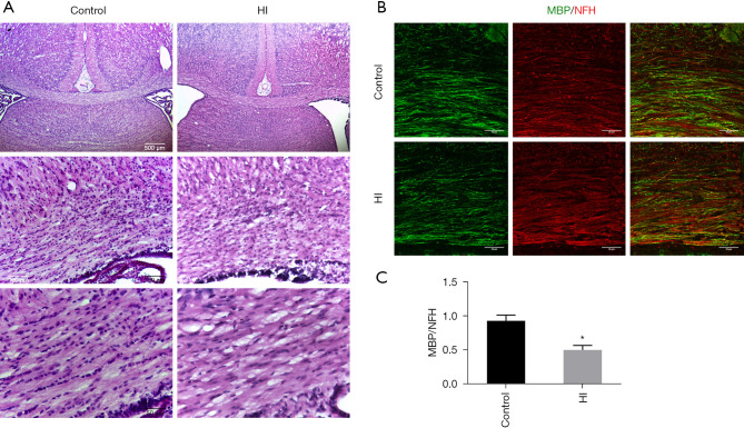
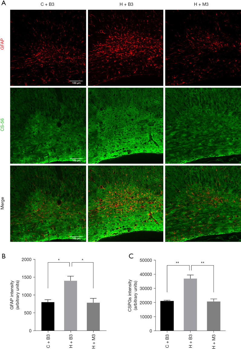
Background: The underlying cause of neurological sequelae after immature cerebral hypoxia-ischaemia (HI) white matter injury is impaired myelination. Previous studies have indicated that astrocyte activation is closely related to impaired myelination. However, the mechanism of reactive gliosis in white matter injury post-HI remains poorly understood.

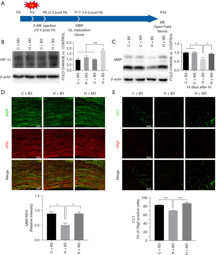
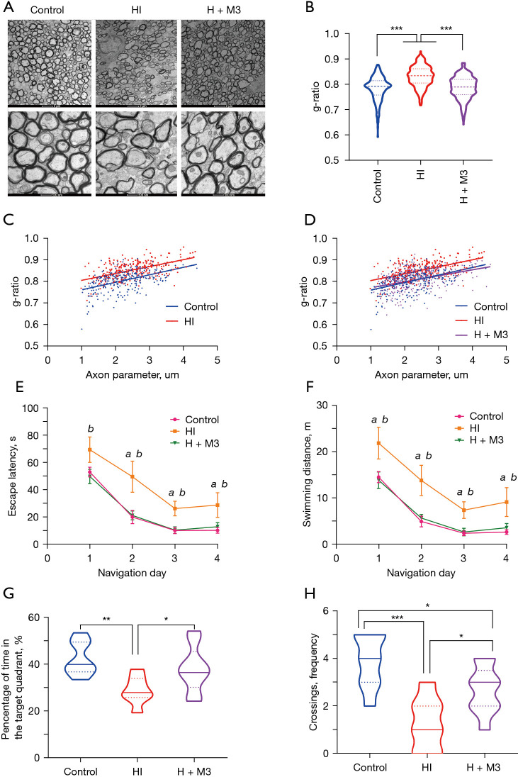
Methods: Studies using adult ischaemic animal models demonstrated that hypoxia inducible factor-1α (HIF-1α) expression was involved in the formation of reactive astrocytes. Here, we investigated the temporal expression of HIF-1α and its impact on reactive gliosis and further myelination using a perinatal HI white matter injury model induced in rats at postnatal day 3. The temporal pattern of HIF-1α expression post-HI injury was tested by western blotting and immunofluorescence. Rats were treated with a HIF-1α inhibitor at 72 hours post-HI injury. Reactive gliosis and myelination were assessed with western blotting, immunofluorescence and electron microscopy, and neurological functions were examined by behavioural testing.

Results: Our results showed that the expression of HIF-1α was upregulated in neurons at 24 hours and in astrocytes at 7 days post-HI. Inhibiting delayed HIF-1α expression post-HI injury could restrain reactive gliosis, ameliorate hypomyelination, and improve the performance of rats in the Morris water maze test.

Conclusions: Our findings suggest that a delayed increase in HIF-1α in astrocytes is involved in glial scar formation and leads to arrested oligodendrocyte maturation, impaired myelination, and long-term neurological function after experimental white matter injury in immature rats.

Keywords: White matter injury; astrogliosis; hypoxia inducible factor-1α (HIF-1α); myelination; oligodendrocytes.


Conflict of interest statement

Conflicts of Interest: All authors have completed the ICMJE uniform disclosure form (available at https://tp.amegroups.com/article/view/10.21037/tp-21-407/coif). The authors have no conflicts of interest to declare.

Figures

Figure 1
Figure 1
Brain tissue injury and abnormal myelination in the P3 HI injury rat model. (A) Representative pictures of H-E staining in the ipsilateral corpus callosum at 14 d post injury (P17). Scale bar: 500 µm (top), 100 µm (middle), 50 µm (bottom). (B) Representative pictures of immunofluorescence stained with MBP (green) and NFH (red) in the ipsilateral corpus callosum at 14 d post injury (P17). Scale bar: 50 µm. (C) The ratio of the MBP and NFH signal intensities. Values were shown as mean ± SEM; n=3–4/group; *, P<0.05 by t-test.
Figure 2
Figure 2
Temporal pattern of HIF-1α expression in the ipsilateral corpus callosum after HI injury. (A) Western blot quantification of HIF-1α protein in the ipsilateral corpus callosum at different time points post HI injury. n=6–8/group. (B) Representative pictures of immunofluorescence of HIF-1α in the ipsilateral corpus callosum at 24 h, 72 h, 7 d, and 14 d post HI injury. (C,D) Quantification of HIF-1α intensities at 24 h and 7 d post HI injury. (E-G) Representative pictures of immunofluorescence stained with anti-HIF-1α (green) and anti-Olig2 (red), anti-GFAP (red) or anti-NeuN (red) antibodies in the ipsilateral corpus callosum at 24 h, 72 h, and 7 d post HI injury. Scale bars in (B): 100 µm; in (E): main images, 30 µm, insets 5 µm; in (F) and (G): main images, 100 µm, insets, 10 µm; n=3–4/group. Values were shown as mean ± SEM; *, P<0.05; **, P<0.01; ***, P<0.001 by t-test.
Figure 3
Figure 3
Effect of late-onset HIF-1α increase on myelination. (A) Timeline of the experimental paradigm of HI injury, intervention, and detection. (B) Representative western blot image and quantification of HIF-1α expression at 7 days post injury after 2-ME injection. (C) Western blot images and quantification of MBP on 14 d post injury after late HIF-1α inhibition. n=8-10/group. (D) Representative pictures of immunofluorescence stained with MBP (green) and NFH (red) in the ipsilateral corpus callosum and the ratio of MBP and NFH signal intensities at 14 d post injury after late HIF-1α inhibition. (E) Representative pictures of immunofluorescence stained with CC1 (mature oligodendrocyte marker) (green) and Olig2 (oligodendrocyte marker) (red) in the ipsilateral corpus callosum and the percentage of CC1 (+) Olig2 (+) cells of Olig2 (+) cells at 14 d post injury after late HIF-1α inhibition. Scale bar: 100 µm; n=3–4/group. Values were shown as mean ± SEM; *, P<0.05; ***, P<0.001 by ANOVA followed by Tukey’s post hoc test. C+B3: control with blank injection group, C+M3: control with 2-ME injection group, H+B3: HI injury with blank injection group, H+M3: HI injury with 2-ME injection group.
Figure 4
Figure 4
Effect of late-onset HIF-1α increase on astrocyte activation and gliosis. (A) Representative picture of immunofluorescence stained with CS-56 (CSPGs marker) (green) and GFAP (red) in the ipsilateral corpus callosum. (B) Quantification of GFAP intensity at 14 d post injury after late HIF-1α inhibition. (C) Quantification of CSPGs intensity at 14 d post injury after late HIF-1α inhibition. Scale bar: 100 µm; n=3–4/group. Values were shown as mean ± SEM; *, P<0.05; **, P<0.01 by ANOVA followed by Tukey’s post hoc test.
Figure 5
Figure 5
Electron microscopy and behavior function tests. (A) Representative pictures of electron microscopy of corpus callosum. (B) Quantification of G-ratio of myelin. (C and D) Linear regression of G-ratio and axon parameter. (C) The intercepts rather than slopes between control and HI group were significantly different (P<0.001). (D) The slopes and intercepts between control and late inhibition were not significantly different (P>0.05), while the intercepts between HI and late inhibition group were significantly different (P<0.001). n=4–6 rats/group and at least 50 myelinated axons/rat. Values were shown as mean ± SEM; ***, P<0.001 by ANOVA followed by Tukey’s post hoc test. (E,F) Escape latency and swimming distances of each training day in navigation trail. a, significant difference between HI and control; b, significant difference between HI and late inhibition group. (G,H) Percentage in the target quadrant and frequency of platform crossings in probe trail. n=8/group; values were shown as mean ± SEM; *, P<0.05; **, P<0.01; ***, P<0.001 by ANOVA followed by LSD post hoc test.

References

    1. Adams-Chapman I, Heyne RJ, DeMauro SB, et al. Neurodevelopmental Impairment Among Extremely Preterm Infants in the Neonatal Research Network. Pediatrics 2018;141:e20173091. 10.1542/peds.2017-3091 - DOI - PMC - PubMed
    1. GBD 2017 Causes of Death Collaborators. Global, regional, and national age-sex-specific mortality for 282 causes of death in 195 countries and territories, 1980-2017: a systematic analysis for the Global Burden of Disease Study 2017. Lancet 2018;392:1736-88. 10.1016/S0140-6736(18)32203-7 - DOI - PMC - PubMed
    1. Gano D, Andersen SK, Partridge JC, et al. Diminished white matter injury over time in a cohort of premature newborns. J Pediatr 2015;166:39-43. 10.1016/j.jpeds.2014.09.009 - DOI - PMC - PubMed
    1. Duerden EG, Halani S, Ng K, et al. White matter injury predicts disrupted functional connectivity and microstructure in very preterm born neonates. Neuroimage Clin 2019;21:101596. 10.1016/j.nicl.2018.11.006 - DOI - PMC - PubMed
    1. Wheelock MD, Austin NC, Bora S, et al. Altered functional network connectivity relates to motor development in children born very preterm. Neuroimage 2018;183:574-83. 10.1016/j.neuroimage.2018.08.051 - DOI - PMC - PubMed