Review
. 2022 Feb 15:9:792559.
doi: 10.3389/fcvm.2022.792559. eCollection 2022.

Arrhythmias in Patients With Valvular Heart Disease: Gaps in Knowledge and the Way Forward

Affiliations
Review

Arrhythmias in Patients With Valvular Heart Disease: Gaps in Knowledge and the Way Forward

Maciej Kubala et al. Front Cardiovasc Med. .

Abstract

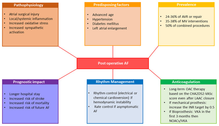
The prevalence of both organic valvular heart disease (VHD) and cardiac arrhythmias is high in the general population, and their coexistence is common. Both VHD and arrhythmias in the elderly lead to an elevated risk of hospitalization and use of health services. However, the relationships of the two conditions is not fully understood and our understanding of their coexistence in terms of contemporary management and prognosis is still limited. VHD-induced left ventricular dysfunction/hypertrophy and left atrial dilation lead to both atrial and ventricular arrhythmias. On the other hand, arrhythmias can be considered as an independent condition resulting from a coexisting ischemic or non-ischemic substrate or idiopathic ectopy. Both atrial and ventricular VHD-induced arrhythmias may contribute to clinical worsening and be a turning point in the natural history of VHD. Symptoms developed in patients with VHD are not specific and may be attributable to hemodynamical consequences of valve disease but also to other cardiac conditions including arrhythmias which are notably prevalent in this population. The issue how to distinguish symptoms related to VHD from those related to atrial fibrillation (AF) during decision making process remains challenging. Moreover, AF is a traditional limit of echocardiography and an important source of errors in assessment of the severity of VHD. Despite recent progress in understanding the pathophysiology and prognosis of postoperative AF, many questions remain regarding its prevention and management. Furthermore, life-threatening ventricular arrhythmias can predispose patients with VHD to sudden cardiac death. Evidence for a putative link between arrhythmias and outcome in VHD is growing but available data on targeted therapies for VHD-related arrhythmias, including monitoring and catheter ablation, is scarce. Despite growing evidences, more research focused on the prognosis and optimal management of VHD-related arrhythmias is still required. We aimed to review the current evidence and identify gaps in knowledge about the prevalence, prognostic considerations, and treatment of atrial and ventricular arrhythmias in common subtypes of organic VHD.

Keywords: aortic stenosis; arrhythmic mitral valve prolapse; atrial arrhythmia; postoperative atrial fibrillation; valvular heart disease; ventricular arrhythmia.


Conflict of interest statement

The authors declare that the research was conducted in the absence of any commercial or financial relationships that could be construed as a potential conflict of interest.

Figures

Figure 1
Figure 1
Representative of bileaflet mitral valve thickening and prolapse coexisting with mitral annular disjunction and myocardial arrhythmogenic substrate and 12-lead surface ECG illustrating multifocal ventricular arrhythmias in the same patient. Sixty-nine year old man without history of coronary artery disease referred for work-up of episodes of syncope. Bidimensional echocardiography, parasternal long-axis view at end-systole (upper panels) showed a bileaflet mitral valve thickening and annular disjunction (red arrow). Doppler evaluation showed a mild mitral regurgitation due to a bileaflet myxomatous mitral valve prolapse. Cardiac MRI cine imaging in two-chamber view (right bottom) identified a 11 mm large mitral annular disjunction localized to the posterior left ventricular wall and basal short-axis late gadolinium—enhanced (LGE) cardiac MRI (middle bottom) demonstrated midwall LGE most prominent at basal inferior and inferoseptal wall consistent with intramural myocardial fibrosis. Sustained monomophic ventricular tachycardia (on the right) with a right bundle branch block and superior axis coresponding to the posterobasal left ventricular wall origin was induced at the time of electrophysiology study with programmed electrical stimulation. Frequent multifocal premature ventricular complexes of two different morphologies were also recorded: the first one manifesting right bundle branch block configuration, V5 transition and D2/D3 negative/positive discordance pointing toward the anterolateral papillary muscle and the second one with left bundle branch block morphology, V3 transition and inferior axis consistent with an outflow tract origin.
Figure 2
Figure 2
Twelve-lead surface ECG of ventricular tachycardia following aortic valve replacement and representative of CT scan and electroanatomic substrate: (A) In a 23 year-old man with congenital aortic stenosis treated with balloon aortic valvotomy followed by surgical aortic valve replacement with mechanical Carbomedics 21 prosthesis at the age of 7 years and implanted with a double chamber pacemaker for post-operative complete atrio-ventricular heart block. At the age of 23 years, he was admitted for a sustained symptomatic ventricular tachycardia leading to upgrading his pacemaker to implantable cardioverter defibrillator. Cardiac CT scan (on the top : coronal oblique left ventricular outflow tract and basal short-axis views) performed prior to electrophysiology study showed a periaortic left ventricular myocardial thinning localized to anteroseptal, inferoseptal and inferobasal LV segments (yellow arrows) consistent with a periaortic scar. Bipolar endocardial electroanatomic map (on the bottom, antero-posterior view) displayed periaortic inferoseptal low-voltage abnormal area (in gray). The clinical monomophic VT with a right bundle branch block and superior axis configuration was induced at the time of electrophysiology study with programmed electrical stimulation. The earliest recorded activation signal (50 ms to the onset of the surface QRS recorded on distal tip of the ablation catheter, red arrow) identified at the edge of the low voltage inferoseptal area was targeted with ablation (black dots) allowing for VT interruption. (B) A 18 year-old man with congenital severe bicuspid aortic valve stenosis leading to aortic valvuloplasty at the age of 5 and 16 years followed by surgical aortic valve replacement with mechanical 21 St Jude prosthesis at the age of 17 years. One year after aortic valve replacement an asymptomatic non-sustained monomorphic VT was recorded on a 24 h Holter and exercise treadmill test. 12-lead ECG displayed a Rs morphology in D1, qR pattern in V1 and a lack of precordial transition suggesting aortomitral continuity source.
Figure 3
Figure 3
Representative of the interaction between AF and VHD in terms of diagnostic challenges and prognostic and therapeutic considerations. AF, atrial fibrillation; AR, aortic regurgitation; AS, aortic stenosis; INR, international normalized ratio; MR, mitral regurgitation; MS, mitral stenosis; NOAC, non-vitamin K oral anticoagulants; PMC, percutaneous mitral commissurotomy; VHD, valvular heart disease; VKA, vitamin-K antagonists.
Figure 4
Figure 4
Examples of difficult cases of echocardiographic evaluation of aortic valve disease in patients with AF. (A) A case of paradoxical low-flow low-gradient AS in a 71-year-old female patient with AF. The left ventricle is small with concentric remodeling and preserved LVEF and the aortic valve presents moderate to severe calcifications. There is a clear variation of the aortic flow according to the cardiac cycles. The MPG is low, calculated at 30 mm Hg and the SVi is averaged at 32 ml/m2, in favor of a paradoxical low-flow, low-gradient AS. After electrical cardioversion, and the restoration of SR, the cycles no longer fluctuate, the SVi has normalized to 46 ml/m2 and the MPG is high at 43 mmHg. The AVA remained stable at around 0.7cm2 before and after cardioversion. (B) A case of classical low-flow low-gradient AS in a 75-year-old male patient with AF. There is a left ventricular dysfunction, with a LVEF estimated at 33%. The flows vary slightly according to the cardiac cycles. The MPG is low, averaged at 28 mmHg as well as the SVi which is estimated at 25 mml/m2. The cardiac CT-scan is in favor of a severe AS because the calcium score is very high at 3,220 and the aortic valve planimetry finds 0.95cm2. (C) A case of aortic regurgitation in a 67-year-old male patient with AF. It is difficult to distinguish between a moderate and a severe AR on echocardiography because of the marked variation of the parameters (vena contracta, LVOT VTI and PHT) between cardiac cycles. Evaluation by phase-contrast CMR, which is less disturbed by AF than echocardiography, leads to the conclusion of severe AR with a calculated regurgitation fraction of 45%. AF, atrial fibrillation; AR, aortic regurgitation; AS, aortic stenosis; AVA, aortic valve area; CMR, cardiac magnetic resonance; LVEF, left ventricular ejection fraction; LVOT VTI, left ventricular outflow tract velocity time integral; MPG, mean pressure gradient; PHT, pressure half time; SR, sinus rhythm; SVi, stroke volume index.
Figure 5
Figure 5
Pathophysiology, prognostic implications and management of postoperative atrial fibrillation. AF, atrial fibrillation; AVR, aortic valve replacement; INR, international normalized ratio; LAAC, left atrial appendage closure; MV, mitral valve; NOAC, non-vitamin K oral anticoagulants; OAC, oral anticoagulation therapy; VKA, vitamin-K antagonists.

References

    1. Iung B, Delgado V, Rosenhek R, Price S, Prendergast B, Wendler O, et al. Contemporary presentation and management of valvular heart disease: the EURObservational Research Programme Valvular Heart Disease II Survey. Circulation. (2019) 140:1156–69. 10.1161/CIRCULATIONAHA.119.041080 - DOI - PubMed
    1. Vahanian A, Iung B, Himbert D, Nataf P. Changing demographics of valvular heart disease and impact on surgical and transcatheter valve therapies. Int J Cardiovasc Imaging. (2011) 27:1115–22. 10.1007/s10554-011-9804-7 - DOI - PubMed
    1. Benjamin EJ, Wolf PA, D'Agostino RB, Silbershatz H, Kannel WB, Levy D. Impact of atrial fibrillation on the risk of death: the Framingham Heart Study. Circulation. (1998) 98:946–52. 10.1161/01.cir.98.10.946 - DOI - PubMed
    1. Banerjee A, Allan V, Denaxas S, Shah A, Kotecha D, Lambiase PD, et al. Subtypes of atrial fibrillation with concomitant valvular heart disease derived from electronic health records: phenotypes, population prevalence, trends and prognosis. Europace. (2019) 21:1776–84. 10.1093/europace/euz220 - DOI - PMC - PubMed
    1. Hiss RG, Lamb LE. Electrocardiographic findings in 122,043 individuals. Circulation. (1962) 25:947–61. 10.1161/01.cir.25.6.947 - DOI - PubMed