

Osteochondral allograft in the treatment of an extruded osteoarticular segment of the distal tibia: A case report
- PMID: 35242989
- PMCID: PMC8881710
- DOI: 10.1016/j.tcr.2022.100627
Osteochondral allograft in the treatment of an extruded osteoarticular segment of the distal tibia: A case report
Abstract
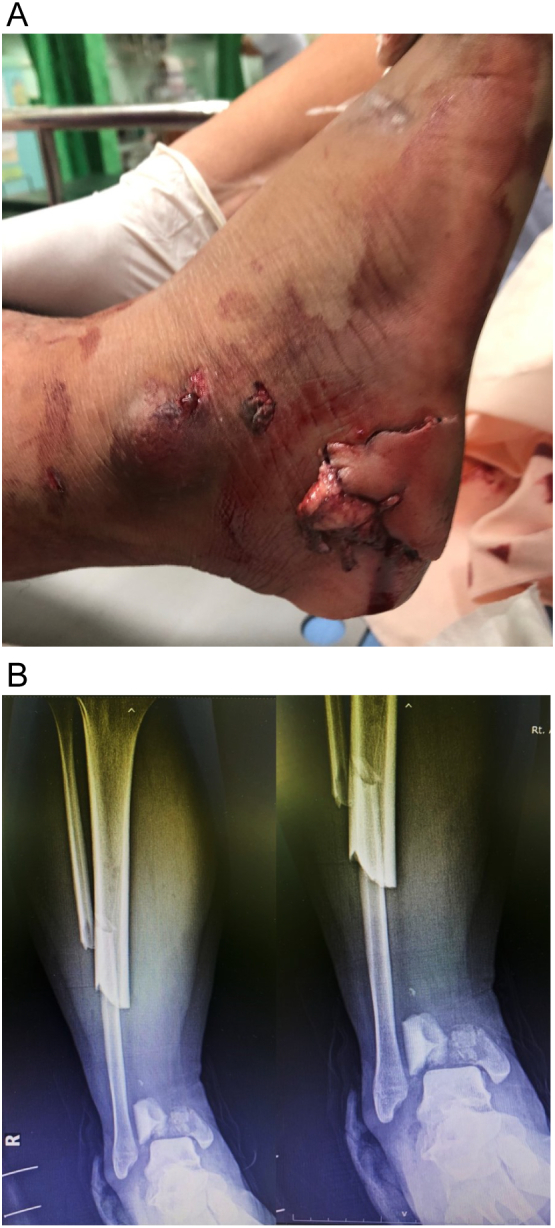
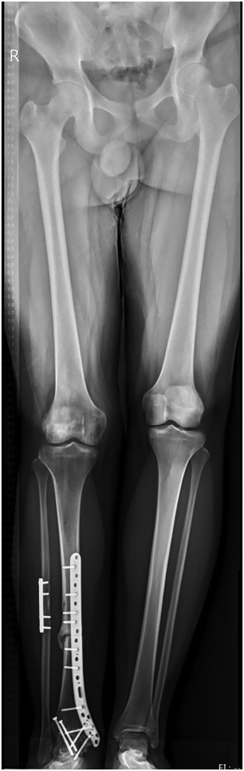
Open tibial plafond fracture with massive bone loss presents a challenge for orthopedic surgeons. Particularly unusual is extrusion of an osteoarticular segment of the distal tibia. Here we present the case of such a patient, who was treated using osteochondral allograft (OCA) and fusion procedures. The patients can regain independent walking without pain within 6 months, indicating that OCA may be a sensible option for the treatment of open tibial plafond fracture with extrusion of the osteoarticular distal tibia in cases in which the surrounding soft tissue is intact but bone reimplantation is not possible due to severe damage and contamination.
Keywords: Ankle joint; Limb salvage; Massive bone loss; Osteochondral allograft; Tibial plafond fracture.
© 2022 The Authors.
Conflict of interest statement
There are no conflicts of interest for any of the authors.
Figures


References
-
- Bourne R.B. Pylon fractures of the distal tibia. Clin. Orthop. Relat. Res. 1989:42–46. - PubMed
-
- Muller F.J., Nerlich M. Tibial pilon fractures. Acta Chir. Orthop. Traumatol. Cechoslov. 2010;77:266–276. - PubMed
-
- Shin K.H., Park H.J., Yoo J.H., Hahn S.B. Reconstructive surgery in primary malignant and aggressive benign bone tumor of the proximal humerus. Yonsei Med. J. 2000;41:304–311. - PubMed
-
- Lesensky J., Prince D.E. Distraction osteogenesis reconstruction of large segmental bone defects after primary tumor resection: pitfalls and benefits. Eur. J. Orthop. Surg. Traumatol. 2017;27(6):715–727. - PubMed
Publication types
LinkOut - more resources
Full Text Sources
