

Inter- and intrafraction motion assessment and accumulated dose quantification of upper gastrointestinal organs during magnetic resonance-guided ablative radiation therapy of pancreas patients
- PMID: 35243032
- PMCID: PMC8861831
- DOI: 10.1016/j.phro.2022.02.007
Inter- and intrafraction motion assessment and accumulated dose quantification of upper gastrointestinal organs during magnetic resonance-guided ablative radiation therapy of pancreas patients
Abstract
Background and purpose: Stereotactic body radiation therapy (SBRT) of locally advanced pancreatic cancer (LAPC) is challenging due to significant motion of gastrointestinal (GI) organs. The goal of our study was to quantify inter and intrafraction deformations and dose accumulation of upper GI organs in LAPC patients.
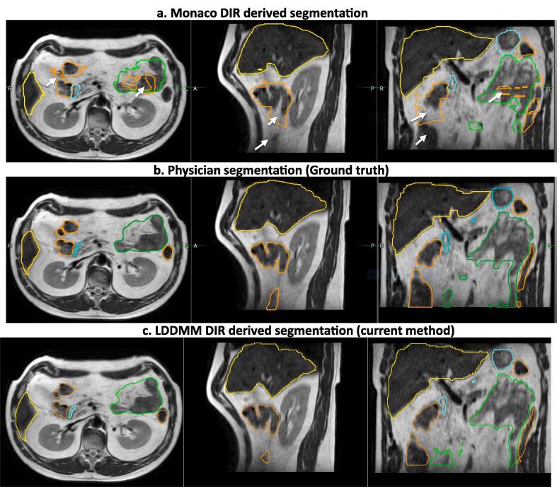
Materials and methods: Five LAPC patients undergoing five-fraction magnetic resonance-guided radiation therapy (MRgRT) using abdominal compression and daily online plan adaptation to 50 Gy were analyzed. A pre-treatment, verification, and post-treatment MR imaging (MRI) for each of the five fractions (75 total) were used to calculate intra and interfraction motion. The MRIs were registered using Large Deformation Diffeomorphic Metric Mapping (LDDMM) deformable image registration (DIR) method and total dose delivered to stomach_duodenum, small bowel (SB) and large bowel (LB) were accumulated. Deformations were quantified using gradient magnitude and Jacobian integral of the Deformation Vector Fields (DVF). Registration DVFs were geometrically assessed using Dice and 95th percentile Hausdorff distance (HD95) between the deformed and physician's contours. Accumulated doses were then calculated from the DVFs.
Results: Median Dice and HD95 were: Stomach_duodenum (0.9, 1.0 mm), SB (0.9, 3.6 mm), and LB (0.9, 2.0 mm). Median (max) interfraction deformation for stomach_duodenum, SB and LB was 6.4 (25.8) mm, 7.9 (40.5) mm and 7.6 (35.9) mm. Median intrafraction deformation was 5.5 (22.6) mm, 8.2 (37.8) mm and 7.2 (26.5) mm. Accumulated doses for two patients exceeded institutional constraints for stomach_duodenum, one of whom experienced Grade1 acute and late abdominal toxicity.
Conclusion: LDDMM method indicates feasibility to measure large GI motion and accumulate dose. Further validation on larger cohort will allow quantitative dose accumulation to more reliably optimize online MRgRT.
Keywords: 1.5 T magnetic field; Deformable image registration (DIR); LDDMM; MR-guided radiation therapy; MR-linac.
© 2022 The Authors.
Conflict of interest statement
The authors declare the following financial interests/personal relationships which may be considered as potential competing interests: NT has received honorarium and travel support from Philips and Elekta healthcare. CHC have received travel support from Elekta healthcare.
Figures


References
-
- Siegel R.L., Miller K.D., Fuchs H.E., Jemal A. Cancer Statistics, 2021. CA Cancer J Clin. 2021;71:7–33. - PubMed
-
- Tempero M.A., Malafa M.P., Al-Hawary M., Behrman S.W., Benson A.B., Cardin D.B., et al. Pancreatic adenocarcinoma, Version 2.2021, NCCN clinical practice guidelines in oncology. J Natl Compr Canc Netw. 2021;19:439–457. - PubMed
-
- Hidalgo M. Pancreatic cancer. N Engl J Med. 2010;362:1605–1617. - PubMed
Grants and funding
LinkOut - more resources
Full Text Sources
