Case Reports
. 2022 Feb 17:26:101434.
doi: 10.1016/j.ajoc.2022.101434. eCollection 2022 Jun.

The multifaceted presentation of syphilitic chorioretinitis examined by multimodal imaging: A case series

Affiliations
Case Reports

The multifaceted presentation of syphilitic chorioretinitis examined by multimodal imaging: A case series

Lorenzo Ferro Desideri et al. Am J Ophthalmol Case Rep. .

Abstract

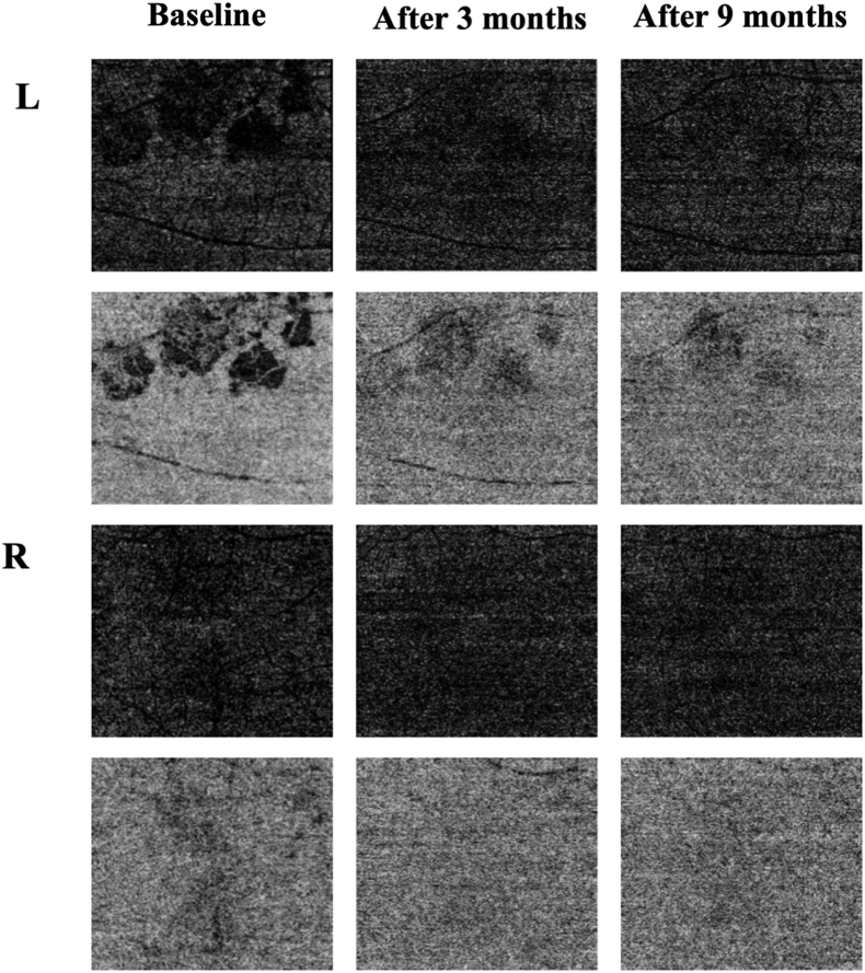
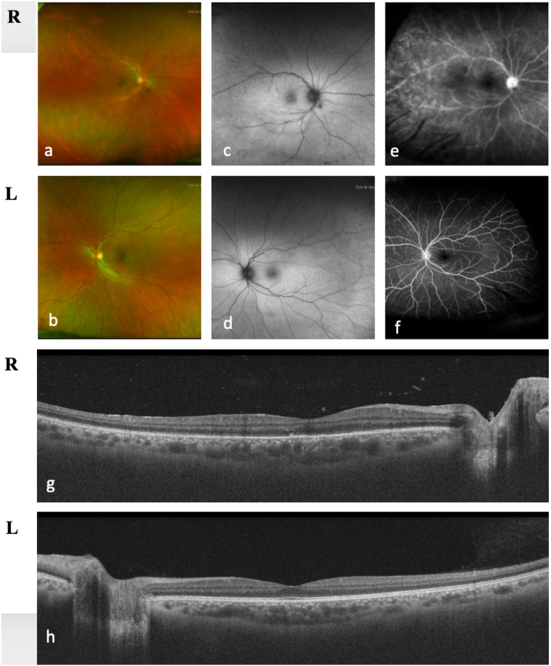
Ocular syphilis is also known as the 'great masquerader' for the wide variety of clinical features associated with this infection. Although chorioretinitis represents the most frequent manifestation in the posterior pole, other clinical entities can be described, including retinal vasculitis, optic disc disorders, necrotizing vasculitis and acute syphilitic posterior placoid chorioretinopathy (ASPPC). This latter is an infrequent ocular manifestation of syphilis, whose pathophysiology remains still unknown; however, multimodal imaging, including optical coherence tomography angiography (OCTA), has enabled us to better describe its pathophysiology and clinical course. In this study we report a case series of 3 different patients with syphilis-related chorioretinopathies; in this regard, the role of multimodal imaging has emerged has an extremely useful approach in order to better understand the pathophysiology of syphilitic chorioretinopathies. This could help clinicians (both ophthalmologist and infectious disease specialists) to early treat and prevent the severe ocular complications related to this fearsome disease.

Keywords: Chorioretinitis; Multimodal imaging; Ocular syphilis; Uveitis; oct.


Figures

Fig. 1
Fig. 1
Case 1- Fundus examination reveals the presence of moderate vitreitis and a yellowish round lesion in the temporal region outside the vascular arcades in the left eye (a), whereas in the right eye only mild vitreitis was detectable (b). Fundus autofluorescence shows a hyperfluorescent round area in correspondence of the lesion in the left eye (c), while the right one was normal (d). At wide-field fluorescein angiography examination, the presence of mild vascular leakage in the mid-periphery was observed in both eyes as sign of retinal vasculitis (e, f) with a hypofluorescent ischemic area in the temporal region in correspondence of the lesion in the left eye (e). Swept source optical coherence tomography imaging shows the presence of vitreitis with a normal foveal profile (i,l)however, in the temporal region of the left eye a hyperreflective intraretinal lesion suggesting active retinitis (l).
Fig. 2
Fig. 2
Case 1- Follow-up images after 3 and 9 months by swept-source optical coherence tomography angiography. At baseline it is evident the presence of nodular areas of flow void in the choriocapillaris in association with the placoid lesion in the left eye and also in the right eye some areas of reduced vascular density were found in the choriocapillaris. After 9 months, regression of the nodular areas of fluid void was only partial in the choriocapillaris of the left eye, while they were there was just a slightly decreased vascular density in the right one.
Fig. 3
Fig. 3
Case 1- After 9 months, the macular profile is preserved at fundus examination (a, b) in both eyes. Fundus autofluorescence images shows patchy hyperfluorescent areas in the perifoveal region in the left eye (c) and a normal profile in the right eye (d). Structural optical coherence tomography is preserved (e, f).
Fig. 4
Fig. 4
Case 2- Baseline images of the left eye. At wide-field fluorescein angiography the presence of mild vascular leakage in the mid-periphery is present combined with a hypofluorescent ischemic area in the temporal region in correspondence of the lesion (a). Fundus autofluorescence reveals a hyperfluorescent round area in correspondence of the lesion (b). Fundus examination shows moderate vitreitis and a yellowish round lesion in the temporal region outside the vascular arcades (c). At swept-source optical coherence tomography, vitreitis is evident and the foveal profile is preserved; however, in the temporal region, a hyperreflective intraretinal lesion suggesting active retinitis is present (d).
Fig. 5
Fig. 5
Case 3- Baseline images. At fundus examination mild vitreitis is present in the right eye, while the macular region had no significant alterations are detectable in both eyes (a, b). Fundus autofluorescence is unremarkable (c, d). At fluorescein angiography, moderate vasculitis in the mid-periphery and mild papillitis are present in the right eye, whereas the left eye was normal (e, f). At optical coherence tomography, there is vitreitis and some scattered hyperreflective granular spots and the disruption of the ellipsoid zone in the right eye, with a preserved macular profile in the left one (g, h).

References

    1. Hook E.W., 3rd Syphilis. Lancet. 2017;389(10078):1550–1557. - PubMed
    1. Kiss S., Damico F.M., Young L.H. Ocular manifestations and treatment of syphilis. Semin Ophthalmol. 2005;20(3):161–167. - PubMed
    1. Dutta Majumder P., Chen E.J., Shah J., et al. Ocular syphilis: an update. Ocul Immunol Inflamm. 2019;27(1):117–125. - PubMed
    1. Ormerod L.D., Puklin J.E., Sobel J.D. Syphilitic posterior uveitis: correlative findings and significance. Clin Infect Dis. 2001;32(12):1661–1673. - PubMed
    1. Gass J.D., Braunstein R.A., Chenoweth R.G. Acute syphilitic posterior placoid chorioretinitis. Ophthalmology. 1990;97(10):1288–1297. - PubMed

Publication types

LinkOut - more resources