

Case Reports
doi: 10.1016/j.case.2021.11.007.
eCollection 2022 Feb.
Closure of an Aortocardiac Fistula in a Horse
Affiliations
- PMID: 35243200
- PMCID: PMC8883141
- DOI: 10.1016/j.case.2021.11.007
Item in Clipboard
Case Reports
Closure of an Aortocardiac Fistula in a Horse
CASE (Phila).
.
No abstract available
Keywords: Aortocardiac fistula; Echocardiography; Equine; Horse; Subendocardial dissection.
Figures

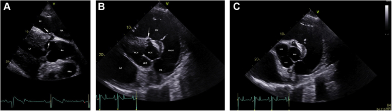
Two-dimensional echocardiograms performed immediately after ACF diagnosis, illustrating the ACF (white arrows) extending from the right sinus of Valsalva to the right ventricle, underneath the septal leaflet of the tricuspid valve. (A) Long-axis view of the left ventricular outflow tract obtained from the right parasternal window. The right ventricular inlet is displayed in the near field. (B) Short-axis view of the aorta obtained from the right parasternal window, highlighting the ACF (white arrow). (C) Short-axis view of the aorta obtained from the right parasternal window, illustrating the site of the rupture (R) and LCA. The right coronary artery is not imaged in this figure but is visible in Video 1. Ao, Aorta; LA, left atrium; LCA, left coronary artery; LCC, left coronary cusp; LV, left ventricle; NCC, noncoronary cusp; PA, pulmonary artery; RA, right atrium; RCC, right coronary cusp; RPA, right pulmonary artery (cross-section); RV, right ventricle; RVOT, right ventricular outflow tract.
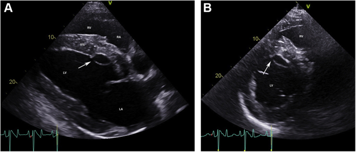
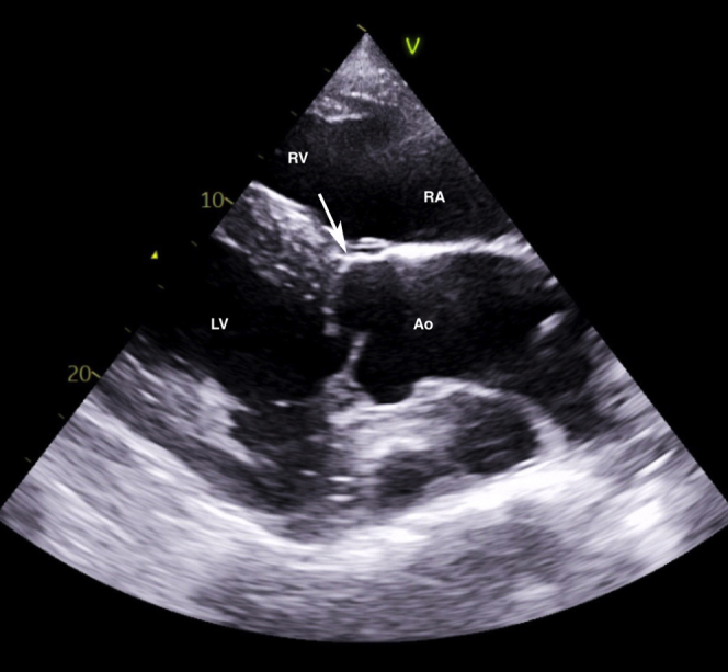
Two-dimensional echocardiograms performed immediately after ACF diagnosis, illustrating subendocardial dissection of blood along the left side of the interventricular septum, resulting in a large anechoic subendocardial pouch. The site of endocardial rupture is indicated (arrow). (A) Long-axis four-chamber view of the heart obtained from the right parasternal window. The right ventricular inlet is displayed in the near field. (B) Short-axis view of the left ventricle at the chordal level obtained from the right parasternal window. IVS, Interventricular septum; LA, left atrium; LV, left ventricle; RA, right atrium; RV, right ventricle.
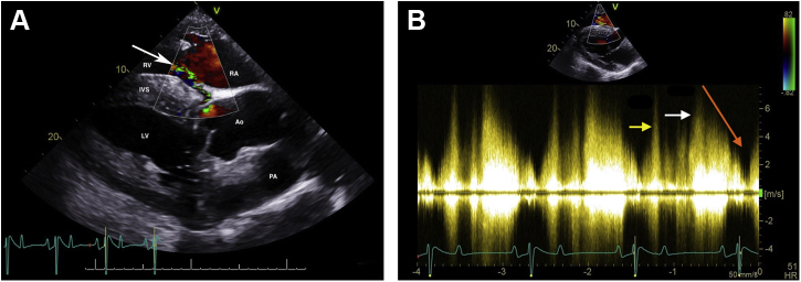
Color flow Doppler (A) and continuous-wave Doppler echocardiography (B) of the left-to-right shunt through the ACF from the right sinus of Valsalva into the right ventricle. All images were obtained from the right parasternal window immediately after ACF diagnosis. (A) Color Doppler two-dimensional echocardiogram of the left-to-right shunt through the ACF from the right sinus of Valsalva into the right ventricle, underneath the septal leaflet of the tricuspid valve (arrow), in the left ventricular outflow tract view. The right ventricular inlet is displayed in the near field. (B) Continuous-wave Doppler echocardiography demonstrating a peak shunt velocity of 4.2 m/sec in systole (yellow arrow) and 5.2 m/sec in early diastole (white arrow), with a rapid decline in maximal velocity (steep diastolic runoff; orange arrow). Ao, Aorta; IVS, interventricular septum; LV, left ventricle; PA, pulmonary artery; RA, right atrium; RV, right ventricle.
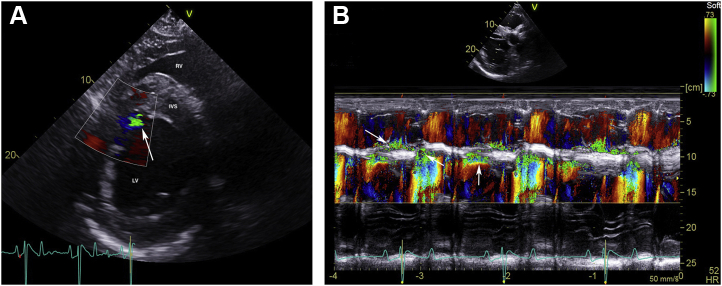
Color flow Doppler (A) and color M-mode echocardiography (B) of the left-to-right shunt through the ACF with subendocardial dissection and a small jet extending into the left ventricle (arrow). All images were obtained from the right parasternal window immediately after ACF diagnosis. (A) Color Doppler two-dimensional echocardiogram of the shunt into the left ventricle along the caudal edge of the subendocardial dissection (arrow) in the short-axis view of the left ventricle, just below the mitral valve. (B) Color M-mode echocardiogram of the left-to right shunt through the ACF and turbulent flow in the subendocardial dissection obtained from the short-axis view between the aorta and mitral valve. The vertical arrow demonstrates turbulence in the subendocardial dissection, while the diagonal arrows represent turbulence from the right sinus of Valsalva into the right ventricle. IVS, interventricular septum; LV, left ventricle; RV, right ventricle.
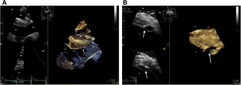
Three-dimensional (3D) echocardiograms of the ACF (A) and the subendocardial dissection (B) obtained from the right parasternal window immediately after ACF diagnosis. (A) Modified long-axis 3D image of the left ventricular outflow tract with slight cranial and dorsal obliquity. The right ventricular inlet is displayed in the near field. Arrows are pointing to the ACF. (B) Short-axis 3D image of the left ventricle, just below the mitral valve. The right ventricle is displayed in the near field. Arrows are pointing to the subendocardial dissection. Ao, Aorta; IVS, interventricular septum. LV, left ventricle; RV, right ventricle.
Two-dimensional echocardiogram of the left ventricular outflow tract view obtained from a right parasternal window, similar to Figure 1, performed 2 weeks after discharge from the hospital with identical findings on day 634 post discharge. The right ventricular inlet is displayed in the near field. Notice that the previously detected ACF (arrow) is no longer visible. Ao, Aorta; LV, left ventricle; RA, right atrium; RV, right ventricle.
References
-
- Marr C.M., Reef V.B., Brazil T.J., Thomas W.P., Knottenbelt D.C., Kelly D.F., et al. Aorto-cardiac fistulas in seven horses. Vet Radiol Ultrasound. 1998;39:22–31. - PubMed
-
- Fennich H., Doghmi N., Rim F., Belhaj S., Cheikhi F., Cherti M. Spontaneous rupture of right aortic sinus of Valsalva leading to massive cystic dissection of interventricular septum and complete heart block. Echocardiography. 2018;35:2109–2112. - PubMed
-
- Rooney J.R., Prickett M.E., Crowe M.W. Aortic ring rupture in stallions. Pathol Vet. 1967;4:268–274. - PubMed
-
- Sleeper M.M., Durando M.M., Miller M., Habecker P.L., Reef V.B. Aortic root disease in four horses. J Am Vet Med Assoc. 2001;15:491–496. - PubMed
-
- Roby K.A., Reef V.B., Shaw D.P., Sweeney C.R. Rupture of an aortic sinus in a 15-year-old broodmare. J Am Vet Med Assoc. 1986;189:305–308. - PubMed
Publication types
LinkOut - more resources
Full Text Sources
