. 2022 Feb 5;25(3):103877.
doi: 10.1016/j.isci.2022.103877. eCollection 2022 Mar 18.

A plant receptor domain with functional analogies to animal malectin disables ER stress responses upon infection

Affiliations

A plant receptor domain with functional analogies to animal malectin disables ER stress responses upon infection

Laïla Giordano et al. iScience. .

Abstract

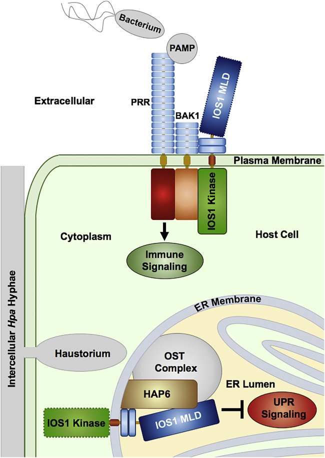
Malectins from the oligosaccharyltransferase (OST) complex in the endoplasmic reticulum (ER) of animal cells are involved in ER quality control and contribute to the Unfolded Protein Response (UPR). Malectins are not found in plant cells, but malectin-like domains (MLDs) are constituents of many membrane-bound receptors. In Arabidopsis thaliana, the MLD-containing receptor IOS1 promotes successful infection by filamentous plant pathogens. We show that the MLD of its exodomain retains IOS1 in the ER of plant cells and attenuates the infection-induced UPR. Expression of the MLD in the ios1-1 knockout background is sufficient to complement infection-related phenotypes of the mutant, such as increased UPR and reduced disease susceptibility. IOS1 interacts with the ER membrane-associated ribophorin HAP6 from the OST complex, and hap6 mutants show decreased pathogen-responsive UPR and increased disease susceptibility. Altogether, this study revealed a previously uncharacterized role of a plant receptor domain in the regulation of ER stress during infection.

Keywords: Molecular biology; Plant biology; Plant pathology.


Conflict of interest statement

The authors declare no competing interests.

Figures

None
Graphical abstract
Figure 1
Figure 1
The MLD retains IOS1 in the ER (A) Laser-scanning confocal micrographs showing partial co-localization of RFP-tagged IOS1 with the 2xPLC citrine-tagged PM protein and the GFP-HDEL ER marker upon transient co-expression in N. benthamiana. Signals from the Citrine channel were colored in green. (B) The RFP-labeled IOS1 variant ED-TM with the signal peptide (SP), MLD, LRRs, and membrane-spanning region (TM), but without kinase domain (left panel) co-localizes with green HDEL-GFP, as revealed by yellow-orange color in merged images. The IOS1 variant LRR-TM-Kinase with SP, LRRs, TM, and kinase domain, but without the MLD (right panel) localizes to the PM but does not co-localize with the ER marker. (C and D) Arabidopsis cotyledons expressing the GFP-fused IOS1 variants consisting of the complete extracellular domain, but devoid of the TM and the kinase domain (C), or of the MLD alone (D). Both variants reside in the ER. Bars represent 10 μM p35S, 35S promoter; CF, cytoplasmic filament; N, nucleus; pER, perinuclear ER.
Figure 2
Figure 2
Localization of IOS1 variants in roots of Arabidopsis marker lines after transgenic expression by the native IOS1 promoter Genes encoding the IOS1 variants shown above the micrographs were transformed into Arabidopsis marker lines encoding either the citrine-labeled Tubby-C protein for PM and nuclear localization, or the ER-localized GFP-HDEL protein. Confocal laser-scanning micrographs show the synthesis of the RFP-labeled IOS1 variants in the root cell elongation zone for which developmentally regulated IOS1 promoter activity was previously reported (Hok et al., 2014). (A) Expression of RFP-tagged native IOS1 in the PM and ER marker lines. (B) Expression of the RFP-tagged IOS1 variant lacking the kinase domain in the ER marker line. (C) Expression of the RFP-tagged IOS1 variant lacking the transmembrane (TM) and kinase domains in the ER marker line. (D) Expression of the RFP-tagged IOS1 variant lacking the LRRs, the TM, and the kinase domain in the ER marker line. (E) Expression of the RFP-tagged IOS1 variant lacking the MLD in the PM and ER marker lines. Bars represent 10 μM pIOS1, native IOS1 promoter; N, nucleus; pER, perinuclear ER.
Figure 3
Figure 3
The IOS1 MLD attenuates pathogen-induced ER stress and promotes susceptibility to Hpa (A) The accumulation of UPR gene transcripts is increased in Hpa-infected ios1-1 mutant seedlings when compared to the wild-type (wt), as analyzed by RT-qPCR. Samples were collected 4 days after control treatment (H2O) or infection (Hpa). (B) Hpa-inoculated ios1-1 mutants produce spliced bZIP60 mRNA. RT-PCR was performed with selective primers on transcripts extracted from H2O-treated and Hpa-infected wt and ios1-1 mutant plantlets. M, 100 bp DNA ladder. (C) Chemical inhibition and induction of the UPR promotes and represses plant susceptibility to Hpa, respectively. Seedlings were treated with DMSO (Control), 4-PBA, DTT, or Tunicamycin (Tm). (D) Enhanced ER stress in ios1-1 upon Hpa inoculation is attenuated to wt levels by the MLD. Accumulation of ARF1 and bZIP60 gene transcripts was analyzed by RT-qPCR in wt and ios1-1 mutant seedlings, and in those from the complemented mutant lines ED3.6 and ML10.2, and expressed as the ratio between inoculated and uninoculated plants. (E) The MLD restores susceptibility in the ios1-1 mutant. RT-qPCR data in (A) and (D) are from three biological replicates each involving two technical replicates. Data were analyzed using ACT8 and UBQ10 as reference genes. Sporulation rates in (C) and (E) were determined seven dpi as a parameter for disease susceptibility. Shown are the amounts of conidiospores per g fresh weight (FW) from two biological replicates each consisting of 20 samples. Statistically significant differences for RT-qPCR data and sporulation assays were determined by the nonparametric Mann-Whitney test and the paired t test, respectively. Significance groups are represented by stars above the boxplots in the respective graphs (∗, p < 0.05; ∗∗, p < 0.01; ∗∗∗, p < 0.001). Biological replicates are represented in all Box Whisker plots by different gray tones. See also Figures S1, S2, S3, and S4.
Figure 4
Figure 4
The IOS1 exodomain associates with the ribophorin HAP6 in the ER (A) OST1A, OST1B, and HAP6 transcript accumulations upon infection of wt plants with Hpa, as quantified by RT-qPCR using expression of the Actin8-encoding gene as internal standard for normalization. RT-qPCR data are from three biological replicates, each involving two technical replicates. Data were analyzed using ACT8 and UBQ10 as reference genes. Asterisks indicate statistically significant differences as determined by nonparametric Mann-Whitney tests (∗∗, p < 0.01). Biological replicates are represented by dots in different gray tones. (B) Co-expression of the extracellular IOS1 domain (ED-TM) bait with the HAP6 prey in the mb-SUS Y2H system allows cells to grow on synthetic complete (SC) medium in the absence of adenine (A) and histidine (H), indicative for physical association between the proteins. Growth on SC medium containing A and H, but neither tryptophan (W) nor leucine (L) confirmed accomplished mating. The empty pNX32 prey vector shows absence of autoactivation by the ED-TM bait. The pNubWt-Xgate vector positively controls yeast growth. (C) Co-immunoprecipitation of IOS1 domains with HAP6 upon transient expression in N. benthamiana. Production of the RFP-labeled IOS1 variants and GFP-labeled HAP6 in protein preparations was controlled on Western blots (Wb; Input, top panels). Precipitated proteins with anti-GFP and anti-RFP beads were revealed by Wb with anti-RFP and anti-GFP antibodies, respectively (IP, bottom panels). An antibody against the plant photosystem II PsbO protein was used for Input normalization. Please note that the apparent molecular mass of the MLD-LRR-TM-RFP variant is higher than the expected one (84 kDa) because of N-glycosylations of the IOS1 exodomain. See also Figures S5, S6, and S7.
Figure 5
Figure 5
HAP6 promotes ER stress and attenuates susceptibility to Hpa (A) Expression of ARF1 and bZIP60 in wt and the allelic rpn2-1 and rpn2-2 knock-down mutants, 4 days after water treatment (-) or inoculation with Hpa (+). (B) Hpa sporulation on wt, rpn2-1, and rpn2-2 seedlings, as determined 7 days after inoculation. Shown are the amounts of conidiospores per g fresh weight (FW) from two biological replicates, each consisting of 20 samples. (C) Accumulation of transcripts from the downy mildew Ppat5 gene, 4 days after inoculation of wt, rpn2-1, and rpn2-2 seedlings.RT-qPCR data in (A) and (C) were analyzed using ACT8 and UBQ10 as reference genes and are from three biological replicates each consisting of two technical replicates. Statistically significant differences for RT-qPCR data and sporulation assays were determined by the nonparametric Mann-Whitney test and the paired t test, respectively. Significance groups are represented by stars above the boxplots in the respective graphs (∗, p < 0.05; ∗∗, p < 0.01; ∗∗∗, p < 0.001). Biological replicates are represented in all Box Whisker plots by different gray tones. See also Figures S2 and S8.
Figure 6
Figure 6
Proposed roles for individual domains of the IOS1 receptor during the interaction with bacteria and filamentous biotrophs As previously reported (Yeh et al., 2016) IOS1 stabilizes the complex between the pattern-recognition receptors (PRR) FLS2 and EFR with their co-receptor BAK1 at the PM. Stabilization involves the IOS1 kinase domain and fortifies pathogen-associated molecular pattern (PAMP)-triggered immune responses. Knockout mutants for the IOS1 kinase are thus more susceptible to bacterial infection. Filamentous biotrophic fungi and oomycetes establish feeding sites called haustoria inside living host cells. Subsequent increased metabolic activity and protein synthesis in the host cell generates ER stress and triggers the unfolded protein response (UPR). The UPR is positively regulated by the ribophorin HAP6 and probably other compounds of the Oligosaccharyltransferase (OST) complex. The infection stimulates synthesis of IOS1 that transits to the ER, where the MLD disables the infection-related UPR, and promotes the development of the pathogen. The knockout mutant for the IOS1 MLD is thus less susceptible to infection by filamentous biotrophs. Receptor domains with dotted outlines appear dispensable for the indicated functions at the PM or the ER.

Similar articles

Cited by

References

    1. Angelos E., Ruberti C., Kim S.J., Brandizzi F. Maintaining the factory: the roles of the unfolded protein response in cellular homeostasis in plants. Plant J. 2017;90:671–682. - PMC - PubMed
    1. Bittner-Eddy P.D., Allen R.L., Rehmany A.P., Birch P., Beynon J.L. Use of suppression subtractive hybridization to identify downy mildew genes expressed during infection of Arabidopsis thaliana. Mol. Plant Pathol. 2003;4:501–507. - PubMed
    1. Boisson-Dernier A., Roy S., Kritsas K., Grobei M.A., Jaciubek M., Schroeder J.I., Grossniklaus U. Disruption of the pollen-expressed FERONIA homologs ANXUR1 and ANXUR2 triggers pollen tube discharge. Development. 2009;136:3279–3288. - PMC - PubMed
    1. Carvalho H.H., Silva P.A., Mendes G.C., Brustolini O.J., Pimenta M.R., Gouveia B.C., Valente M.A., Ramos H.J., Soares Ramos J.R., Fontes E.P. The endoplasmic reticulum binding protein BiP displays dual function in modulating cell death events. Plant Physiol. 2014;164:654–670. - PMC - PubMed
    1. Castillo K., Rojas-Rivera D., Lisbona F., Caballero B., Nassif M., Court F.A., Schuck S., Ibar C., Walter P., Sierralta J., et al. BAX inhibitor-1 regulates autophagy by controlling the IRE1α branch of the unfolded protein response. EMBO. J. 2011;30:4465–4478. - PMC - PubMed

LinkOut - more resources