

Artificial intelligence in liver diseases: Improving diagnostics, prognostics and response prediction
- PMID: 35243281
- PMCID: PMC8867112
- DOI: 10.1016/j.jhepr.2022.100443
Artificial intelligence in liver diseases: Improving diagnostics, prognostics and response prediction
Abstract
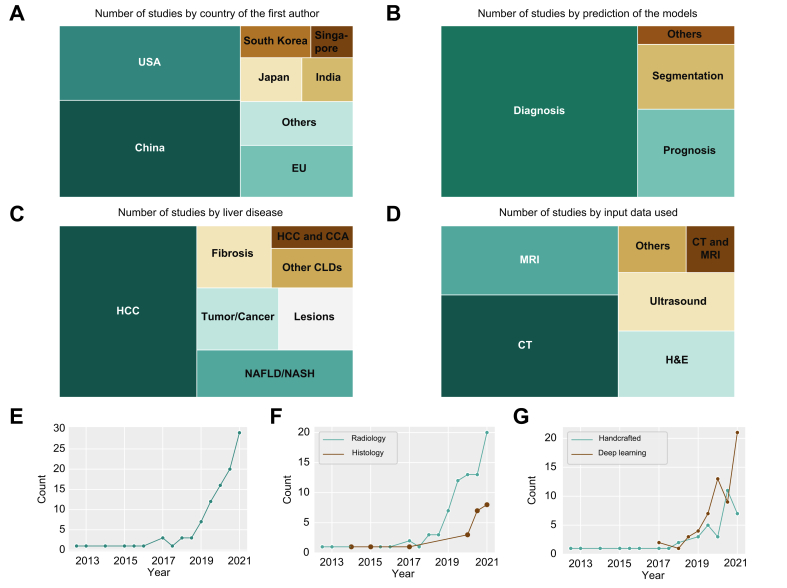
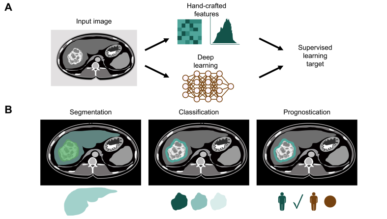
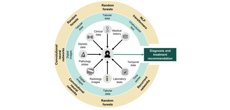
Clinical routine in hepatology involves the diagnosis and treatment of a wide spectrum of metabolic, infectious, autoimmune and neoplastic diseases. Clinicians integrate qualitative and quantitative information from multiple data sources to make a diagnosis, prognosticate the disease course, and recommend a treatment. In the last 5 years, advances in artificial intelligence (AI), particularly in deep learning, have made it possible to extract clinically relevant information from complex and diverse clinical datasets. In particular, histopathology and radiology image data contain diagnostic, prognostic and predictive information which AI can extract. Ultimately, such AI systems could be implemented in clinical routine as decision support tools. However, in the context of hepatology, this requires further large-scale clinical validation and regulatory approval. Herein, we summarise the state of the art in AI in hepatology with a particular focus on histopathology and radiology data. We present a roadmap for the further development of novel biomarkers in hepatology and outline critical obstacles which need to be overcome.
Keywords: AI, artificial intelligence; Artificial intelligence; CNN, convolutional neural network; DICOM, Digital Imaging and Communications in Medicine; HCC, hepatocellular carcinoma; ML, machine learning; MVI, microvascular invasion; NAFLD, non-alcoholic fatty liver disease; NASH, non-alcoholic steatohepatitis; TACE, transarterial chemoembolisation; TRIPOD, Transparent Reporting of a multivariable prediction model for Individual Prognosis or Diagnosis; WSIs, whole slide images; deep learning; diagnostic support system; imaging; machine learning; multimodal data integration.
© 2022 The Authors.
Conflict of interest statement
JNK declares consulting services for Owkin (France) and Panakeia (UK) and has received honoraria for scientific talks and participation in advisory boards by MSD, Eisai and Bayer. JC is a consultant for Guerbet, Bayer and Philips. VP is involved in a collaborative study with Owkin, France. DN and TPS declare no conflicts of interest. Please refer to the accompanying ICMJE disclosure forms for further details.
Figures
References
-
- Winkfield B., Aubé C., Burtin P., Calès P. Inter-observer and intra-observer variability in hepatology. Eur J Gastroenterol Hepatol. 2003;15:959–966. - PubMed
-
- Russell S., Norvig P. 2002. Artificial intelligence: a modern approach.
-
- Pearce C.B., Gunn S.R., Ahmed A., Johnson C.D. Machine learning can improve prediction of severity in acute pancreatitis using admission values of APACHE II score and C-reactive protein. Pancreatology. 2006;6:123–131. - PubMed
-
- Waljee A.K., Joyce J.C., Wang S., Saxena A., Hart M., Zhu J., et al. Algorithms outperform metabolite tests in predicting response of patients with inflammatory bowel disease to thiopurines. Clin Gastroenterol Hepatol. 2010;8:143–150. - PubMed
Publication types
LinkOut - more resources
Full Text Sources
Miscellaneous
