. 2022 Feb 23;4(2):fcac043.
doi: 10.1093/braincomms/fcac043. eCollection 2022.

Paediatric intracranial dural arteriovenous shunts: types, clinical presentation and therapeutic management

Affiliations

Paediatric intracranial dural arteriovenous shunts: types, clinical presentation and therapeutic management

Stanislas J Smajda et al. Brain Commun. .

Abstract

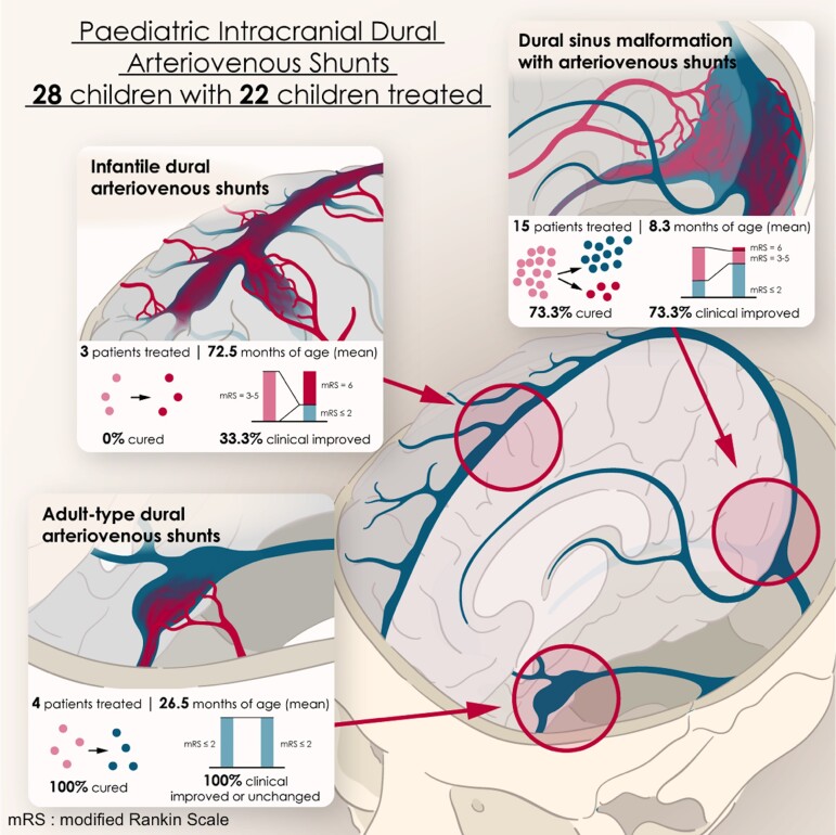
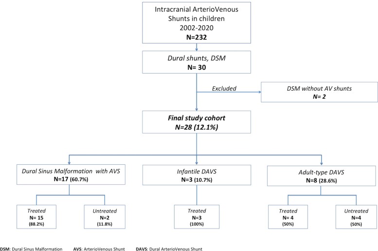
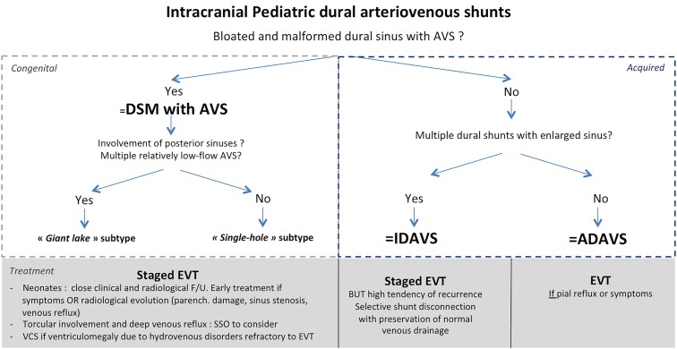
Paediatric intracranial dural arteriovenous shunts have clinical presentations and evolutions, with angiographic characteristics that differ from those described in adults. We report our experience concerning their therapeutic management, emphasizing the relevance of early diagnosis and appropriate treatment for satisfactory neurocognitive development. Using a prospective database, we reviewed the clinical and radiological data of all children with dural arteriovenous shunts managed between 2002 and 2020. Dural shunts were categorized into three types: dural sinus malformations with arteriovenous shunts; infantile dural arteriovenous shunts; and adult-type dural arteriovenous shunts. Therapeutic strategies and outcomes were analysed depending on lesional subtypes. Modified Rankin Scale for the paediatric population was assessed pre-treatment and at last follow-up. Twenty-eight patients [16 girls (57.1%); 12 boys (42.9%)] were included: 17 dural sinus malformation [10 boys (58.8%); seven girls (41.2%)], three infantile shunts [three girls (100%)], eight adult-type shunts [four girls (50%)]; four boys (50%)], with a mean age of 19.2 ± 36.6 months at presentation. Twelve (42.9%) had a modified Rankin Scale score of 0-2, four (14.3%) had a score of 3, three (10.7%) had a score of 4 and eight (28.6%) had a score of 5. Embolization was performed in 22 children [78.6%; 12 girls (54.5%); 10 boys (45.5%)]. Fifteen patients could be cured (68.2%): 11 dural sinus malformations (73.3%), four adult-type lesions (100%) but no infantile shunt. Mean post-treatment follow-up was 39.5 months (max. 139 months): 14 patients (63.6%) presented a modified Rankin Scale score of 0-2 and eight (36.4%) had a score ≥3. In the dural sinus malformation group, the modified Rankin Scale score was improved in 11 patients (73.3%) and unchanged in three (20%). Only one patient with infantile subtype (33.3%) improved clinically. In the adult-subtype group, all children (100%) improved. Of six untreated patients [four girls (66.7%); two boys (33.3%)], four with adult-subtype shunts showed uneventful evolutions, one with dural sinus malformation died, and therapeutic abortion was conducted in an antenatally diagnosed dural sinus malformation. Paediatric dural fistulas comprise different subtypes with variable clinical courses. Proper diagnosis is mandatory for optimal therapeutic strategies within appropriate therapeutic windows.

Keywords: congenital; dural shunts; fistula; paediatrics; vascular malformation.


Figures

Graphical Abstract
Graphical Abstract
Figure 1
Figure 1
Flowchart for study cohort selection. DSM, dural sinus malformation; AVS, arteriovenous shunt; DAVS, dural arteriovenous shunt.
Figure 2
Figure 2
Illustrative case. Midline DSM involving the torcular in a 3-year-old child presenting chronic headaches and severe behavioural disorders with anorexia requiring gastrostomy. MRI showed (A, axial T2) a bloated torcular (white arrowhead). MRA (B, TOF sequence) diagnosed an arterialization of the torcular (white arrowhead) vascularized by both middle meningeal arteries (white arrows). Multiple shunts (black arrows) in the malformed torcular were confirmed by a six-vessel angiography with super-selective injections of both external carotid artery (ECA) branches (AP and lateral views of the right ECA (C and D); left ECA not shown). Staged trans-arterial EVT obtained complete obliteration of the AVS. Control MRI (E, axial T2) and MRA (not shown) showed remodelling of the torcular without any parenchymal damage. Control angiography of both internal and ECA branches confirmed the obliteration of the DSM and the remodelling of the torcular (F, right internal carotid injection AP view; right maxillary artery in AP (G), and lateral (H) views). The patient improved clinically with complete resolution of the headaches and behavioural disorders.
Figure 3
Figure 3
Illustrative case. Single-hole fistula subtype DSM involving the left sigmoid sinus in a 21-month-old infant with seizures, right hemiparesis and neurocognitive delay. Since the infant was referred from a foreign country, there was a time interval of 4 months between the diagnosis in the home country and the treatment in our institution. MRI (A, axial view, FLAIR sequences), showed a bloated aspect of the left sigmoid sinus (white arrowhead) with thrombosis of the right transverse sinus (white arrow) confirmed on T1 post-gadolinium MRI (B, axial view) white arrow. Congestion of infratentorial veins was diagnosed, associated with pathological enhancement of the pons (asterisk). Angiography diagnosed a single-hole fistula (double white arrows) vascularized mainly by the mastoid branch of the left occipital artery (C, lateral view). Because of proximal and distal occlusion of the sinus, the high-flow shunt was responsible for reflux in the Labbe vein (black arrow) and thus poor ipsilateral hemispheric venous drainage (D, venous phase of the left ICA, long white arrow). The patient was cured after trans-arterial EVT with glue. MRI realized a few months after treatment (E, axial T2) demonstrated, however, global brain atrophy related to the long-lasting consequences of the venous congestion. Axial T1 post-gadolinium (F) showed regression of the pathological enhancement of the pons and disappearance of the posterior fossa congested veins. A selective occipital artery control angiography failed to show any residual shunts (G). The venous phase of the left ICA (H) normalized.
Figure 4
Figure 4
Illustrative case. Infantile DVASs in a 1-year-old child with seizures and neurocognitive delay. MRI (T2 axial view, A) and MRA (B, lateral view) showed atrophic changes in the right cerebral hemisphere and dilated vascular structures suspected of correspondence to arterialized veins (white arrow) bilaterally in the fronto-insular area. A DVA was suspected in the left hemisphere (asterisk). Angiography of the right (C, AP view) and left (D, AP view) external carotid arteries diagnosed bilateral dural arteriovenous shunts (black arrows) located at the level of the convexity accessory sinuses. A deep complex DVA of the left cerebral hemisphere (E, left internal carotid artery, AP view) was confirmed by left ICA angiography. Both dural shunts were embolized: trans-arterially with glue on the left side (long arrow) and combining trans-arterial and transvenous approaches with glue and coils for the right side (F, skull X-ray, AP view), which allowed complete obliteration of the shunts. A 6-month control angiography of the right (G, AP view) and left (H, AP view) external carotid arteries demonstrated, however, recurrence of the right shunt (black arrow) but stable complete obliteration of the left shunt. The patient was planned for a complementary embolization that occluded the right shunt; angiography confirmed, however, the occurrence of other dural shunts, whilst MRI showed development of intracerebral cavernomas (not shown).
Figure 5
Figure 5
Illustrative case. Adult-type DAVS in a 1-year-old child presenting a left chemosis with exophtalmia (without oculomotor palsy) for 3 months. Arterialization of the left cavernous sinus (white arrow) was suspected on MRA (A, axial TOF); Doppler ultrasonography (B) showed an arterialization of the superior ophthalmic vein. The diagnosis of dural cavernous arteriovenous fistula (black arrow) was confirmed by ECA angiography (C and D, AP and lateral views), the shunt being vascularized by the cavernous branch of the middle meningeal artery (asterisk) and responsible for reflux in the superior ophthalmic vein (long arrow) and the inferior petrosal sinus. The patient was cured after trans-arterial EVT with glue and improved clinically with complete resolution of the symptoms. Control angiography failed to show any residual shunt (Left maxillary angiography in AP (E) and lateral (F) views).
Figure 6
Figure 6
Decisional tree for the management of DAVS. AVS, arteriovenous shunt; DAVS, dural arteriovenous shunt; DSM, dural sinus malformation; IDAVS, infantile dural arteriovenous shunt; ADAVS, adult-type dural arteriovenous shunt.

References

    1. Hetts SW, Moftakhar P, Maluste N, et al. Pediatric intracranial dural arteriovenous fistulas: Age-related differences in clinical features, angioarchitecture, and treatment outcomes. J Neurosurg Pediatr. 2016;18:602–610. - PubMed
    1. Garcia-Monaco R, Rodesch G, Terbrugge K, Burrows P, Lasjaunias P. Multifocal dural arteriovenous shunts in children. Childs Nerv Syst. 1991;7:425–431. - PubMed
    1. Lasjaunias P, Magufis G, Goulao A, et al. Anatomoclinical aspects of dural arteriovenous shunts in children. Review of 29 cases. Interv Neuroradiol. 1996;2:179–191. - PubMed
    1. Sanchez-Mejia RO, Chennupati SK, Gupta N, Fullerton H, Young WL, Lawton MT. Superior outcomes in children compared with adults after microsurgical resection of brain arteriovenous malformations. J Neurosurg. 2006;105(2 Suppl):82–85. - PubMed
    1. Barbosa M, Mahadevan J, Weon YC, et al. Dural Sinus Malformations (DSM) with Giant Lakes, in neonates and infants. Review of 30 consecutive cases. Interv Neuroradiol. 2003;9:407–424. - PMC - PubMed

LinkOut - more resources