. 2022 Jan 28;3(2):100508.
doi: 10.1016/j.xcrm.2022.100508. eCollection 2022 Feb 15.

Autologous NK cells as consolidation therapy following stem cell transplantation in multiple myeloma

Affiliations

Autologous NK cells as consolidation therapy following stem cell transplantation in multiple myeloma

Hareth Nahi et al. Cell Rep Med. .

Abstract

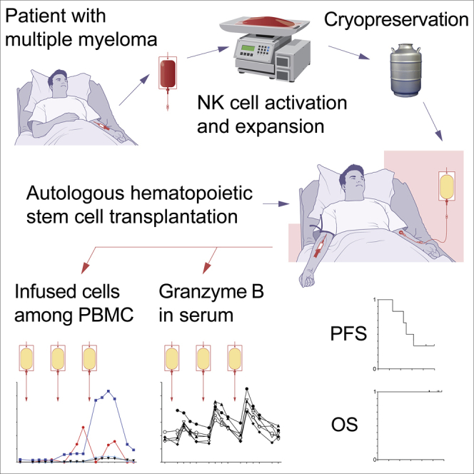
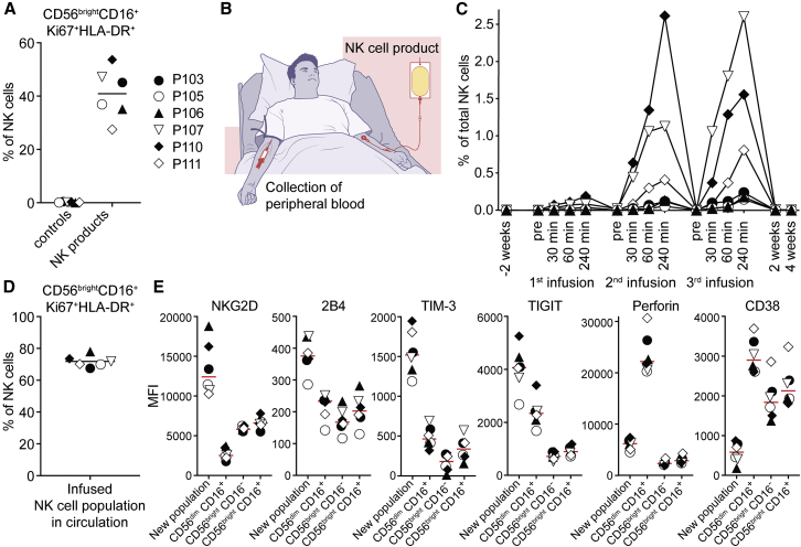
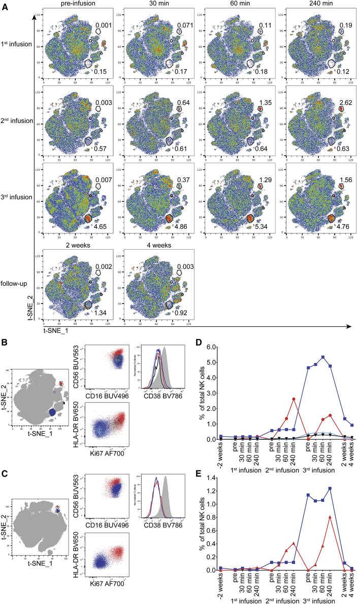
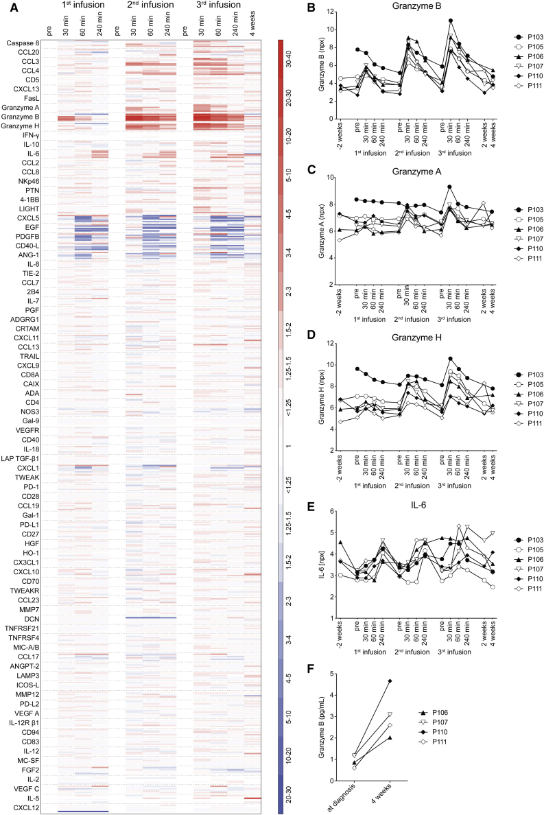
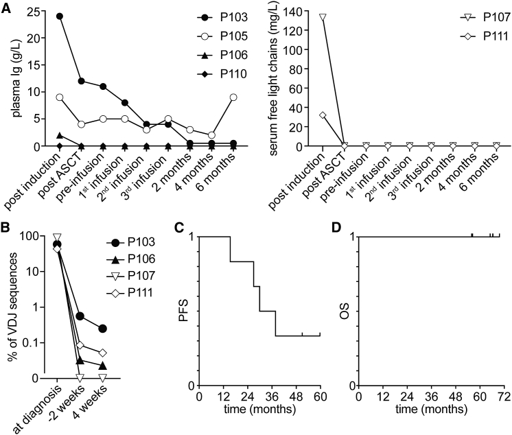
Few approaches have been made toward exploring autologous NK cells in settings of cancer immunotherapy. Here, we demonstrate the feasibility of infusing multiple doses of ex vivo activated and expanded autologous NK cells in patients with multiple myeloma (MM) post-autologous stem cell transplantation. Infused NK cells were detected in circulation up to 4 weeks after the last infusion. Elevations in plasma granzyme B levels were observed following each consecutive NK cell infusion. Moreover, increased granzyme B levels were detected in bone marrow 4 weeks after the last infusion. All measurable patients had objective, detectable responses after NK cell infusions in terms of reduction in M-component and/or minimal residual disease. The present study demonstrates that autologous NK cell-based immunotherapy is feasible in a setting of MM consolidation therapy. It opens up the possibility for usage of autologous NK cells in clinical settings where patients are not readily eligible for allogeneic NK cell-based immunotherapies.

Keywords: NK cells; adoptive cell therapy; consolidation; granzyme B; hematology; immunotherapy; immunotyping; myeloma.


Conflict of interest statement

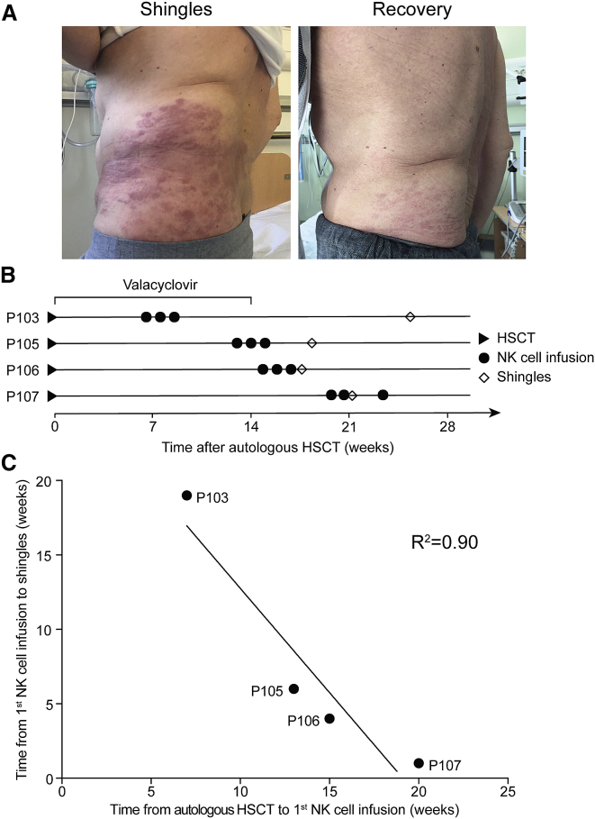
J. Liwing and P.-H.H. are employed by XNK Therapeutics (XNK); H.N., S.M., and M.C. are consulting for XNK; H.-G.L., is a board member of XNK; M.G., B.S., L.W.-J., K.M., G.G., H.-G.L., and E.A. are minority shareholders of XNK. A patent application pertaining to the use of antiviral prophylaxis in the context of autologous NK cell infusions has been filed (WO 2019/211310 A1). The remaining authors have declared that no competing interests exist.

Figures

None
Graphical abstract
Figure 1
Figure 1
Flow-cytometry-based tracking of infused autologous NK cell product in patients (A) Percentage of ex vivo activated and expanded NK cells with the CD56brightCD16+Ki67+HLA-DR+ phenotype in the NK cell products. Controls represent study subject peripheral blood NK cells before the first infusion. Lines represent the mean values. Symbols represent patients in all panels displayed (n = 6). (B) Strategy employed to detect ex vivo activated and expanded autologous NK cells among peripheral blood NK cells directly following the infusion. (C) Relative size of a defined subset of the infused NK cell population as detected in the circulation of study subjects after infusion of the NK cell product. Infused NK cells were identified by their distinct phenotype by using t-SNE analysis. (D) Percentage of NK cells with the CD56brightCD16+Ki67+HLA-DR+ phenotype within the infused cell populations followed in (C). Data shown are pooled from all time points. Line represents the mean value. (E) Median fluorescence intensities of selected markers on the infused NK cell populations followed in (C) compared with other NK cell subpopulations. Data shown are pooled over all time points. Lines represent the median values.
Figure 2
Figure 2
Temporal appearance and phenotype of infused populations within study subject peripheral blood NK cells (A) The temporal appearance of infused populations within the study subject peripheral blood NK cells. Representative t-SNE analysis based on 19 markers of 1 study subject (P110) is shown. The numbers next to the gates represent the percentage of that population within total NK cells at the respective time point. Two populations with different kinetics of appearance are marked (the population on the top right is included in Figure 1C). (B–E) (B and C) Comparison of the phenotypes. (D and E) The relative sizes of the infused NK cell populations detected in the circulation after infusion of the NK cell product for study subject P110 (B and D) and study subject P111 (C and E). The color coding in the t-SNE plots on the left represents the populations in the graphs to the right.
Figure 3
Figure 3
Assessment of the plasma proteome in conjunction with each NK cell product infusion (A) A total of 92 plasma proteins were assessed by proximity extension assay. As described in STAR Methods, 8 were excluded from analysis. The heat map shows the log2-based npx values from each of the 6 study subjects translated into fold change related to the value of the pre-infusion sample of each infusion. Red indicates fold increase; blue indicates fold decrease. (B–E) Assessment of peripheral blood plasma granzyme B (B), granzyme A (C), granzyme H (D), and IL-6 (E) in relation to infusion of ex vivo activated and expanded NK cells. The relative concentration was measured by proximity extension assay and is presented in arbitrary log2-based units npx for all study subjects (n = 6). (F) Assessment of bone marrow plasma granzyme B at diagnosis and after infusion of ex vivo activated and expanded NK cells by ELISA. Data shown from 4 study subjects; p = 0.021, paired t test.
Figure 4
Figure 4
Outcome of autologous NK cell-based immunotherapy for consolidation treatment of patients with multiple myeloma (MM) (A) Dynamics of plasma M-component (left, n = 4) and serum-free light chains (right, n = 2) in study subjects in the course of the clinical study. ASCT, autologous stem cell transplant. (B) IgH variability, diversity, and joining(VDJ) rearrangement analysis of BM samples taken at diagnosis and respective MRD values 2 weeks before the first and 4 weeks after the last infusion of the NK cell product. Percentages of the clonal IgH VDJ sequence (as identified in MM diagnosis samples) out of total IgH VDJ sequences are displayed. Data from 4 of 6 study subjects are shown. (C and D) (C) PFS and (D) OS of all study subjects (n = 6) during the course of the study calculated from the time of inclusion.
Figure 5
Figure 5
Development of shingles in the first 4 patients following NK cell product infusion (A) Shingles in 1 study subject following NK cell product infusion (left) and recovery after antiviral treatment (right). (B) Development of shingles in relation to NK cell product infusion. Diamonds mark the first appearance of shingles. Period of post-HSCT valacyclovir treatment is indicated (n = 4). (C) Correlation analysis between the time from HSCT to the NK cell product infusion and the time from NK cell product infusion to development of shingles. Regression calculated by Pearson correlation.

Comment in

References

    1. Miller J.S., Soignier Y., Panoskaltsis-Mortari A., McNearney S.A., Yun G.H., Fautsch S.K., McKenna D., Le C., Defor T.E., Burns L.J., et al. Successful adoptive transfer and in vivo expansion of human haploidentical NK cells in patients with cancer. Blood. 2005;105:3051–3057. doi: 10.1182/blood-2004-07-2974. - DOI - PubMed
    1. Bachanova V., Cooley S., Defor T.E., Verneris M.R., Zhang B., McKenna D.H., Curtsinger J., Panoskaltsis-Mortari A., Lewis D., Hippen K., et al. Clearance of acute myeloid leukemia by haploidentical natural killer cells is improved using IL-2 diphtheria toxin fusion protein. Blood. 2014;123:3855–3863. doi: 10.1182/blood-2013-10-532531. - DOI - PMC - PubMed
    1. Romee R., Rosario M., Berrien-Elliott M.M., Wagner J.A., Jewell B.A., Schappe T., Leong J.W., Abdel-Latif S., Schneider S.E., Willey S., et al. Cytokine-induced memory-like natural killer cells exhibit enhanced responses against myeloid leukemia. Sci. Transl Med. 2016;8:357ra123. doi: 10.1126/scitranslmed.aaf2341. - DOI - PMC - PubMed
    1. Ciurea S.O., Schafer J.R., Bassett R., Denman C.J., Cao K., Willis D., Rondon G., Chen J., Soebbing D., Kaur I., et al. Phase 1 clinical trial using mbIL21 ex vivo-expanded donor-derived NK cells after haploidentical transplantation. Blood. 2017;130:1857–1868. doi: 10.1182/blood-2017-05-785659. - DOI - PMC - PubMed
    1. Bjorklund A.T., Carlsten M., Sohlberg E., Liu L.L., Clancy T., Karimi M., Cooley S., Miller J.S., Klimkowska M., Schaffer M., et al. Complete remission with reduction of high-risk clones following haploidentical NK-cell therapy against MDS and AML. Clin. Cancer Res. 2018;24:1834–1844. doi: 10.1158/1078-0432.CCR-17-3196. - DOI - PubMed

Publication types