Review
. 2022 Feb 2;3(2):100214.
doi: 10.1016/j.xinn.2022.100214. eCollection 2022 Mar 29.

Nanotechnology for cardiovascular diseases

Affiliations
Review

Nanotechnology for cardiovascular diseases

Qinqin Hu et al. Innovation (Camb). .

Abstract

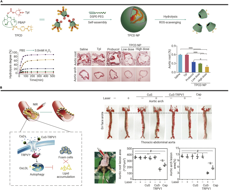
Cardiovascular diseases have become the major killers in today's world, among which coronary artery diseases (CADs) make the greatest contributions to morbidity and mortality. Although state-of-the-art technologies have increased our knowledge of the cardiovascular system, the current diagnosis and treatment modalities for CADs still have limitations. As an emerging cross-disciplinary approach, nanotechnology has shown great potential for clinical use. In this review, recent advances in nanotechnology in the diagnosis of CADs will first be elucidated. Both the sensitivity and specificity of biosensors for biomarker detection and molecular imaging strategies, such as magnetic resonance imaging, optical imaging, nuclear scintigraphy, and multimodal imaging strategies, have been greatly increased with the assistance of nanomaterials. Second, various nanomaterials, such as liposomes, polymers (PLGA), inorganic nanoparticles (AuNPs, MnO2, etc.), natural nanoparticles (HDL, HA), and biomimetic nanoparticles (cell-membrane coating) will be discussed as engineered as drug (chemicals, proteins, peptides, and nucleic acids) carriers targeting pathological sites based on their optimal physicochemical properties and surface modification potential. Finally, some of these nanomaterials themselves are regarded as pharmaceuticals for the treatment of atherosclerosis because of their intrinsic antioxidative/anti-inflammatory and photoelectric/photothermal characteristics in a complex plaque microenvironment. In summary, novel nanotechnology-based research in the process of clinical transformation could continue to expand the horizon of nanoscale technologies in the diagnosis and therapy of CADs in the foreseeable future.

Keywords: cardiovascular diseases; drug delivery system; molecular imaging; multimodal imaging; nanotechnology.


Conflict of interest statement

The authors declare no competing interests.

Figures

None
Graphical abstract
Figure 1
Figure 1
Schemes of nanotechnology-based biosensors for detection of CADs biomarkers (A) CdTe@IRMOF-3@CdTe nanocomposites to enlarge ECL signals. The strong ECL emission was achieved from isoreticular metal-organic framework (IRMOF) accelerator enriched quantum dots (CdTe), which were applied as an efficient ECL signal tag for trace cTnI detection. IRMOF allowed for encapsulating large amounts of CdTe, and functioned as a novel coreactant accelerator for promoting the conversion of S2O82− into SO4●_, further boosting the ECL emission of CdTe. IRMOF@CdTe-based immunosensor eventually performed a wide response range from 1.1 fg/mL to 11 ng/mL and a very low detection limit (0.46 fg/mL) (copyright American Chemical Society, 2018). (B) HsGDY@NDs-based aptasensors for detecting Myo and cTnI. The large surface and porous structure of HsGDY@NDs could absorb larger amounts of aptamer strands, giving low detection limits of 6.29 and 9.04 fg/mL for Myo and cTnI, respectively (copyright Elsevier Ltd., 2021). (C) Construction of RMSNs-based ECL-LFI strip for rapid, portable, and sensitive diagnosis. The RMSNs-based ECL-lateral flow immunosensor (ECL-LFI) enabled highly sensitive detection of cTnI-spiked human serum within 20 min at femtomolar levels (≈0.81 pg/mL) (copyright Wiley-VCH, 2020). (D) Ratiometric ECL-RET double-model detection of cTnI. Based on nanomaterials' features, a dual-wavelength ratiometric ECL resonance energy transfer (ECL-RET) sensing platform was developed for the detection of cTnI, showing high stability and low LOD (3.94 fg/mL) (copyright American Chemical Society, 2019). (E) uPAD for multi-target detection. With advantages of stability and sensitivity, the nanomaterials, including AuNPs, AgNPs, and gold urchin NPs, were used as optical labels to provide visible color signals (copyright Elsevier Ltd. 2019). (F) Increase of catalytic activity with G4/MOFzymes for POCT of target miRNAs. The interfaced G4 DNAzymes on MOFs (G4/MOFzymes) were produced by targeting miRNA-triggered rolling circle amplification (RCA) reactions, which displayed an about 100-fold higher catalytic activity than those in solution. By using the G4/MOFzyme catalysts in the luminol/H2O2 CL system, sensitive detection of myocardial infarction (AMI)-related two miRNAs (low to 1 fM seen with naked eyes) was achieved in human serum with a smartphone as a portable imaging detector (copyright American Chemical Society, 2020).
Figure 2
Figure 2
Various nanotechnology-based molecular imaging methods (A) PP/PS@MIONs used in MRI imaging of MI.With external magnetic field-induced targeting and PS targeting, the PP/PS@MIONs nanosystem enhanced the accumulation in infarcted area, showing accurate MRI-based visualization of MI at an early stage. (This is an open access article distributed under the terms of the Creative Commons Attribution [CC BY-NC] license.) (B) Platelet membrane-coated nanoparticles for magnetic resonance imaging activated endothelium, collagen, and form cells in plaques. Biomimetic PNPs could not only bind to advanced plaques but also probe the pre-atherosclerotic lesions (copyright American Chemical Society, 2018). (C) T1/T2 dual-mode MRI for detecting thrombus. cRGD@MLP-Gd exhibits a T2 contrast enhancement at 1 h after intravenous administration, followed by a visibly larger T1 contrast enhancement at the thrombus site (copyright Royal Society of Chemistry, 2020). (D) “Off-on” nanoprobe P-ICG2-PS-Lip for optical imaging of macrophages in vulnerable plaques. Note that the peptide-ICG2 was optically silent under normal conditions but activated in the presence of the lysosomal enzyme, cathepsin B. The NIRF fluorescent signal of P-ICG2-PS-Lip was successfully observed at the plaques on the artery walls (copyright Elsevier Ltd., 2020). (E) NIR-II nanoprobe used in optical imaging of MI. With the analysis of time course experiments, the AngII-Ag2S NDs could specifically accumulate at the ischemic myocardial tissues after intravenous injection within a few minutes, which opens a new avenue toward cost-effective, fast, and accurate in vivo imaging of the ischemic myocardium after AMI (copyright Wiley-VCH, 2020). (F) 99mTC-HFn nanotracer for PET imaging of vulnerable plaques. The specific uptake of 99mTc-HFn in plaques enabled quantitative measuring of the vulnerable and early active plaques as well as dynamic changes of inflammation during plaque progression (copyright American Chemical Society, 2018). (G) OPN/Ti3C2/ICG nanoprobe for accurate PAI of vulnerable plaques. OPN/Ti3C2/ICG possessed enhanced PA performance and high specificity to foam cells in vulnerable atherosclerotic plaques (copyright Wiley-VCH, 2020).
Figure 3
Figure 3
Multimode imaging strategies for specific and accurate detection of atherosclerotic plaques and thrombi (A) Marcoflor nanotracer for PET/MRI to visualize atherosclerosis. In PET/MRI experiment, 18F-Macroflor PET imaging detected changes in macrophage population size, while molecular MRI reported on increasing or resolving inflammation (copyright Springer Nature, 2017). (B) 89Zr-19F-HDL nanotracer to monitor myeloid cell dynamic in atherosclerotic mice with myocardial infarction with PET/MRI. With the 89Zr label, the short-term dynamics and biodistribution of myeloid cells in vivo could be monitored at high levels of sensitivity by PET. Optical imaging could be used to study the associated cell subsets at a cellular level. The incorporated fluorine core allowed the nanotracer to quantify (by MRI) the myeloid cell dynamics up to 28 days post-injection, which remedied the physical decay of PET signals. With the integrative strengths of multimodal imaging, in atherosclerotic mice with myocardial infarction, the nanotracer displayed rapid myeloid cell egress from the spleen and bone marrow and their accumulation in atherosclerotic plaques and at the myocardial infarct site (∗P < 0.05, ∗∗P < 0.01 and NS, no significance, two-sided Mann–Whitney U-test) (copyright Springer Nature, 2020). (C) Folate-conjugated 2D Pd@Au nanomaterials (Pd@Au-PEG-FA) for SPECT, CT, and PA imaging in heavy atherosclerotic plaques. CT helped to restrict the pathological depiction more accurately. With synergistic effects from high sensitivity of SPECT and high resolution of CT, Pd@Au-PEG-FA produced strong PA signals that could provide structural imaging information of cardiac vasculature with high temporal and spatial precision (copyright Springer Nature, 2020). (D) Thrombin-activatable scintillating nanoprobes for NIR-XEL imaging of in vivo thrombosis. Such nanoprobes showed XEL-off originally and enabled robust thrombin-activated turn-on XEL, which conferred XEL imaging background-free attribute and allowed it for detecting the early thrombosis on the basis of in situ elevated thrombin levels (copyright Wiley-VCH, 2021).
Figure 4
Figure 4
Multiple smart nanoplatforms targeting the lesion in the progression of atherosclerosis The surface modification of nanoplatforms via peptides, antibodies, ligands, and cell membranes could target different cells or components in the plaque to achieve precise delivery of chemicals, proteins, peptides, or nucleic acids and finally release these cargos to exert therapeutic effects.
Figure 5
Figure 5
Nanocarriers to deliver chemical drugs (A) Cargo-switching CSNP to deliver statins, showing antiatherogenic effects and regression of atherosclerotic plaques. The scale bar indicates 200 μm (∗∗∗P < 0.001, one-way ANOVA and Tukey’s multiple comparison test) (copyright American Chemical Society, 2020). (B) MM/RAPNP fabrication and its treatment for atherosclerosis in ApoE−/− mice. MM/RAPNP could effectively suppress macrophage phagocytosis and atherosclerosis progression in vivo (∗∗P < 0.01, ∗∗∗P < 0.001 and ns, no significance) (This is an open access article distributed under the terms of the Creative Commons Attribution [CC BY-NC] license). (C) rHDL-6877002 for reducing plaque volumes and the number of macrophages. Such CD4-TRAF6 blocking nanoimmunotherapy strategy could effectively inhibit monocyte recruitment and decrease plaque inflammation, as well as avoid the immune toxicity. Scale bar, 100 μm (∗P < 0.05) (copyright Elsevier Ltd., 2018).
Figure 6
Figure 6
Nanocarriers for delivering therapeutic nucleic acids (A) Fabrication and silencing efficacy of S2P50-siCamk2g NPs. The lipid-PEG surface was used to stabilize the NPs, achieve an increased circulating lifetime, and avoid rapid clearance. Besides, the plaque macrophage-targeting peptide (S2P) was incorporated on the lipid-PEG layer, which further increases the specificity of targeting; treatment of WD-fed Ldlr−/− mice with S2P50-siCamk2g-loaded NPs lowers plaque necrosis and increases lesional efferocytosis. The scale bar indicates 200 μm (∗P < 0.05, one-way ANOVA) (copyright American Association for the Advancement of Science, 2020). (B) Schematic illustration of miRNA mimic-loaded chitosan nanoparticles prepared using the ionic gelation method; in vivo treatment with chitosan nanoparticles containing miR-33 inhibits RCT. The injection of miR-33 NP resulted in the reduction of cholesterol efflux to apoA1 and reverse cholesterol transport (RCT) (∗P < 0.05, ∗∗P < 0.01, Student’s t test)(copyright American Chemical Society, 2019).
Figure 7
Figure 7
Nanomaterial themselves act as therapeutic drugs (A) Engineering of a broad-spectrum ROS-scavenging TPCD nanoparticle for targeted therapy of atherosclerosis. After intravenous administration, TPCD NPs accumulated in atherosclerotic lesions in ApoE−/− mice by passive targeting, significantly inhibited the development of atherosclerosis, as well as stabilized advanced plaques. Scale bar, 200 μm (∗P < 0.05, ∗∗∗P < 0.001) (copyright American Chemical Society, 2018). (B) CuS-TRPV1 switch for photothermal activation of TRPV1 signaling to reduce atherosclerotic lesions. With the photothermal property of CuS NPs, the TRPV1 channels opened and triggered calcium ions (Ca2+) influx after NIR irradiation, leading to autophagy activation, cholesterol efflux, and impede foam cell formation (∗∗P < 0.01 for CuS-TRPV1 + Laser vs. PBS, #P < 0.05 for Cap vs. PBS, &P < 0.05 for CuS-TRPV1 + Laser vs. Cap, Student’s t test) (copyright Springer Nature, 2018).

References

    1. Benjamin E.J., Muntner P., Alonso A., et al. Heart disease and stroke statistics-2019 update: a report from the American Heart Association. Circulation. 2019;139:e56–e528. - PubMed
    1. Celermajer D.S., Chow C.K., Marijon E., et al. Cardiovascular disease in the developing world: prevalences, patterns, and the potential of early disease detection. J. Am. Coll. Cardiol. 2012;60:1207–1216. - PubMed
    1. World Health Organization . World Health Organization; Geneva: 2021. Cardiovascular Diseases (CVDs)
    1. Virani S.S., Alonso A., Aparicio H.J., et al. Heart disease and stroke statistics-2021 update. Circulation. 2021;143:e254–e743. - PubMed
    1. Linde J.J., Kelbæk H., Hansen T.F., et al. Coronary CT angiography in patients with non-ST-segment elevation acute coronary syndrome. J. Am. Coll. Cardiol. 2020;75:453–463. - PubMed