. 2022 Apr 4;220(4):iyac020.
doi: 10.1093/genetics/iyac020.

The wing imaginal disc

Affiliations

The wing imaginal disc

Bipin Kumar Tripathi et al. Genetics. .

Abstract

The Drosophila wing imaginal disc is a tissue of undifferentiated cells that are precursors of the wing and most of the notum of the adult fly. The wing disc first forms during embryogenesis from a cluster of ∼30 cells located in the second thoracic segment, which invaginate to form a sac-like structure. They undergo extensive proliferation during larval stages to form a mature larval wing disc of ∼35,000 cells. During this time, distinct cell fates are assigned to different regions, and the wing disc develops a complex morphology. Finally, during pupal stages the wing disc undergoes morphogenetic processes and then differentiates to form the adult wing and notum. While the bulk of the wing disc comprises epithelial cells, it also includes neurons and glia, and is associated with tracheal cells and muscle precursor cells. The relative simplicity and accessibility of the wing disc, combined with the wealth of genetic tools available in Drosophila, have combined to make it a premier system for identifying genes and deciphering systems that play crucial roles in animal development. Studies in wing imaginal discs have made key contributions to many areas of biology, including tissue patterning, signal transduction, growth control, regeneration, planar cell polarity, morphogenesis, and tissue mechanics.

Keywords: Drosophila; FlyBook; imaginal disc; wing.


Figures

Fig. 1.
Fig. 1.
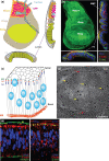
The wing imaginal disc. a) Schematic of late third instar wing disc, with approximate locations of notum, hinge, wing, and pleural regions identified by distinct coloring, and approximate locations of precursors for macrochaete identified by numbers, as indicated by Bryant (1975a): 1, 2—scutellar bristles; 3, 4—dorsocentral bristles; 5, 6—postalar bristles; 7, 8—supraalar bristles; 9, 10—notopleural bristles; 11—presutural bristle. Approximate location of wing margin (red) and longitudinal veins L2–L5 (purple) are also indicated. b) Schematic of adult wing, hinge, and half notum, marked as in (a) to illustrate relationship between wing disc and adult derivatives. c) Schematic of third instar larva with approximate location of the wing disc indicated.
Fig. 2.
Fig. 2.
Embryonic origin of the wing disc. Schematics showing approximate embryonic locations of thoracic imaginal disc primordia above, and signaling inputs that control their specification below, based on Requena et al. (2017). a) At stage 11, formation of the TP (red oval) is promoted by Wg (blue line) signaling near the A–P compartment boundary (dashed line) and suppressed in more dorsal and more ventral cells by Dpp and EGFR signaling, respectively. b) At stage 12, the wing primordia begins to form, comprising cells from both the TP and more dorsal cells (green). The Wg stripe is interrupted by Doc-mediated repression, and formation of the wing primordia is promoted by Dpp and inhibited by Wg and EGFR signaling. At stage 14, the wing primordia migrate dorsally, separating from the leg primordia. c) Images of the thoracic/leg primordia (Dll stain, red) and wing/haltere primordia (snail-DP-lacZ reporter, green) in embryos at the indicated stages (gift of Carlos Estella). Cells of the TP that will become wing/haltere primordia lose expression of Dll. w marks wing primordium, h marks haltere primordium.
Fig. 3.
Fig. 3.
Cell biology of the wing disc. a) Schematics of a horizontal view of the wing disc (top left) and sections across the length and width of the disc to illustrate different cell types, including the squamous PE (gray) columnar cells of the wing (olive), hinge (brown) and notum (orange), and AMP (pink) and tracheal (blue) cells underlying the notum. Red arrows point to the hinge–notum (top), hinge–hinge (middle), and hinge–pouch (bottom) folds. b) Confocal micrographs of late third instar wing disc. Top left panel displays a maximum projection of E-cad staining (green). Panels at right and bottom show slices across the length and width of the disc, and include staining for DNA and F-actin as well as E-cad. c) Schematic section of columnar wing disc epithelia, illustrating pseudostratification, with nuclei in blue, and relative locations of marginal zone (green), adherens junctions (red), and septate junctions (dark blue) indicated. BM is indicated in orange at bottom. d) Extracted surface of confocal micrograph of apical surface of the wing region of the wing disc, with cells outlined by E-cadherin staining. Note the variations in apical cell size. Red arrows highlight the fold at the edge of the wing pouch; yellow arrows highlight a few examples of mitotic cells, which are transiently enlarged as they round up. e) Confocal micrographs of vertical sections through the wing disc, showing DNA (blue), F-actin (red), and at left collagen (encoded by viking, green) and at right E-cad (green).
Fig. 4.
Fig. 4.
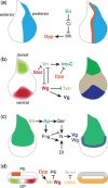
Early wing disc patterning. Schematics of wing discs illustrating key early steps in patterning. a) Expression of En in the posterior of the wing disc creates distinct anterior and posterior compartments. En represses expression of the Hh pathway transcription factor Ci. Ci directly and indirectly represses Hh, restricting Hh expression to posterior cells. The complementary expression of Ci and Hh limits Hh to signaling to anterior cells, where it induces transcription of Dpp. b) The wing disc is subdivided into appendage-body wall regions by the differential expression of Wg in ventral cells and Vn in dorsal cells, which mutually antagonize each other. Vn promotes notum fate by activating expression of Iro-C genes, while Wg promotes wing fate by promoting expression of Vg and suppressing expression of Tsh. c) Expression of Ap in the dorsal half of the wing disc creates distinct dorsal and ventral compartments. Ap regulates activation of Notch (N) along the D–V boundary by promoting transcription of Fng and Ser in dorsal cells. Notch activation along the D–V boundary leads to upregulation of Vg, from its boundary enhancer, as well, in the future wing region, Wg. d) Schematic cross-section illustrating subdivision of the wing disc into DP and PE cell layers. Expression of Vn, Wg, and Lines promotes DP fate and repress PE fate and Bowl.
Fig. 5.
Fig. 5.
Patterning by wing disc morphogens. Patterning of the wing region. a) Dpp spreads from the A–P boundary, forming a protein gradient that leads to graded repression of Brk. Brk represses expression of Sal complex genes and Omb, which are expressed in different domains due to different sensitivities to Brk levels. Several genes expressed in different A–P domains downstream of Dpp ultimately act in concert to position L2 and L5 wing vein primordia. b) Wg spreads from the D–V boundary, forming a protein gradient that promotes expression of Sens at high levels, and Vg and Dll at lower levels. Expression of the proneural gene ac (data not shown) overlaps sens but is only found in anterior cells, where sensory bristles will form. c) Adult wing patterning established by wing disc patterning includes positions of the wing veins and the wing margin bristles and hairs, shown at higher magnification in the boxes at bottom. d) Patterning of the notal region includes subdivision into a medial Pnr-expressing region and a lateral Iro-C-expressing region; differences amongst the different members of the Iro-C also occur during third instar (Ikmi et al. 2008). Multiple members of the odd protein family are expressed in the more anterior region of the notum. The adult notum is formed by fusion of the heminota from the left and right wing discs. Notal patterning defines regions where mechanosensory bristles form; some of the macrochaete are identified as in Fig. 1.
Fig. 6.
Fig. 6.
Proximal-distal patterning in the wing. a) At left, schematics of the wing region depicting expression of Vg, Wg, and Dpp at top left in a disc and at bottom in a wing. The Wg and Dpp compartment boundary stripes intersect in the middle of the wing pouch, which will later correspond to the distal tip of the adult wing (arrows). Right of the disc schematic, the relative expression domains at late third instar of several genes involved in P–D patterning are indicated [adopted from Cho and Irvine (2004)]. Approximate location in the adult of Wg expression in the inner and outer proximal wing rings has been determined by staining lacZ enhancer trap lines (Neumann and Cohen 1996a; Liu et al. 2000). At far right, a mid-third instar disc stained for expression of Vg, Rn, and Nub is shown to illustrate P–D differences in gene expression [reproduced with permission from Cho and Irvine (2004)]. b) Illustration of Vg regulation. Vg expression in the wing pouch is mediated by distinct boundary (BE, responding to Notch) and quadrant (QE, responding to Wg and Dpp) enhancers, as shown in schematic form, and, at far right, staining of reporters. Notch is activated along the D–V boundary throughout third instar and then also along the A–P boundary at late third instar. Vg expression mediated by the quadrant enhancer can spread into more proximal cells through Ds-Fat signaling, illustrated in the box at bottom left. Ds-Fat mediated signaling from wing pouch cells also establishes the inner ring of Wg expression in the proximal wing.
Fig. 7.
Fig. 7.
PCP in the wing disc. a) Schematics illustrating polarization of proteins of the Ds-Fat PCP pathway in the wing disc. Left panel shows schematic cross-section through cells, right panel shows 3D perspective. At pupal stages, wing hairs (brown triangles), pointing distally, will form near the distal vertices of each cell. b) Schematics illustrating polarization of proteins of the Fz PCP pathway in the wing disc. Left panel shows schematic cross-section through cells, right panel shows 3D perspective. The Stan (also known as Flamingo) protein accumulates on both distal and proximal cell membranes. c) Example of PCP in the adult wing, illustrated by distally pointing hairs in wild-type, and misoriented hairs in a prickle mutant. Portions of this figure reproduced from Ambegaonkar and Irvine (2015).
Fig. 8.
Fig. 8.
Wing disc growth. Wing discs stained for Wg are shown at different times after egg laying to illustrate the growth of the wing disc. Embryogenesis, first instar and second instar each take ∼1 day at 25°C, while third instar takes just over 2 days, so the disc shown correspond to mid second instar (64 h after egg laying), late second/early third instar (72 h), mid-third instar (96 h) and late-third instar (120 h). All discs are shown at the same magnification.
Fig. 9.
Fig. 9.
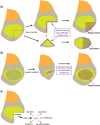
Wing disc regeneration. a) When a portion of a wing disc is excised, the missing part can be regenerated through a process including healing of the epithelium, stimulation of cell proliferation near the cut edges, and tissue repatterning. Smaller fragments typically generate duplicated structures rather than regenerating. b) Wing disc regeneration can also occur after genetically induced ablation of cells in a defined region of the disc. c) The damage response is coordinated by Jnk, which is activated by ROS and triggers multiple responses that enable regeneration.
Fig. 10.
Fig. 10.
Wing disc morphogenesis. a) Schematic illustrating steps in wing disc eversion, which occurs during the first half of prepupal development (the ∼12-h period that serves as a transition between larval and pupal development). (i) The larval wing disc is attached to the cuticle by a stalk connected to the PE. (ii) Part of the PE attaches to the larval cuticle, and the wing pouch begins to elongate and flatten. (iii) The PE over the notum invades the larval cuticle and ruptures, generating an opening for the disc to evert through. The PE over the wing contracts (green arrows), in part through cells becoming columnar rather than squamous. (iv) The disc has everted through the hole created by invasion and rupture of the PE and larval cuticle. Eversion is driven by contraction of the remaining PE. b) Longitudinal sections of discs at (i) 0, (ii) 2, and (iv) 4 h after puparium formation, roughly corresponding to the same stages in the schematic. Green arrowheads point to the edges of the wing pouch area, which will form the distal wing. Images reproduced with permission from Domínguez-Giménez et al. (2007).

References

    1. Adler PN. The frizzled/stan pathway and planar cell polarity in the Drosophila wing. Curr Top Dev Biol. 2012;101:1–31. - PMC - PubMed
    1. Adler PN, Charlton J, Liu J.. Mutations in the cadherin superfamily member gene dachsous cause a tissue polarity phenotype by altering frizzled signaling. Development. 1998;125(5):959–968. - PubMed
    1. Aegerter-Wilmsen T, Aegerter CM, Hafen E, Basler K.. Model for the regulation of size in the wing imaginal disc of Drosophila. Mech Dev. 2007;124(4):318–326. - PubMed
    1. Aegerter-Wilmsen T, Heimlicher MB, Smith AC, de Reuille PB, Smith RS, Aegerter CM, Basler K.. Integrating force-sensing and signaling pathways in a model for the regulation of wing imaginal disc size. Development. 2012;139(17):3221–3231. - PubMed
    1. Agnès F, Suzanne M, Noselli S.. The Drosophila JNK pathway controls the morphogenesis of imaginal discs during metamorphosis. Development. 1999;126(23):5453–5462. - PubMed

Publication types

Substances

LinkOut - more resources