. 2022 Jun;13(3):1717-1730.
doi: 10.1002/jcsm.12953. Epub 2022 Mar 3.

ACSL4 contributes to ferroptosis-mediated rhabdomyolysis in exertional heat stroke

Affiliations

ACSL4 contributes to ferroptosis-mediated rhabdomyolysis in exertional heat stroke

Sixiao He et al. J Cachexia Sarcopenia Muscle. 2022 Jun.

Abstract

Background: Rhabdomyolysis (RM) is a common complication of exertional heat stroke (EHS) and constitutes a direct cause of death. However, the mechanism underlying RM following EHS remains unclear.

Methods: The murine EHS model was prepared by our previous protocol. RNA sequencing is applied to identify the pathological pathways that contribute to RM following EHS. Inhibition of the acyl-CoA synthetase long-chain family member 4 (ACSL4) was achieved by RNA silencing in vitro prior to ionomycin plus heat stress exposure or pharmacological inhibitors in vivo prior to heat and exertion exposure. The histological changes, the iron accumulation, oxidized phosphatidylethanolamines species, as well as histological evaluation and levels of lipid metabolites in skeletal muscle tissues were measured.

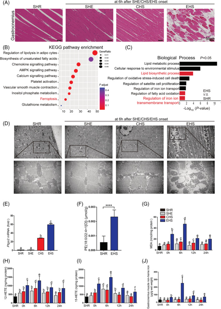
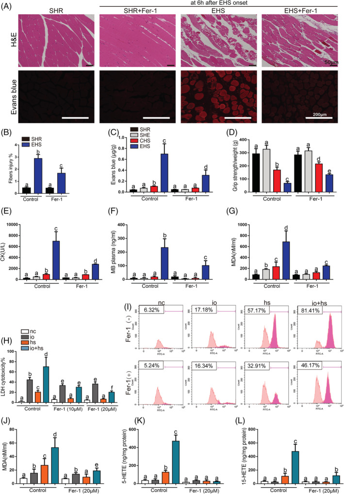
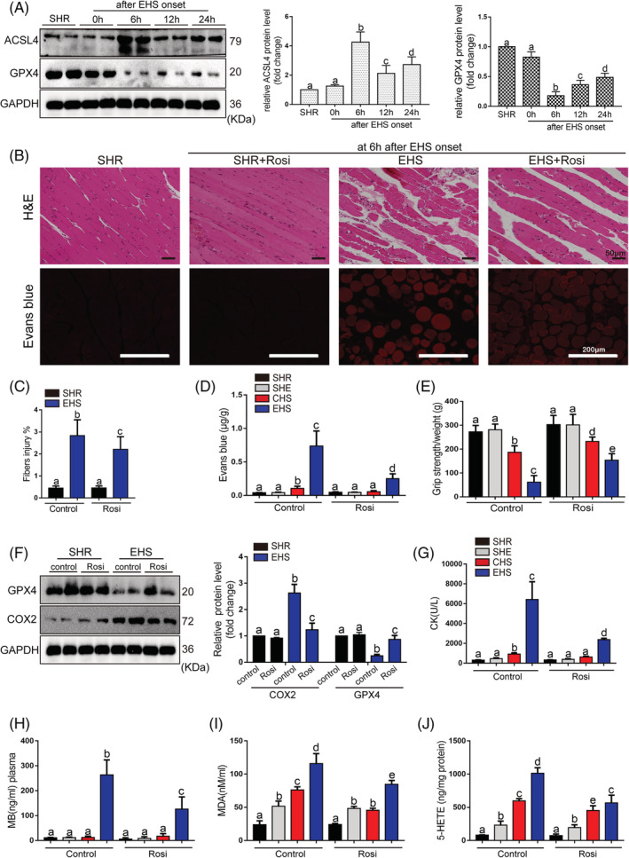
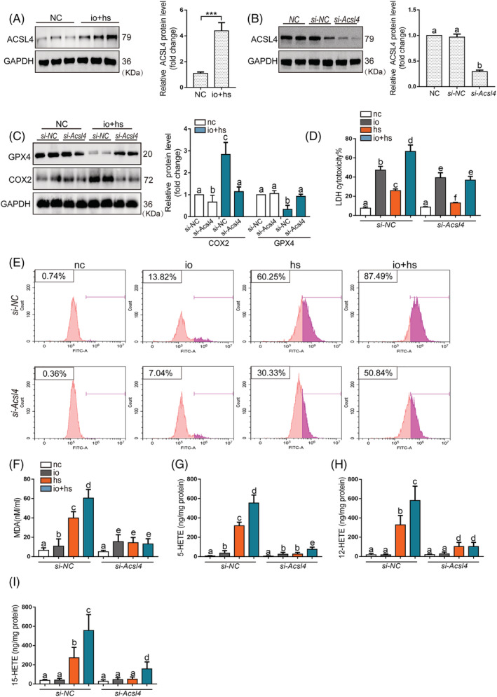
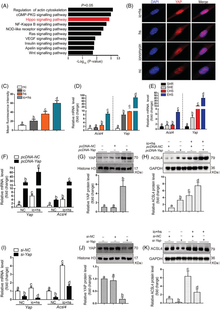
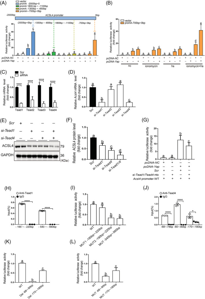
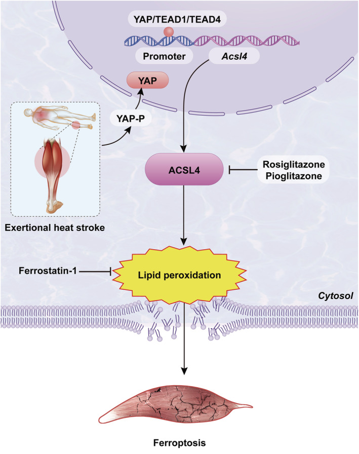
Results: We demonstrated that ferroptosis contributes to RM development following EHS. Ferroptosis inhibitor ferrostatin-1 administration once EHS onset significantly ameliorated the survival rate of EHS mice from 35.357% to 52.288% within 24 h after EHS (P = 0.0028 compared with control) and markedly inhibited RM development induced by EHS. By comparing gene expression of between sham heat rest (SHR) (n = 3) and EHS (n = 3) mice in the gastrocnemius (Gas) muscle tissue, we identified that Acsl4 mRNA expression is elevated in Gas muscle tissue of EHS mice (P = 0.0038 compared with SHR), so as to its protein levels (P = 0.0001 compared with SHR). Followed by increase in creatine kinase (CK) and myoglobin (MB) levels, the labile iron accumulation, decrease in glutathione peroxidase 4 (GPX4) expression, and elevation of lipid peroxidation products. From in vivo and in vitro experiments, inhibition of Acsl4 significantly improves muscle cell death caused by EHS, thereby ameliorating RM development, followed by reduction in CK and MB levels by 30-40% (P < 0.0001; n = 8-10) and 40% (P < 0.0001; n = 8-10), restoration of GPX4 expression, and decrease in lipid peroxidation products. Mechanistically, ACSL4-mediated RM seems to be Yes-associated protein (YAP) dependent via TEA domain transcription factor1/TEA domain transcription factor4.

Conclusions: These findings demonstrate an important role of ACSL4 in mediating ferroptosis activation in the development of RM following EHS and suggest that targeting ACSL4 may represent a novel therapeutic strategy to limit the skeletal muscle cell death and prevent RM after EHS.

Keywords: ACSL4; Exertional heat stroke; Ferroptosis; Lipid peroxidation; Rhabdomyolysis.


Conflict of interest statement

The authors declare that they have no conflict of interest.

Figures

Figure 1
Figure 1
Distinguishing features of ferroptosis in RM after EHS. All samples of muscle tissues were collected at 6 h after SHE, CHS, and EHS onset, SHR as the control group (A–F). (A) Representative H&E staining of Gas muscle (SHR, SHE, CHS, and EHS, scale bar = 50 μm in H&E). (B) KEGG pathway enrichment analysis of upregulated differentially expressed genes (DEGs) identified ferroptosis‐related genes highly expressed in Gas muscle of EHS mice. (C) Gene ontology (GO: biological process) analysis against upregulated genes between SHR and EHS mice. (D) TEM representative images among SHR, SHE, CHS, and EHS groups. (E) Relative levels of Ptgs2 mRNA expression were measured in the Gas muscle suffering from SHE, CHS, EHS, or SHR (n = 6 mice/group). (F) LC–MS/MS assessment of pro‐ferroptotic PEox (PE (18:0/22:4 + 1[O])) (n = 6 mice/group). Gas muscle tissues were collected at intervals of 0, 6, 12, and 24 h following SHE, CHS, and EHS onset, SHR as the control group. (G–I) In parallel, the levels of MDA, 12‐HETE, and 15‐HETE from 0 to 24 h were assayed using the respective kits (n = 6 mice/group). (J) The non‐heme iron level in each group from 0 to 24 h (n = 6 mice/group). Significance in (F) was calculated using the Student's t‐test; ****P < 0.0001. Summary data are presented as the mean ± SEM. Significance was calculated using a one‐way ANOVA with Tukey's post hoc test; groups labelled with different letters differed significantly (*P < 0.05).
Figure 2
Figure 2
The effects of Fer‐1 on the skeletal muscle injury of EHS mice in vivo and on the viability and lipid peroxidation of primary myoblast cells exposed to io + hs in vitro. Mice were treated with ferrostatin‐1 (10 mg/kg) by intraperitoneal injection 2 h before experiment. All samples of Gas muscle were collected at 6 h following SHE, CHS, and EHS onset, SHR as the control. (A) Representative H&E staining and EBD fluorescence of Gas muscle slices were imaged by microscopy (scale bar = 50 μm in H&E, 200 μm in EBD testing image). (B) The fibres injury score in EHS mice with or without Fer‐1 treatment (n = 6 mice/group). (C) The degree of permeability in muscle cells was detected by measuring the quality of EBD permeating into muscle cells under standard concentration curve (n = 6 mice/group). (D) The grip strength test in each group (n = 10 mice/group). (E–G) In parallel, serum creatine kinase (CK) levels and MB level were measured at 6 h after SHE, CHS, and EHS onset, SHR as the control (n = 6 mice/group); before io + hs induction experiment (containing 5 μM of ionomycin in medium, heat stress, 43°C 5% CO2 incubator heat stress for 2 h), primary myoblasts pretreatment with ferropstatin‐1 (20 μM) for 6 h. All samples were collected at 6 h following io + hs exposure. (H) LDH cytotoxicity percent was assayed at 6 h after io + hs induction (n = 6). (I) Cell lipid peroxidation signal was detected by C11 BODIPI 581/591 staining by flow cytometry. (J–L) In parallel, the levels of MDA, 5‐HETE, and 15‐HETE were assayed at 6 h following io + hs exposure (n = 6). Summary data are presented as the mean ± SEM. Significance was calculated using a one‐way ANOVA with Tukey's post hoc test; groups labelled with different letters differed significantly (*P < 0.05).
Figure 3
Figure 3
The effects of pharmacological inhibition of ACSL4 by Rosi on the development of RM after EHS onset. (A) The expression levels of ACSL4 and GPX4 in EHS mice were detected using the western blotting test. The representative western blotting images were from three independent experiments. Rosi (0.4 mg/kg, intravenous injection, 2 h before EHS experiment) was administered to mice. All samples were collected at 6 h after EHS onset. (B) Representative H&E staining and Evans blue fluorescence of Gas muscle slices were imaged by microscopy (scale bar = 50 μm in H&E, 200 μm in Evans blue testing image). (C) The fibre injury score in EHS mice with or without Rosi treatment (n = 6 mice/group). (D) The degree of permeability in muscle cells was detected by measuring the quality of EBD permeating into muscle cells under standard concentration curve (n = 6 mice/group). (E) The grip strength test in each group (n = 10 mice/group). (F) GPX4 and COX2 protein levels were assessed by western blotting, muscle tissues were collected at 6 h after EHS onset, the representative images of western blotting were from three independent experiments. (G–J) In parallel serum CK levels and MB levels, Gas muscles levels of MDA and 5‐HETE were measured in each groups (n = 6 mice/group). Summary data are presented as the mean ± SEM. Significance was calculated using a one‐way ANOVA with Tukey's post hoc test; groups labelled with different letters differed significantly (*P < 0.05).
Figure 4
Figure 4
The effects of genetic inhibition of Acsl4 on the viability and lipid peroxidation of primary myoblasts cells exposed to io + hs in vitro. (A) Western blotting was used to determine ACSL4 expression of primary myoblasts in the presence or absence of io + hs exposure, the representative images of western blotting were from three independent experiments. (B) Primary myoblast were transfected with si‐NC or si‐Acsl4 for 2 days before io + hs induction. All samples were collected at 6 h after io + hs induction. ACSL4 expression level under io + hs induction for 2 h after siRNA transfection was assessed by western blotting, the representative images of western blotting were from three independent experiments. (C) GPX4 and ACSL4 protein levels were determined by western blotting after io + hs induction, the representative images of western blotting were from three independent experiments. (D) The LDH cytotoxicity percent was assayed at 6 h after io + hs induction (n = 6). (E) Cell lipid peroxidation signal was detected by C11 BODIPI 581/591 staining by flow cytometry. (F–I) In parallel, the levels of MDA, 5‐HETE, and 15‐HETE were assayed at 6 h following io + hs exposure (n = 6). Significance was calculated using the Student's t‐test; *P < 0.05. Summary data are presented as the mean ± SEM. Significance was calculated using a one‐way ANOVA with Tukey's post hoc test; groups labelled with different letters differed significantly (*P < 0.05).
Figure 5
Figure 5
The regulation of the transcription and expression of ACSL4 by YAP entry into the nucleus in primary myoblasts. (A) KEGG analyses of RNA‐seq data showing the top 10 enriched pathways in EHS mice compared with SHR mice. (B–C) The location of YAP in nuclear was determined by laser scanning confocal microscopy (n = 6). (D–E) In parallel, QPCR analysis of Acsl4 and Yap mRNA levels in primary myoblast subjected to io + hs and Gas muscle tissues of mice with EHS, samples were collected at 6 h after EHS onset or after io + hs induction during recovery time (n = 6 mice/group or n = 6). (F–K) Primary myoblast cells were transfected with Yap overexpression plasmid or siRNA for 2 days, and then subjected to io + hs induction. The YAP protein levels and the mRNA and protein levels of ACSL4 were determined after io + hs induction. The representative western blotting images were from three independent experiments. Summary data are presented as the mean ± SEM. Significance was calculated using a one‐way ANOVA with Tukey's post hoc test; groups labelled with different letters differed significantly (*P < 0.05).
Figure 6
Figure 6
TEAD1/TEAD4 acting through binding to the ACSL4 promoter region for its expression. (A) Regions of the 2000‐bp proximal promoter sequence of Acsl4 gene via distinct promoter‐luciferase reporters are identified. Luciferase activity measurements of designated promoter‐reporter constructs in C2C12 cells were transfected with Yap plasmids (n = 3). The region −700‐bp upstream of the translation start site of the Acsl4 promoter was essential for YAP‐induced promoter‐reporter induction. (B) C2C12 cells were collected for assay in four conditions. Cells cotransfected with the Yap and WT (−700 bp) plasmids were collected and luciferase activity was analysed (n = 3). (C) siRNA transfection significantly decreased the expression of Tead1, Tead2, Tead3, and Tead4. N = 3 independent experiments (n = 6). (D) C2C12 cells were transfected with Tead1, Tead2, Tead3, and Tead4 siRNA or scramble siRNA for 48 h, respectively. Total RNA was isolated analysis of mRNA expression of Acsl4. (E–F) C2C12 cell were transfected with Tead1, Tead4, and Tead1 + Tead4 siRNA or scrambled siRNA (Scr) for 48 h, and then, western blotting for ACSL4 expression was performed, the representative images of western blotting were from three independent experiments. (G) Luciferase reporter constructs harbouring the WT (0–700 bp) Acsl4 promoter were cotransfected with the Yap overexpression plasmid and scramble siRNA or Tead1/4 siRNA into C2C12 cells. Forty‐eight post‐transfection, the cells were harvested for dual luciferase assays (n = 3). N = 3 independent experiments. (H) TEAD1 binding to the promoter region of Acsl4 was analysed by ChIP monitoring the occupancy of Tead1 on the Acsl4 promoters in two different part of promoter (n = 3). (I) Cells transfected with three Mut plasmid were used in luciferase assay and the results were normalized to those of the WT group (n = 3). (J) TEAD4 binding to the promoter region of Acsl4 was analysed by ChIP monitoring the occupancy of TEAD4 on the Acsl4 promoters in three different part of promoter (n = 3). (K–L) Cells transfected with two Del plasmid or Mut plasmid were used in the luciferase assay and the results were normalized to those of the WT group (n = 3). Significance in (C), (H), and (J) were calculated using the Student's t‐test; *P < 0.05, ****P < 0.0001. Summary data are presented as the mean ± SEM. Significance was calculated using a one‐way ANOVA with Tukey's post hoc test; groups labelled with different letters differed significantly (*P < 0.05).
Figure 7
Figure 7
The mechanism of RM mediated by ferroptosis following EHS.

References

    1. Epstein Y, Yanovich R. Heatstroke. N Engl J Med 2019;380:2449–2459. - PubMed
    1. Laitano O, Oki K, Leon LR. The role of skeletal muscles in exertional heat stroke pathophysiology. Int J Sports Med 2021;42:673–681. - PubMed
    1. Warren JD, Blumbergs PC, Thompson PD. Rhabdomyolysis: a review. Muscle Nerve 2002;25:332–347. - PubMed
    1. Giannoglou GD, Chatzizisis YS, Misirli G. The syndrome of rhabdomyolysis: pathophysiology and diagnosis. Eur J Intern Med 2007;18:90–100. - PubMed
    1. Widner DB, Liu C, Zhao Q, Sharp S, Eber MR, Park SH, et al. Activated mast cells in skeletal muscle can be a potential mediator for cancer‐associated cachexia. J Cachexia Sarcopenia Muscle 2021;12:1079–1097. - PMC - PubMed

Publication types