. 2022 May;12(5):937-958.
doi: 10.1002/2211-5463.13391. Epub 2022 Mar 17.

Epigallocatechin gallate triggers apoptosis by suppressing de novo lipogenesis in colorectal carcinoma cells

Affiliations

Epigallocatechin gallate triggers apoptosis by suppressing de novo lipogenesis in colorectal carcinoma cells

Phuriwat Khiewkamrop et al. FEBS Open Bio. 2022 May.

Abstract

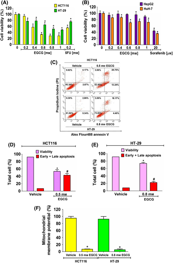
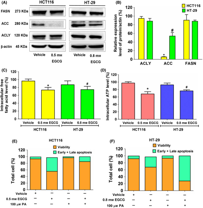
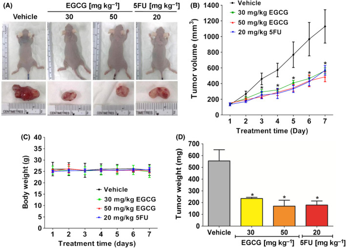
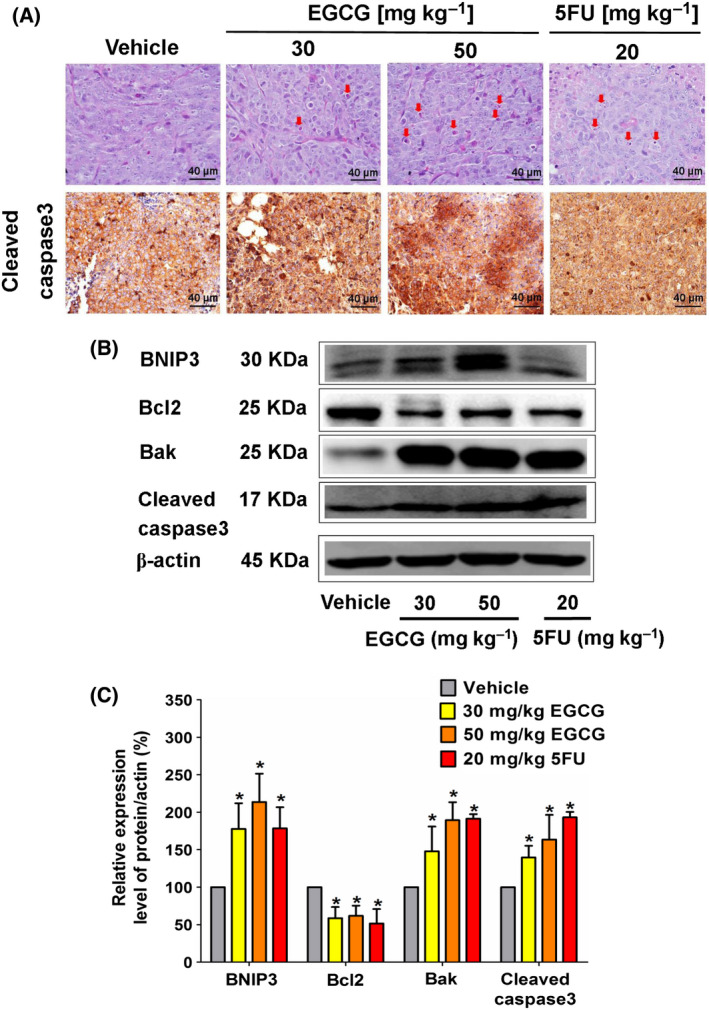
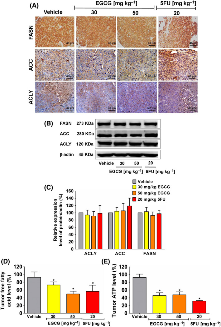
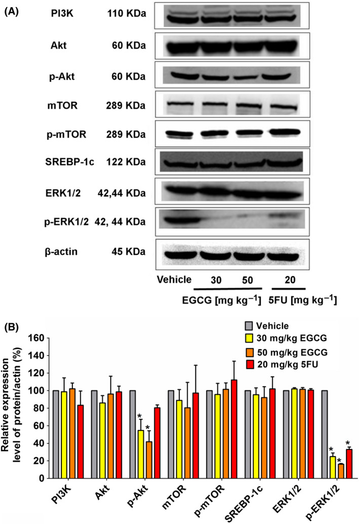
The de novo lipogenesis (DNL) pathway has been identified as a regulator of cancer progression and aggressiveness. Downregulation of key lipogenesis enzymes has been shown to activate apoptosis in cancerous cells. Epigallocatechin gallate (EGCG) inhibits cancer cell proliferation without causing cytotoxicity in healthy cells. The present study aimed to investigate the effects of EGCG on the promotion of apoptosis associated with the DNL pathway inhibition in cancer cells, both in vitro and in vivo. We observed that two colorectal cancer cell lines (HCT116 and HT-29) had a higher cytotoxic response to EGCG treatment than hepatocellular carcinoma cells, including HepG2 and HuH-7. EGCG treatment decreased cell viability and increased mitochondrial damage-triggered apoptosis in both HCT116 and HT-29 cancer cells. Additionally, we treated mice transplanted with HCT116 cells with 30 or 50 mg·kg-1 EGCG for 7 days to evaluate the apoptotic effects of EGCG treatment in a xenograft mouse model of cancer. We observed a decrease in intracellular fatty acid levels, which suggested that EGCG-induced apoptosis was associated with a decrease in fatty acid levels in cancer. Suppression of ATP synthesis by EGCG indicated that cell death induction in cancer cells could be mediated by shared components of the DNL and energy metabolism pathways. In addition, EGCG-induced apoptosis suppressed the expression of the phosphorylation protein kinase B and extracellular signal-regulated kinase 1/2 signaling proteins in tumors from xenografted mice. Cytotoxic effects in unaffected organs and tissues of the mouse xenograft model were absent upon EGCG treatment.

Keywords: PI3K/Akt/mTOR/SREBP-1c signaling; apoptosis; colorectal cancer; de novo lipogenesis; epigallocatechin gallate.


Conflict of interest statement

The authors declare no conflict of interest.

Figures

Fig. 1
Fig. 1
The inhibitory effect of EGCG on cell viability and induction of apoptosis cell death of cancer cells. Cells were incubated with EGCG for 24 h and subjected to a MTT assay. The percentage of cell viability (A) (HCT116 and HT‐29) and (B) (HepG2 and HuH‐7) following treatment was compared with 100% of the vehicle control group. (C) Cells were incubated with EGCG at an IC50 concentration for 24 h and then evaluated by annexin V/PI staining. The quantitative bar graphs show percentages of viability and early and late apoptotic cells of (D) HCT116 and (E) HT‐29 cells. (F) The JC‐1 staining assay was used to investigate EGCG‐induced mitochondrial damage. The histograms show the percentages of the red and green fluorescent intensity ratios. Data from at least three independent triplicated experiments are presented as the mean ± SD, n = 3. *P < 0.05 and # P < 0.05 compared to the control of each cell. All data were analyzed using one‐way ANOVA with Tukey’s post‐hoc test.
Fig. 2
Fig. 2
The effect of EGCG on expression of enzymes in the DNL pathway, free fatty acid and ATP levels in colorectal cancer cells, HCT116 and HT‐29 cells. Cells were incubated with EGCG at an IC50 concentration for 24 h. (A) An immunoblotting assay was used to assess the expression of enzymes in the DNL pathway. (B) A histogram depicts relative protein expression in comparison to β‐actin. (C) The histogram depicts free fatty acid levels and (D) ATP levels compared to the vehicle control. Apoptotic assay in (E) HCT116 and (F) HT‐29 cells using annexin V/PI staining in EGCG, palmitate (PA) at 100 μm, and a combination of EGCG and PA. Data are expressed as the mean ± SD from at least a triplicate of n = 3. *P < 0.05 and # P < 0.05 compared to the control of each cell. All data were analyzed using one‐way ANOVA with Tukey’s post‐hoc test.
Fig. 3
Fig. 3
The effect of EGCG on the proliferation of HCT116 tumor xenografts in nude mice. HCT116 cells were injected subcutaneously at the back area of nude mice. (A) Representative images show tumor formation and tumor nodules in nude mice. (B) Tumor volume and (C) body weigh were measured every day for 7 days and (D) tumor weight was measured at the end of the experiment. Representative data used five tumors in each group, presented as the mean ± SD. *P < 0.05 compared to the control. All data were analyzed using one‐way ANOVA with Tukey’s post‐hoc test.
Fig. 4
Fig. 4
The effect of EGCG on the morphology of the apoptotic characteristics and the expression of proapoptotic proteins in HCT116 tumor xenografts of nude mice. (A) Representative image of tumor hemotoxylin and eosin staining presents the morphology of apoptosis (arrow marks) and immunohistochemistry (scale bar = 40 μm) depicts the intensity of cleaved caspase‐3 expression in tumor tissues following the indicated dose of EGCG treatment. (B) A representative immunoblotting assay shows anti‐apoptotic and proapoptotic protein expression in tumor tissue and (C) a histogram depicts the quantitative protein expression/β‐actin level. Representative data from five tumors in each group were collected and presented as the mean ± SD. *P < 0.05 compared to the control using one‐way ANOVA with Tukey’s post‐hoc analysis.
Fig. 5
Fig. 5
The effect of EGCG on enzyme expression in the DNL pathway, free fatty acid and ATP levels in HCT116 tumor xenografts of nude mice. (A) Representative image of tumor immunohistochemistry staining (scale bar = 40 μm) shows the expression of enzymes in the DNL pathway. (B) ACLY, ACC and FASN expression was measured using an immunoblotting assay and (C) quantitated in a histogram compared to 100% of the control group. (D) The histogram represents free fatty acid and (E) ATP levels. Representative data were collected from five tumors in each group and presented as the mean ± SD. *P < 0.05 compared to the control using one‐way ANOVA with Tukey’s post‐hoc analysis.
Fig. 6
Fig. 6
The effect of EGCG on the upstream signaling PI3K/Akt/mTOR/SREBP‐1c and ERK pathways in HCT116 tumor xenografts of nude mice. (A) Representative immunoblotting shows expression proteins collected from five tumors in each group, which was quantitated in a histogram (B) compared to 100% of the control group. The values are presented as the mean ± SD. *P < 0.05 compared to the control using one‐way ANOVA with Tukey’s post‐hoc analysis.
Fig. 7
Fig. 7
The effect of EGCG on the internal organs of HCT116 tumor xenograft‐bearing mice. Representative images of hemotoxylin and eosin staining from five tumors in each group demonstrate the morphology of internal organs (scale bar = 40 μm) following treatment with the indicated agent and the positive control 5FU.

References

    1. Wang H. MicroRNAs and apoptosis in colorectal cancer. Int J Mol Sci. 2020;21:5353. - PMC - PubMed
    1. Wehler TC, Hamdi S, Maderer A, Graf C, Gockel I, Schmidtmann I, et al. Single‐agent therapy with sorafenib or 5‐FU is equally effective in human colorectal cancer xenograft—no benefit of combination therapy. Int J Colorectal Dis. 2013;28:385–98. - PMC - PubMed
    1. Chandimali N, Sun H‐N, Kong L‐Z, Zhen X, Liu R, Kwon T, et al. Shikonin‐induced apoptosis of colon cancer cells is reduced by peroxiredoxin V expression. Anticancer Res. 2019;39:6115–23. - PubMed
    1. Zhu W, Li MC, Wang FR, Mackenzie GG, Oteiza PI. The inhibitory effect of ECG and EGCG dimeric procyanidins on colorectal cancer cells growth is associated with their actions at lipid rafts and the inhibition of the epidermal growth factor receptor signaling. Biochem Pharmacol. 2020;175:113923. - PMC - PubMed
    1. Park J, Kim H‐D, Lee S‐H, Kwak C‐H, Chang Y‐C, Lee Y‐C, et al. Ascochlorin induces caspase‐independent necroptosis in LPS‐stimulated RAW 264.7 macrophages. J Ethnopharmacol. 2019;239:111898. - PubMed

Grants and funding

LinkOut - more resources