. 2023 Jan 5;33(3):597-611.
doi: 10.1093/cercor/bhac087.

Altered prefrontal signaling during inhibitory control in a salient drug context in cocaine use disorder

Affiliations

Altered prefrontal signaling during inhibitory control in a salient drug context in cocaine use disorder

Ahmet O Ceceli et al. Cereb Cortex. .

Abstract

Introduction: Drug addiction is characterized by impaired response inhibition and salience attribution (iRISA), where the salience of drug cues is postulated to overpower that of other reinforcers with a concomitant decrease in self-control. However, the neural underpinnings of the interaction between the salience of drug cues and inhibitory control in drug addiction remain unclear.

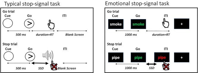
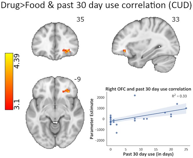
Methods: We developed a novel stop-signal functional magnetic resonance imaging task where the stop-signal reaction time (SSRT-a classical inhibitory control measure) was tested under different salience conditions (modulated by drug, food, threat, or neutral words) in individuals with cocaine use disorder (CUD; n = 26) versus demographically matched healthy control participants (n = 26).

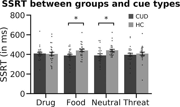
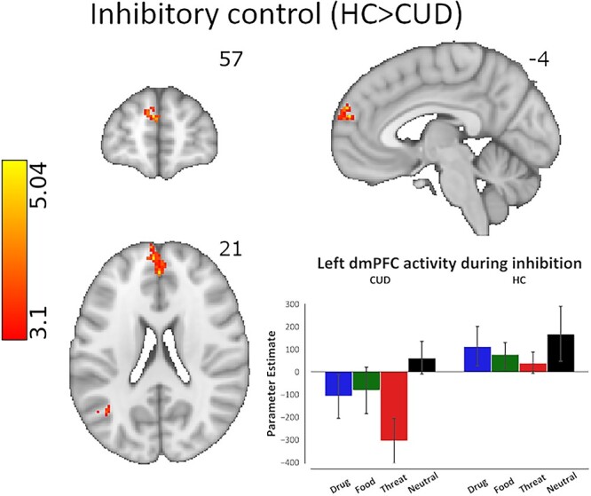
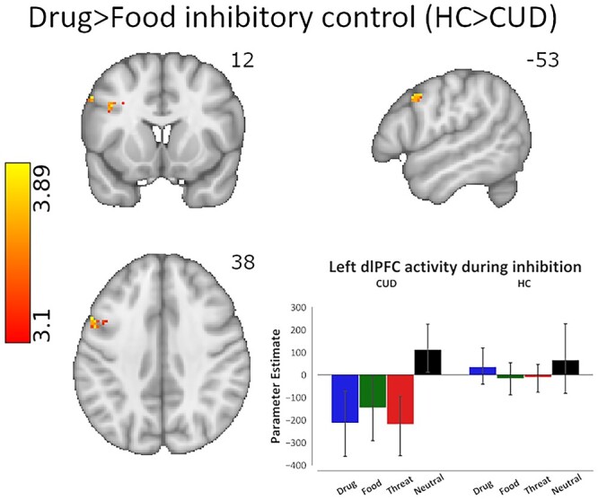
Results: Despite similarities in drug cue-related SSRT and valence and arousal word ratings between groups, dorsolateral prefrontal cortex (dlPFC) activity was diminished during the successful inhibition of drug versus food cues in CUD and was correlated with lower frequency of recent use, lower craving, and longer abstinence (Z > 3.1, P < 0.05 corrected).

Discussion: Results suggest altered involvement of cognitive control regions (e.g. dlPFC) during inhibitory control under a drug context, relative to an alternative reinforcer, in CUD. Supporting the iRISA model, these results elucidate the direct impact of drug-related cue reactivity on the neural signature of inhibitory control in drug addiction.

Keywords: craving; cue reactivity; dorsolateral prefrontal cortex; response inhibition; stop-signal task.


Figures

Fig. 1
Fig. 1
The emotional stop-signal task. Participants are instructed to respond via button presses as quickly and accurately as possible to the change in word color from white to blue/green, and to suppress their responses when the word color turns red after a variable delay (i.e. the SSD). Uniquely, word categories comprised neutral words, as well as drug, food, and threat-related words, permitting the examination of emotional reactivity during inhibitory control. Typical SST figure adapted from Aron and Poldrack 2006.
Fig. 2
Fig. 2
CUD and HC groups’ SSRT in response to each cue type. We found no significant main effects of group or cue but found a significant group × cue interaction (p = 0.027), where HC were significantly slower to stop than the CUD group in response to food and neutral cues. No drug-cue-related group differences were evident in SSRT.
Fig. 3
Fig. 3
Brain activity differences across CUD and HC groups during inhibitory control, regardless of cue type. The analysis of overall inhibitory control revealed significantly lower activity in the left dmPFC in the CUD group compared with HC (peak voxel MNI coordinates: x = −4, y = 57, z = 21). Cluster defining threshold: Z > 3.1 (P < 0.001), cluster corrected at P < 0.05. The bar plot of activation patterns shows relative activation to all cue types within the voxels that display peak activation as a result of the overall inhibitory control contrast. Inhibitory control was modeled via the hallmark Stop_Success > Stop_Fail contrast of task events corresponding to all cues.
Fig. 4
Fig. 4
Brain activity differences across CUD and HC groups during inhibitory control under drug cue reactivity. The linear contrast of drug cue- versus a competing, nondrug positive reinforcer, food cue-related inhibitory control revealed significantly lower activity in the left dlPFC in the CUD group compared with HC (peak voxel MNI coordinates: x = −53, 12, 38). Cluster defining threshold: Z > 3.1 (P < 0.001), cluster corrected at P < 0.05. The bar plot of activation patterns shows relative activation to all cue types within the voxels that display peak activation as a result of the drug > food inhibitory control contrast. Inhibitory control was modeled via the hallmark Stop_Success > Stop_Fail contrast of task events corresponding to drug and food cues.
Fig. 5
Fig. 5
Whole-brain correlational analysis of cocaine use severity as assayed by recent cocaine use frequency in the past month, and its relationship with inhibitory control under drug cue reactivity (drug > food inhibitory control contrast). When entered into the GLM as a covariate in a whole-brain analysis, recent cocaine use frequency showed significant positive correlations with activity in the right OFC, right dmPFC, and left dlPFC in the CUD group, such that the more severe the cocaine use, the higher the activation in these prefrontal regions during inhibitory control under drug cue reactivity (peak voxel MNI coordinates: x = 33, y = 35, z = −9). Cluster defining threshold: Z > 3.1 (P < 0.001), cluster corrected at P < 0.05. Inhibitory control was modeled via the hallmark Stop_Success > Stop_Fail contrast of task events corresponding to drug and food cues.

References

    1. American Psychiatric Association , editors. Diagnostic and statistical manual of mental disorders. 5th ed.: American Psychiatric Association; 2013
    1. Ames SL, Wong SW, Bechara A, Cappelli C, Dust M, Grenard JL, Stacy AW. Neural correlates of a go/NoGo task with alcohol stimuli in light and heavy young drinkers. Behav Brain Res. 2014:0:382–389. - PMC - PubMed
    1. Aron AR, Poldrack RA. Cortical and subcortical contributions to stop signal response inhibition: role of the subthalamic nucleus. J Neurosci. 2006:26:2424–2433. - PMC - PubMed
    1. Aron AR, Behrens TE, Smith S, Frank MJ, Poldrack RA. Triangulating a cognitive control network using diffusion-weighted magnetic resonance imaging (MRI) and functional MRI. J Neurosci. 2007:27:3743–3752. - PMC - PubMed
    1. Avants BB, Epstein CL, Grossman M, Gee JC. 2008. Symmetric diffeomorphic image registration with cross-correlation: evaluating automated labeling of elderly and neurodegenerative brain. Medical Image Analysis, Special Issue on the Third International Workshop on Biomedical Image Registration – WBIR 2006. 12:26–41. - PMC - PubMed

Publication types