. 2022 Mar 4;79(3):173.
doi: 10.1007/s00018-022-04197-2.

An imbalance of the IL-33/ST2-AXL-efferocytosis axis induces pregnancy loss through metabolic reprogramming of decidual macrophages

Affiliations

An imbalance of the IL-33/ST2-AXL-efferocytosis axis induces pregnancy loss through metabolic reprogramming of decidual macrophages

Yan-Ran Sheng et al. Cell Mol Life Sci. .

Abstract

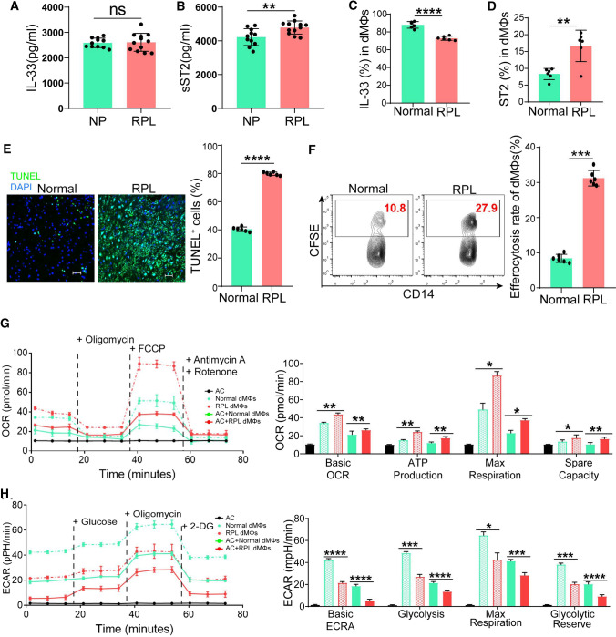
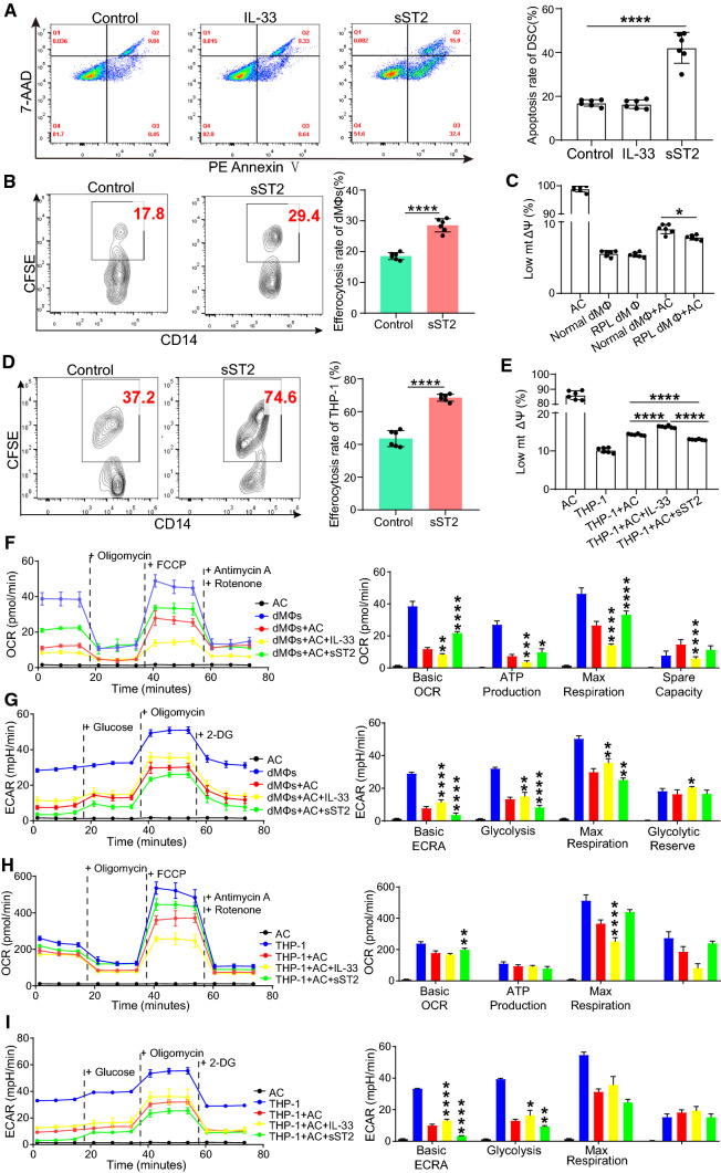
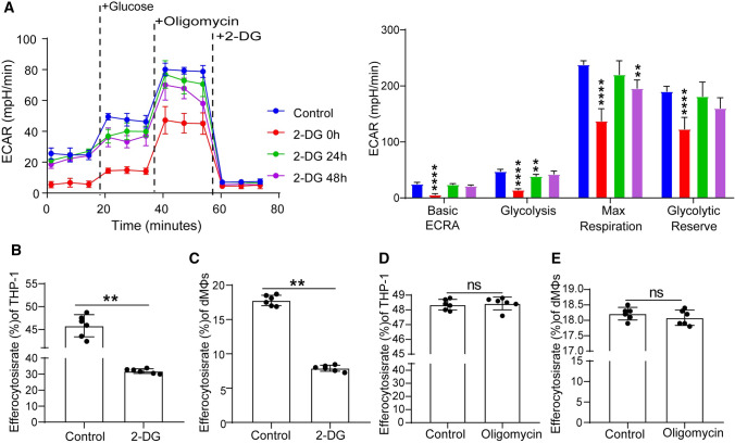
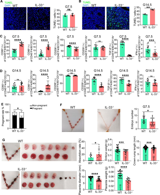
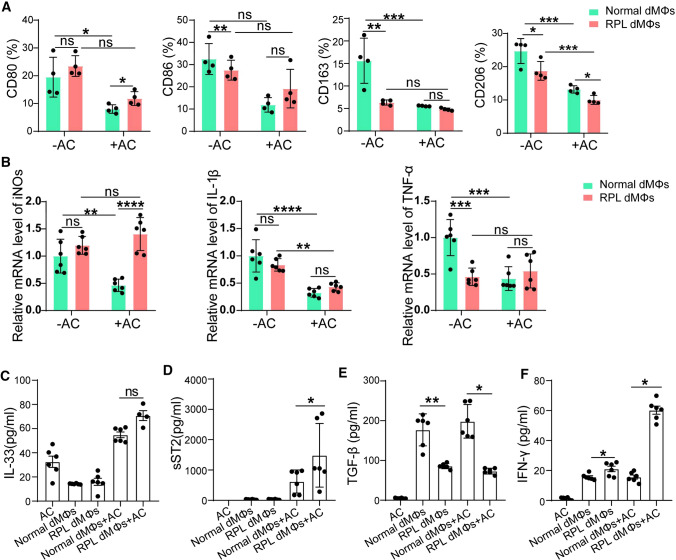
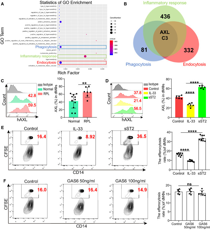
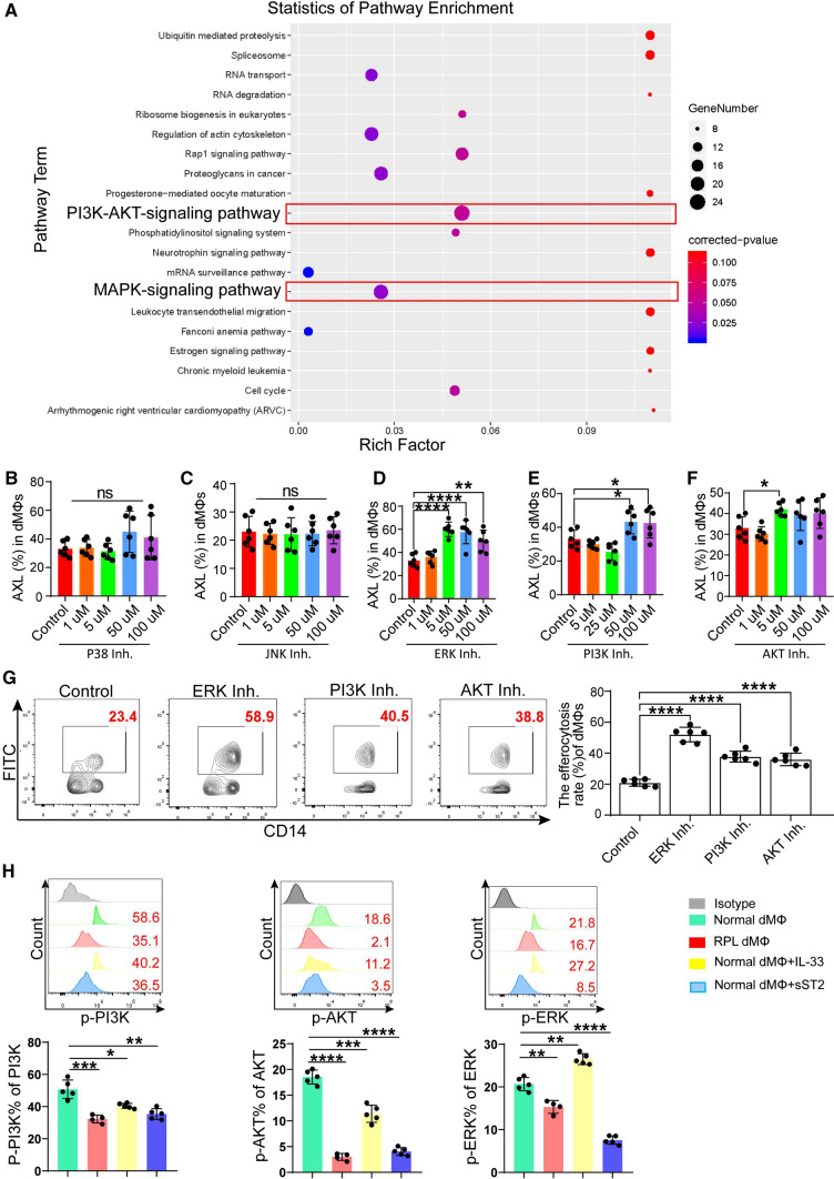
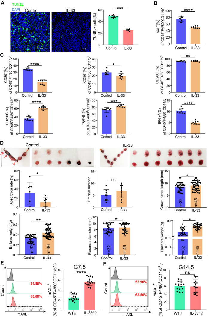
During embryo implantation, apoptosis is inevitable. These apoptotic cells (ACs) are removed by efferocytosis, in which macrophages are filled with a metabolite load nearly equal to the phagocyte itself. A timely question pertains to the relationship between efferocytosis-related metabolism and the immune behavior of decidual macrophages (dMΦs) and its effect on pregnancy outcome. Here, we report positive feedback of IL-33/ST2-AXL-efferocytosis leading to pregnancy failure through metabolic reprogramming of dMΦs. We compared the serum levels of IL-33 and sST2, along with IL-33 and ST2, efferocytosis and metabolism of dMΦs, from patients with normal pregnancies and unexplained recurrent pregnancy loss (RPL). We revealed disruption of the IL-33/ST2 axis, increased apoptotic cells and elevated efferocytosis of dMΦs from patients with RPL. The dMΦs that engulfed many apoptotic cells secreted more sST2 and less TGF-β, which polarized dMΦs toward the M1 phenotype. Moreover, the elevated sST2 biased the efferocytosis-related metabolism of RPL dMΦs toward oxidative phosphorylation and exacerbated the disruption of the IL-33/ST2 signaling pathway. Metabolic disorders also lead to dysfunction of efferocytosis, resulting in more uncleared apoptotic cells and secondary necrosis. We also screened the efferocytotic molecule AXL regulated by IL-33/ST2. This positive feedback axis of IL-33/ST2-AXL-efferocytosis led to pregnancy failure. IL-33 knockout mice demonstrated poor pregnancy outcomes, and exogenous supplementation with mouse IL-33 reduced the embryo losses. These findings highlight a new etiological mechanism whereby dMΦs leverage immunometabolism for homeostasis of the microenvironment at the maternal-fetal interface.

Keywords: Decidual macrophages; Efferocytosis; IL-33/ST2 axis; Metabolic immune reprogramming; Recurrent pregnancy loss.


Conflict of interest statement

The authors declare no conflict of interest.

Figures

Fig. 1
Fig. 1
sST2 is increased in the serum of patients with unexplained RPL, accompanied by increased apoptotic cells, enhanced efferocytosis and a mitochondrial bias during efferocytosis. Serum samples from 10 patients with normal pregnancies and 12 patients with unexplained RPL were collected to determine the levels of IL-33 (A) and sST2 (B) by ELISAs. The expression of IL-33 (C) and ST2 (D) in dMΦs from the patients with normal pregnancies and unexplained RPL was detected by flow cytometry. (E) The number of apoptotic cells at the maternal–fetal interface of the patients with normal pregnancies and unexplained RPL tested by TUNEL staining. (F) The efferocytosis ability of dMΦs from the patients with normal pregnancies and unexplained RPL. (G) The oxidative phosphorylation (OXPHOS) level of efferocytosis-related metabolism when normal (green solid line) and RPL (red solid line) dMΦs were cocultured with or without AC (indicated by the green dashed and red dashed line, respectively). (H) The glycolytic level of efferocytosis-related metabolism when normal (green solid line) and RPL (red solid line) dMΦs were cocultured with or without AC (indicated by the green dashed and red dashed line, respectively). All experiments were performed in triplicate. The data were analyzed by unpaired Student’s two-tailed t test for comparison of two groups and by two-way ANOVA followed by Tukey’s post-hoc test for multiple groups. The data are shown as the mean ± SD. *P < 0.05, **P < 0.01, ***P < 0.001, ****P < 0.0001, ns no statistically significant difference
Fig. 2
Fig. 2
Dysfunction of the IL-33/ST2 axis promotes DSC apoptosis and dMΦ/THP-1 efferocytosis and leads to an OXPHOS bias during efferocytosis. (A) The impact of IL-33 (2 ng/ml, 48 h) and sST2 (200 ng/ml, 48 h) on the apoptosis rate of normal DSCs. (B) The impact of sST2 (200 ng/ml, 48 h) on the efferocytosis of normal dMΦs. (C) The mitochondrial membrane potential (mtΔΨ) of the normal and RPL dMΦs conducting efferocytosis. (D) The impact of sST2 (200 ng/ml, 48 h) on the efferocytosis of THP-1 cells. (E) The impact of IL-33 (2 ng/ml, 48 h) and sST2 (200 ng/ml, 48 h) on the mtΔΨ of THP-1 processing efferocytosis. (F) The OXPHOS level of efferocytosis-related metabolism in normal dMΦs affected by IL-33 (2 ng/ml, 48 h) and sST2 (200 ng/ml, 48 h). (G) The glycolytic level of efferocytosis-related metabolism in normal dMΦs affected by IL-33 (2 ng/ml, 48 h) and sST2 (200 ng/ml, 48 h). The effect of IL-33 (2 ng/ml, 48 h) and sST2 (200 ng/ml, 48 h) on the OXPHOS level (H) and glycolytic level (I) of the THP-1 cell line processing efferocytosis. All experiments were performed in triplicate. The data were analyzed by two-way ANOVA followed by Tukey’s post-hoc test for multiple comparisons. The data are shown as the mean ± SD. *P < 0.05, **P < 0.01, ***P < 0.001, ****P < 0.0001
Fig. 3
Fig. 3
Efferocytosis is more dependent on glycolysis than oxidative phosphorylation. (A) The inhibitory efficiency of different treatment durations of 2-DG (10 µM) on glycolytic metabolism of THP-1 cells compared with the controls. The impact of 2-DG (10 µM) on the efferocytosis of THP-1 cells (B) and dMΦs (C). The impact of oligomycin (1 µM) on the efferocytosis of THP-1 cells (D) and dMΦs (E). All experiments were performed at least three times. The data were analyzed by paired Student’s two-tailed t test for comparison of two groups and by two-way ANOVA with Tukey’s post-hoc test for multiple comparisons for more than two groups. The data are shown as the mean ± SD. *P < 0.05, **P < 0.01, ***P < 0.001, ****P < 0.0001, ns no statistically significant difference
Fig. 4
Fig. 4
IL-33 deficiency increases the risk of pregnancy failure. The number of apoptotic cells at the maternal–fetal interface of pregnant WT and IL-33−/− mice at G7.5 days (A) and G14.5 days (B) tested by TUNEL staining. The expression of CD80, CD86, CD206, CD209, TGF-β and IFN-γ in CD45+F4/80+CD11/b+ uMΦs from the pregnant WT and IL-33−/− mice at G7.5 days (C) and G14.5 days (D). (E) The pregnancy rate of the pregnant WT and IL-33−/− mice at G7.5 and G14.5 days. (F) The implantation number of the pregnant WT and IL-33−/− mice at G7.5 days. (G) The embryo absorption rate, weight and crown-rump length of embryos and the diameter and weight of placentas of pregnant WT and IL-33−/− mice at G14.5 days. Each experiment was performed at least three times. The data were analyzed by unpaired Student’s two-tailed t test for comparison. The data are shown as the mean ± SD. *P < 0.05, **P < 0.01, ***P < 0.001, ****P < 0.0001, ns no statistically significant difference
Fig. 5
Fig. 5
DMΦs from patients with RPL are prone to persistent M1 imbalance and high sST2 after efferocytosis. (A) The expression of CD80, CD86, CD163, and CD206 in normal and RPL dMΦs before and after efferocytosis. (B) Quantitative RT-PCR analysis of the expression levels of iNOS, IL-1β and TNF-α in normal/RPL dMΦs before and after efferocytosis. The concentrations of IL-33 (C), sST2 (D), TGF-β (E), and IFN-γ (F) in the supernatant of dMΦs before and after efferocytosis. All experiments were conducted at least three times. The data of (A) and (B) were analyzed by two-way ANOVA followed by Sidak’s multiple comparisons test. The data in (CF) were analyzed by one-way ANOVA with Tukey’s post-hoc test for multiple comparisons. The data are shown as the mean ± SD. *P < 0.05, **P < 0.01, ***P < 0.001, ****P < 0.0001, ns no statistically significant difference
Fig. 6
Fig. 6
IL-33 suppresses efferocytosis of dMΦs by downregulating the expression of the efferocytosis-related receptor AXL. (A) The GO enrichment analysis diagram of the genes with downregulated expression (HIST1H4C, HIST1H1E, HIST1H2BE, SMIM11A, PLEC, KMT2D, SOGA1, SIGLEC1, C3, EVPL, AXL, GP1BB, PTGDS, HIST2H3A, HIST2H3C, HSPG2). (B) The genes involved in the inflammatory response, phagocytosis and endocytosis pathways were intersected in the form of a Venn diagram. (C) The expression level of human AXL protein in normal and RPL dMΦs. The impact of IL-33 (2 ng/ml, 48 h) and sST2 (200 ng/ml, 48 h) on the expression of AXL (D) and the efferocytosis efficiency (E) of normal dMΦs. (F) The effect of GAS6 (50 ng/ml, 100 ng/ml, 2 h) on the efferocytosis of normal dMΦs. All experiments were performed at least three times. The data were analyzed by unpaired Student’s two-tailed t test for comparison of two groups and by one-way ANOVA with Tukey’s post-hoc test for multiple comparisons for more than two groups. The data are shown as the mean ± SD. *P < 0.05, **P < 0.01, ***P < 0.001, ****P < 0.0001, ns no statistically significant difference
Fig. 7
Fig. 7
The IL-33/ST2 axis inhibits AXL expression in dMΦs by activating the PI3K/AKT and ERK1/2 signaling pathways. (A) The GO enrichment analysis diagram of the genes with upregulated expression in ST2-OE THP-1 cells treated with IL-33 for 48 h and the NC group. The effects of the P38 inhibitor (B), JNK inhibitor (C), ERK inhibitor (D), PI3K inhibitor (E), and AKT inhibitor (F) at different concentrations on the AXL expression of normal dMΦs. (G) The impact of ERK, PI3K, and AKT inhibitors on the efferocytosis efficiency of normal dMΦs. All inhibitors were cocultured with CD14+ dMΦs for 24 h, and during the following 48 h, 2 ng/ml IL-33 was added in the presence of inhibitors. (H) Normal dMΦs were treated with IL-33 or sST2 for 48 h, PI3K, AKT and ERK phosphorylation was analyzed by flow cytometry (n = 5), and normal and RPL dMΦs without treatment served as controls. Each experiment was performed at least three times. The data were analyzed by one-way ANOVA with Tukey’s post-hoc test for multiple comparisons. The data are presented as the mean ± SD after analysis with unpaired t tests. *P < 0.05, **P < 0.01, ***P < 0.001, ****P < 0.0001, ns no statistically significant difference
Fig. 8
Fig. 8
Exogenous IL-33 prevents pregnancy failure in IL-33−/− pregnant mice. (A) The number of apoptotic cells at the maternal–fetal interface of IL-33−/− pregnant mice with or without intraperitoneal injection of IL-33 at G14.5 days tested by TUNEL staining. (B) The expression of AXL on CD45+F4/80+CD11/b+ uMΦs from the IL-33−/− pregnant mice with or without exogenous peritoneal injection of IL-33 (200 ng/100 µl, G3.5, G7.5, G11.5) on G14.5 days. (C) The expression of CD80, CD86, CD206, CD209, TGF-β, and IFN-γ in CD45+F4/80+CD11/b+ uMΦs from the IL-33−/− pregnant mice with or without exogenous peritoneal injection of IL-33 on G14.5 days. (D) The embryo absorption rate, embryo number, crown-rump length and weight of the embryos, and diameter and weight of the placentas of the IL-33−/− pregnant mice with or without exogenous peritoneal injection of IL-33 on G14.5 days. Every experiment was repeated independently at least three times. The expression of AXL on CD45+F4/80+CD11/b+ uMΦs from the WT and IL-33−/− pregnant mice on G7.5 days (E) and G14.5 days (F). The data of AD were analyzed by paired Student’s two-tailed t test for comparison. The data of EF were analyzed by unpaired Student’s two-tailed t test for comparison. The data are shown as the mean ± SD. *P < 0.05, **P < 0.01, ***P < 0.001, ****P < 0.0001, ns no statistically significant difference

References

    1. Doran AC, Yurdagul A, Tabas I. Efferocytosis in health and disease. Nat Rev Immunol. 2020;20(4):254–267. doi: 10.1038/s41577-019-0240-6. - DOI - PMC - PubMed
    1. Boada-Romero E, Martinez J, Heckmann BL, et al. The clearance of dead cells by efferocytosis. Nat Rev Mol Cell Biol. 2020;21(7):398–414. doi: 10.1038/s41580-020-0232-1. - DOI - PMC - PubMed
    1. Zhou Y, Yao Y, Deng Y, et al. Regulation of efferocytosis as a novel cancer therapy. Cell Commun Signal. 2020;18(1):71. doi: 10.1186/s12964-020-00542-9. - DOI - PMC - PubMed
    1. Von Rango U, Krusche CA, Kertschanska S, et al. Apoptosis of extravillous trophoblast cells limits the trophoblast invasion in uterine but not in tubal pregnancy during first trimester. Placenta. 2003;24(10):929–940. doi: 10.1016/s0143-4004(03)00168-1. - DOI - PubMed
    1. Hoijman E, Häkkinen H-M, Tolosa-Ramon Q, et al. Cooperative epithelial phagocytosis enables error correction in the early embryo. Nature. 2021;590(7847):618–623. doi: 10.1038/s41586-021-03200-3. - DOI - PubMed

MeSH terms