. 2022 May;27(3):223-239.
doi: 10.1007/s12192-022-01263-3. Epub 2022 Mar 4.

Canine osteosarcoma cells exhibit basal accumulation of multiple chaperone proteins and are sensitive to small molecule inhibitors of GRP78 and heat shock protein function

Affiliations

Canine osteosarcoma cells exhibit basal accumulation of multiple chaperone proteins and are sensitive to small molecule inhibitors of GRP78 and heat shock protein function

Daphne R Mattos et al. Cell Stress Chaperones. 2022 May.

Abstract

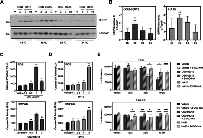
Osteosarcoma is the most common type of bone cancer in dogs and humans, with significant numbers of patients experiencing treatment failure and disease progression. In our search for new approaches to treat osteosarcoma, we previously detected multiple chaperone proteins in the surface-exposed proteome of canine osteosarcoma cells. In the present study, we characterized expression of representative chaperones and find evidence for stress adaptation in canine osteosarcoma cells relative to osteogenic progenitors from normal bone. We compared the cytotoxic potential of direct (HA15) and putative (OSU-03012) inhibitors of Grp78 function and found canine POS and HMPOS osteosarcoma cells to be more sensitive to both compounds than normal cells. HA15 and OSU-03012 increased the thermal stability of Grp78 in intact POS cells at low micromolar concentrations, but each induced distinct patterns in Grp78 expression without significant change in Grp94. Both inhibitors were as effective alone as carboplatin and showed little evidence of synergy in combination treatment. However, HMPOS cells with acquired resistance to carboplatin were sensitive to inhibition of Grp78 (by HA15; OSU-03012), Hsp70 (by VER-155008), and Hsp90 (by 17-AAG) function. These results suggest that multiple nodes within the osteosarcoma chaperome may be relevant chemotherapeutic targets against platinum resistance.

Keywords: AR-12; BiP; Chaperome; HA15; HSP5A; OSU-03012; Proteostasis.


Conflict of interest statement

The authors declare no competing interests.

Figures

Fig. 1
Fig. 1
Localization of Grp78 in canine osteosarcoma cells. A Immunoblot analysis of Grp78, Grp94, Hsp90, Hsp70, Hsp40, and α-tubulin expression in whole cell lysates prepared from normal osteogenic progenitor cells (OPCs) from normal bone, POS, and HMPOS canine osteosarcoma cancer cells. B Immunoblot analysis of Grp78 and representative control proteins in cytoplasmic (C), membrane (M), and nuclear (N) fractions from canine POS and HMPOS osteosarcoma cells. Samples were probed with anti-Grp78 and primary antibodies for detection of cytosolic (Hsp90, GAPDH), membrane (EGFR), and nuclear (HDAC2) proteins as indicated. Whole cell lysates (panel A) or cell fractions (panel B) were prepared at the same time; images are representative of three independent comparisons of all cell lines. C Immunocytochemical analysis of Grp78 distribution in canine POS osteosarcoma cells. Top row: nonpermeabilized or permeabilized cells were stained with anti-Grp78 (green) or anti-Grp78 plus DAPI to visualize nuclei stained (blue), respectively. Bottom row: nonpermeabilized cells were stained with anti-integrin β-1 (red) or anti-integrin β-1 plus anti-Grp78 (merged). D Quantification of corrected total cell fluorescence (CTCF) in cells co-stained for integrin β-1 plus Grp78 measured from three independent comparisons (n = 29 cells)
Fig. 2
Fig. 2
Comparative analysis of basal and stress-induced Grp78 expression in canine and human cancer cells. A Immunoblot analysis of Grp78 and α-tubulin expression in (from left to right) human SAOS-2 osteosarcoma, human SK-ES-1 Ewing’s sarcoma, canine osteosarcoma (D17, COS, POS), human glioblastoma (SF-295, U87-MG, U251-MG), human breast (MDA-MB-231, MCF-7), human DU145 prostate, human SAOS-2 osteosarcoma, human HCT116 colon, and human SKOV-3 ovarian cancer cells. Whole cell lysates, for each panel, were prepared at the same time and processed for Western blot analysis with the antibodies indicated. Lysates were analyzed on separate gels with human SAOS-2 osteosarcoma cells common to the 5- and 9-panel comparison; images are representative of three independent comparisons of each panel. B Immunoblot analysis of Grp78, CHOP, and α-tubulin expression in the absence (0.1% DMSO) or presence of thapsigargin (3 µM) in human SAOS-2 osteosarcoma, canine D17 or COS osteosarcoma cells, and human SK-ES-1 Ewing’s sarcoma, canine POS, or HMPOS osteosarcoma cells. Cells were grown under standard conditions and treated with, or without, thapsigargin for 24 h. Whole cell lysates were probed with antibodies against Grp78, CHOP, and α-tubulin (loading control) as indicated. Images are representative of a comparison that was repeated three times
Fig. 3
Fig. 3
Analysis of Grp78 and Grp94 expression in response to treatment with OSU-03012, apratoxin A, or HA15. A Expression of Grp78 and Grp94, relative to calreticulin and α-tubulin, in canine POS osteosarcoma cells in the absence (0.1% DMSO) or presence of OSU-03012 (3 µM), apratoxin A (300 nM), and HA15 (10 µM) after 8 h or 24 h. At the end of treatment, whole cell lysates were processed for immunoblot analysis and probed with antibodies against Grp78, Grp94, calreticulin, and α-tubulin as indicated. B Histograms show quantification of Grp78 and Grp94 expression from three independent experiments. Signals were normalized to α-tubulin and statistical significance in treated versus untreated cells indicated as *p < 0.05, **p < 0.01, ***p < 0.001, or ****p < 0.0001
Fig. 4
Fig. 4
Concentration–response analysis of OSU-03012-, apratoxin A-, and HA15-induced changes in cell viability in normal canine osteoblasts relative to representative human and canine osteosarcoma cells. Cell viability of A canine osteogenic progenitor cells (OPCs), B human SAOS-2 osteosarcoma, C canine POS osteosarcoma, and D canine HMPOS osteosarcoma cells exposed at the same time to increasing concentrations of OSU-03012, apratoxin A, HA15, or vehicle (0.1% DMSO) for 72 h. Cell viability was determined by the MTT assay. The viability of vehicle-treated cells was defined as 100%. Graphs show a single comparison of three compounds across four cell types that was repeated three times. Points refer to average viability (n = 3 wells) and curves represent the fit of data points by nonlinear regression analysis to a logistic equation
Fig. 5
Fig. 5
OSU-03012 and HA15 stabilize Grp78 in intact canine osteosarcoma cells and induce cell death. A Analysis of Grp78 thermal stability in the presence of 0.1% DMSO (V), 3 µM OSU-03012 (OSU), or 3 µM and 10 µM HA15. Melting curves were generated by exposing intact POS cells (4 × 107) to each treatment as indicated for 1 h. Cells were collected by gentle centrifugation, divided into equal aliquots, and subjected to a single fixed temperature (ranging from 45 to 54 °C). Immunoblot analysis of Grp78 and α-tubulin expression is representative of a single melting curve that was repeated three times with similar results. B Quantification of melting curve data (at highest concentration tested) from three independent studies. C OSU-03012 and D HA15 induce concentration-dependent increases in caspase-3,7 activity in POS and HMPOS osteosarcoma cells. Bars represent mean luminescence values (n = 3 wells per treatment) determined after the 24-h exposure to each compound, as indicated, and are expressed as relative light units ± SE from three independent determinations. E Comparison of the ability of a pan-caspase inhibitor to rescue POS or HMPOS cells from OSU-03012- or HA15-induced cytotoxicity. Cells were exposed to vehicle (0.1% DMSO), OSU-03012 (1 µM, 3 µM, or 10 µM), and HA15 (1 µM, 3 µM, or 10 µM), with or without V-ZAD-fmk (50 µM), and cell viability was assessed using the MTT assay after 24 h. The viability of vehicle-treated cells was defined as 100%. Data points show mean viability ± SE (n = 3 wells per treatment) from a representative comparison that was repeated in three independent experiments. Statistical significance of change in treated, relative to vehicle-treated (0.1% DMSO), cells is indicated as *p < 0.05, **p < 0.01, and ***p < 0.001
Fig. 6
Fig. 6
Analysis of the cytotoxic efficacy of carboplatin in combination with OSU-03012 or HA15 treatment. A POS and B HMPOS osteosarcoma cell viability in response to carboplatin alone (3 nM to 100 µM) and in combination with a fixed concentration of OSU-03012 (1 µM) or HA15 (1 µM). Cells were exposed to single or combination treatments, as indicated, and cell viability was determined at 72 h with the viability of vehicle-treated (0.1% DMSO) cells defined as 100% viability. Data points represent mean viability ± SE (n = 3 wells per treatment), and curves represent the fit of data points by nonlinear regression analysis to a logistic equation. Cell viability curves are representative of a comparison that was repeated three times. Combination index plots for C POS and D HMPOS osteosarcoma cells for fixed combination treatments of carboplatin plus HA15 or OSU-03012 using the Chou-Talalay combination index method. Points below 1 (indicated by the dashed line) indicate synergy
Fig. 7
Fig. 7
Carboplatin-resistant canine osteosarcoma cells remain sensitive to Grp78 inhibitors and small molecular inhibitors of other molecular chaperones. A Characterization of chemoresistant HMPOS canine osteosarcoma cell lines. Parental canine HMPOS osteosarcoma and two carboplatin-resistant variants of HMPOS cell lines (resistant 2.5 CB HMPOS and resistant 10 CB HMPOS) were exposed to increasing concentrations of carboplatin for 72 h. BE Concentration–response analysis of parental and chemoresistant canine osteosarcoma cells in the presence of B OSU-03012, C HA15, D Hsp70 inhibitor VER-155008, and E Hsp90 inhibitor 17-AAG. Cells were treated at the same time, and cell viability was assessed using a MTT assay. Data points represent mean viability ± SE (n = 3 wells per treatment) relative to vehicle control (0.1% DMSO). Each panel is representative of a comparison that was repeated three times in independent determinations

Similar articles

Cited by

References

    1. Aljubran AH, Griffin A, Pintilie M, Blackstein M. Osteosarcoma in adolescents and adults: survival analysis with and without lung metastases. Ann Oncol. 2009;20(6):1136–1141. doi: 10.1093/annonc/mdn731. - DOI - PubMed
    1. Bailly C, Waring MJ. Pharmacological effectors of GRP78 chaperone in cancers. Biochem Pharmacol. 2019;163:269–278. doi: 10.1016/j.bcp.2019.02.038. - DOI - PubMed
    1. Balch WE, Morimoto RI, Dillin A, Kelly JW. Adapting proteostasis for disease intervention. Science. 2008;319(5865):916–919. doi: 10.1126/science.1141448. - DOI - PubMed
    1. Bhattacharjee R, Devi A, Mishra S. Molecular docking and molecular dynamics studies reveal structural basis of inhibition and selectivity of inhibitors EGCG and OSU-03012 toward glucose regulated protein-78 (GRP78) overexpressed in glioblastoma. J Mol Model. 2015;21(10):272. doi: 10.1007/s00894-015-2801-3. - DOI - PubMed
    1. Booth L, Cazanave SC, Hamed HA, Yacoub A, Ogretmen B, Chen CS, Grant S, Dent P. OSU-03012 suppresses GRP78/BiP expression that causes perk-dependent increases in tumor cell killing. Cancer Biol Ther. 2012;13(4):224–236. doi: 10.4161/cbt.13.4.18877. - DOI - PMC - PubMed

Publication types