. 2022 Mar 4;3(3):CD010088.
doi: 10.1002/14651858.CD010088.pub3.

Routine vaginal examinations compared to other methods for assessing progress of labour to improve outcomes for women and babies at term

Affiliations

Routine vaginal examinations compared to other methods for assessing progress of labour to improve outcomes for women and babies at term

Gill Moncrieff et al. Cochrane Database Syst Rev. .

Abstract

Background: Routine vaginal examinations are undertaken at regular time intervals during labour to assess whether labour is progressing as expected. Unusually slow progress can be due to underlying problems, described as labour dystocia, or can be a normal variation of progress. Evidence suggests that if mother and baby are well, length of labour alone should not be used to decide whether labour is progressing normally. Other methods to assess labour progress include intrapartum ultrasound and monitoring external physical and behavioural cues. Vaginal examinations can be distressing for women, and overdiagnosis of dystocia can result in iatrogenic morbidity due to unnecessary intervention. It is important to establish whether routine vaginal examinations are effective, both as an accurate measure of physiological labour progress and to distinguish true labour dystocia, or whether other methods for assessing labour progress are more effective. This Cochrane Review is an update of a review first published in 2013.

Objectives: To compare the effectiveness, acceptability, and consequences of routine vaginal examinations compared with other methods, or different timings, to assess labour progress at term.

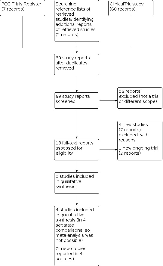
Search methods: For this update, we searched Cochrane Pregnancy and Childbirth Trials Register (which includes trials from CENTRAL, MEDLINE, Embase, CINAHL, and conference proceedings) and ClinicalTrials.gov (28 February 2021). We also searched the reference lists of retrieved studies.

Selection criteria: We included randomised controlled trials (RCTs) of vaginal examinations compared with other methods of assessing labour progress and studies assessing different timings of vaginal examinations. Quasi-RCTs and cluster-RCTs were eligible for inclusion. We excluded cross-over trials and conference abstracts.

Data collection and analysis: Two review authors independently assessed all studies identified by the search for inclusion in the review. Four review authors independently extracted data. Two review authors assessed risk of bias and certainty of the evidence using GRADE.

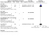
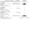
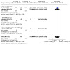
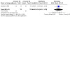
Main results: We included four studies that randomised a total of 755 women, with data analysed for 744 women and their babies. Interventions used to assess labour progress were routine vaginal examinations, routine ultrasound assessments, routine rectal examinations, routine vaginal examinations at different frequencies, and vaginal examinations as indicated. We were unable to conduct meta-analysis as there was only one study for each comparison. All studies were at high risk of performance bias due to difficulties with blinding. We assessed two studies as high risk of bias and two as low or unclear risk of bias for other domains. The overall certainty of the evidence assessed using GRADE was low or very low. Routine vaginal examinations versus routine ultrasound to assess labour progress (one study, 83 women and babies) Study in Turkey involving multiparous women with spontaneous onset of labour. Routine vaginal examinations may result in a slight increase in pain compared to routine ultrasound (mean difference -1.29, 95% confidence interval (CI) -2.10 to -0.48; one study, 83 women, low certainty evidence) (pain measured using a visual analogue scale (VAS) in reverse: zero indicating 'worst pain', 10 indicating no pain). The study did not assess our other primary outcomes: positive birth experience; augmentation of labour; spontaneous vaginal birth; chorioamnionitis; neonatal infection; admission to neonatal intensive care unit (NICU). Routine vaginal examinations versus routine rectal examinations to assess labour progress (one study, 307 women and babies) Study in Ireland involving women in labour at term. We assessed the certainty of the evidence as very low. Compared with routine rectal examinations, routine vaginal examinations may have little or no effect on: augmentation of labour (risk ratio (RR) 1.03, 95% CI 0.63 to 1.68; one study, 307 women); and spontaneous vaginal birth (RR 0.98, 95% CI 0.90 to 1.06; one study, 307 women). We found insufficient data to fully assess: neonatal infections (RR 0.33, 95% CI 0.01 to 8.07; one study, 307 babies); and admission to NICU (RR 1.32, 95% CI 0.47 to 3.73; one study, 307 babies). The study did not assess our other primary outcomes: positive birth experience; chorioamnionitis; maternal pain. Routine four-hourly vaginal examinations versus routine two-hourly examinations (one study, 150 women and babies) UK study involving primiparous women in labour at term. We assessed the certainty of the evidence as very low. Compared with routine two-hourly vaginal examinations, routine four-hourly vaginal examinations may have little or no effect, with data compatible with both benefit and harm, on: augmentation of labour (RR 0.97, 95% CI 0.60 to 1.57; one study, 109 women); and spontaneous vaginal birth (RR 1.02, 95% CI 0.83 to 1.26; one study, 150 women). The study did not assess our other primary outcomes: positive birth experience; chorioamnionitis; neonatal infection; admission to NICU; maternal pain. Routine vaginal examinations versus vaginal examinations as indicated (one study, 204 women and babies) Study in Malaysia involving primiparous women being induced at term. We assessed the certainty of the evidence as low. Compared with vaginal examinations as indicated, routine four-hourly vaginal examinations may result in more women having their labour augmented (RR 2.55, 95% CI 1.03 to 6.31; one study, 204 women). There may be little or no effect on: • spontaneous vaginal birth (RR 1.08, 95% CI 0.73 to 1.59; one study, 204 women); • chorioamnionitis (RR 3.06, 95% CI 0.13 to 74.21; one study, 204 women); • neonatal infection (RR 4.08, 95% CI 0.46 to 35.87; one study, 204 babies); • admission to NICU (RR 2.04, 95% CI 0.63 to 6.56; one study, 204 babies). The study did not assess our other primary outcomes of positive birth experience or maternal pain.

Authors' conclusions: Based on these findings, we cannot be certain which method is most effective or acceptable for assessing labour progress. Further large-scale RCT trials are required. These should include essential clinical and experiential outcomes. This may be facilitated through the development of a tool to measure positive birth experiences. Data from qualitative studies are also needed to fully assess whether methods to evaluate labour progress meet women's needs for a safe and positive labour and birth, and if not, to develop an approach that does.


Conflict of interest statement

Gill Moncrieff: NIHR Fellowship ‐ payment was made to my institution. I am a midwife (currently non‐clinical).

Gillian ML Gyte: received royalties from John Wiley & Sons with regard to A Cochrane Pocketbook ‐ Pregnancy and Childbirth (Hofmeyr and colleagues, 2008). Gill is a member of the Cochrane Pregnancy and Childbirth Editorial Board, but was not involved in the editorial process for this update.

Hannah G Dahlen: I have published on vaginal examination, and undertook the first Cochrane Review on this topic. I am a professor of Midwifery at Western Sydney University.

Gill Thomson: none known.

Mandisa Singata‐Madliki: none known.

Andrew Clegg: none known.

Soo Downe: I was lead author on the previous Cochrane Review on this topic, and on two associated commentary papers. I am a practising midwife. I am not currently in active practice, but am undertaking research in intrapartum care.

Figures

1
1
Study flow diagram.
2
2
Applying the Trustworthiness Screening Tool (TST) criteria.
3
3
Risk of bias summary: review authors' judgements about each risk of bias item for each included study.
1.1
1.1. Analysis
Comparison 1: Routine vaginal examination versus ultrasound (subgroup by parity), Outcome 1: Positive birth experience (primary outcome)
1.2
1.2. Analysis
Comparison 1: Routine vaginal examination versus ultrasound (subgroup by parity), Outcome 2: Augmentation of labour (primary outcome)
1.3
1.3. Analysis
Comparison 1: Routine vaginal examination versus ultrasound (subgroup by parity), Outcome 3: Spontaneous vaginal birth (primary outcome)
1.4
1.4. Analysis
Comparison 1: Routine vaginal examination versus ultrasound (subgroup by parity), Outcome 4: Chorioamnionitis (primary outcome)
1.5
1.5. Analysis
Comparison 1: Routine vaginal examination versus ultrasound (subgroup by parity), Outcome 5: Neonatal infection (primary outcome)
1.6
1.6. Analysis
Comparison 1: Routine vaginal examination versus ultrasound (subgroup by parity), Outcome 6: Admission to NICU (primary outcome)
1.7
1.7. Analysis
Comparison 1: Routine vaginal examination versus ultrasound (subgroup by parity), Outcome 7: Maternal pain (primary outcome)
1.8
1.8. Analysis
Comparison 1: Routine vaginal examination versus ultrasound (subgroup by parity), Outcome 8: Physiological labour and birth
1.9
1.9. Analysis
Comparison 1: Routine vaginal examination versus ultrasound (subgroup by parity), Outcome 9: Caesarean birth
1.10
1.10. Analysis
Comparison 1: Routine vaginal examination versus ultrasound (subgroup by parity), Outcome 10: Operative vaginal birth
1.11
1.11. Analysis
Comparison 1: Routine vaginal examination versus ultrasound (subgroup by parity), Outcome 11: Length of labour (in hours)
1.12
1.12. Analysis
Comparison 1: Routine vaginal examination versus ultrasound (subgroup by parity), Outcome 12: Epidural for pain relief
1.13
1.13. Analysis
Comparison 1: Routine vaginal examination versus ultrasound (subgroup by parity), Outcome 13: Narcotics for pain relief
1.14
1.14. Analysis
Comparison 1: Routine vaginal examination versus ultrasound (subgroup by parity), Outcome 14: Maternal infection
1.15
1.15. Analysis
Comparison 1: Routine vaginal examination versus ultrasound (subgroup by parity), Outcome 15: Postpartum haemorrhage (≥ 1000 mL)
1.16
1.16. Analysis
Comparison 1: Routine vaginal examination versus ultrasound (subgroup by parity), Outcome 16: Postpartum haemorrhage (≥ 500 mL)
1.17
1.17. Analysis
Comparison 1: Routine vaginal examination versus ultrasound (subgroup by parity), Outcome 17: Severe perineal damage
1.18
1.18. Analysis
Comparison 1: Routine vaginal examination versus ultrasound (subgroup by parity), Outcome 18: Maternal incontinence at 6 weeks
1.19
1.19. Analysis
Comparison 1: Routine vaginal examination versus ultrasound (subgroup by parity), Outcome 19: Breatsfeeding/mixed feeding at 6 weeks postpartum
1.20
1.20. Analysis
Comparison 1: Routine vaginal examination versus ultrasound (subgroup by parity), Outcome 20: Postpartum depression/birth trauma/PTSD
1.21
1.21. Analysis
Comparison 1: Routine vaginal examination versus ultrasound (subgroup by parity), Outcome 21: Women's preference for the intervention in future
1.22
1.22. Analysis
Comparison 1: Routine vaginal examination versus ultrasound (subgroup by parity), Outcome 22: Maternal mortality or severe morbidity
1.23
1.23. Analysis
Comparison 1: Routine vaginal examination versus ultrasound (subgroup by parity), Outcome 23: Apgar < 7 at 5 minutes
1.24
1.24. Analysis
Comparison 1: Routine vaginal examination versus ultrasound (subgroup by parity), Outcome 24: Neonatal resuscitation
1.25
1.25. Analysis
Comparison 1: Routine vaginal examination versus ultrasound (subgroup by parity), Outcome 25: Neonatal fits/seizures
1.26
1.26. Analysis
Comparison 1: Routine vaginal examination versus ultrasound (subgroup by parity), Outcome 26: Hypoxic ischaemic encephalopathy
1.27
1.27. Analysis
Comparison 1: Routine vaginal examination versus ultrasound (subgroup by parity), Outcome 27: Perinatal mortality
1.28
1.28. Analysis
Comparison 1: Routine vaginal examination versus ultrasound (subgroup by parity), Outcome 28: Severe perinatal morbidity
1.29
1.29. Analysis
Comparison 1: Routine vaginal examination versus ultrasound (subgroup by parity), Outcome 29: Maternal anxiety ‐ not prespecified
1.30
1.30. Analysis
Comparison 1: Routine vaginal examination versus ultrasound (subgroup by parity), Outcome 30: Maternal comfort ‐ not prespecified
2.1
2.1. Analysis
Comparison 2: Routine vaginal examination versus ultrasound (subgroup by country income), Outcome 1: Positive birth experience (primary outcome)
2.2
2.2. Analysis
Comparison 2: Routine vaginal examination versus ultrasound (subgroup by country income), Outcome 2: Augmentation of labour (primary outcome)
2.3
2.3. Analysis
Comparison 2: Routine vaginal examination versus ultrasound (subgroup by country income), Outcome 3: Spontaneous vaginal birth (primary outcome)
2.4
2.4. Analysis
Comparison 2: Routine vaginal examination versus ultrasound (subgroup by country income), Outcome 4: Chorioamnionitis (primary outcome)
2.5
2.5. Analysis
Comparison 2: Routine vaginal examination versus ultrasound (subgroup by country income), Outcome 5: Neonatal infection (primary outcome)
2.6
2.6. Analysis
Comparison 2: Routine vaginal examination versus ultrasound (subgroup by country income), Outcome 6: Admission to NICU (primary outcome)
2.7
2.7. Analysis
Comparison 2: Routine vaginal examination versus ultrasound (subgroup by country income), Outcome 7: Maternal pain (primary outcome)
5.1
5.1. Analysis
Comparison 5: Routine vaginal examination versus rectal examination (subgroup by parity), Outcome 1: Positive birth experience (primary outcome)
5.2
5.2. Analysis
Comparison 5: Routine vaginal examination versus rectal examination (subgroup by parity), Outcome 2: Augmentation of labour (primary outcome)
5.3
5.3. Analysis
Comparison 5: Routine vaginal examination versus rectal examination (subgroup by parity), Outcome 3: Spontaneous vaginal birth (primary outcome)
5.4
5.4. Analysis
Comparison 5: Routine vaginal examination versus rectal examination (subgroup by parity), Outcome 4: Chorioamnionitis (primary outcome)
5.5
5.5. Analysis
Comparison 5: Routine vaginal examination versus rectal examination (subgroup by parity), Outcome 5: Neonatal infection (primary outcome)
5.6
5.6. Analysis
Comparison 5: Routine vaginal examination versus rectal examination (subgroup by parity), Outcome 6: Admission to NICU
5.7
5.7. Analysis
Comparison 5: Routine vaginal examination versus rectal examination (subgroup by parity), Outcome 7: Maternal pain (primary outcome)
5.8
5.8. Analysis
Comparison 5: Routine vaginal examination versus rectal examination (subgroup by parity), Outcome 8: Physiological labour and birth
5.9
5.9. Analysis
Comparison 5: Routine vaginal examination versus rectal examination (subgroup by parity), Outcome 9: Caesarean birth
5.10
5.10. Analysis
Comparison 5: Routine vaginal examination versus rectal examination (subgroup by parity), Outcome 10: Operative vaginal birth
5.11
5.11. Analysis
Comparison 5: Routine vaginal examination versus rectal examination (subgroup by parity), Outcome 11: Length of labour (in hours)
5.12
5.12. Analysis
Comparison 5: Routine vaginal examination versus rectal examination (subgroup by parity), Outcome 12: Epidural for pain relief
5.13
5.13. Analysis
Comparison 5: Routine vaginal examination versus rectal examination (subgroup by parity), Outcome 13: Narcotics for pain relief
5.14
5.14. Analysis
Comparison 5: Routine vaginal examination versus rectal examination (subgroup by parity), Outcome 14: Maternal infection
5.15
5.15. Analysis
Comparison 5: Routine vaginal examination versus rectal examination (subgroup by parity), Outcome 15: Postpartum haemorrhage (≥ 1000 mL)
5.16
5.16. Analysis
Comparison 5: Routine vaginal examination versus rectal examination (subgroup by parity), Outcome 16: Postpartum haemorrhage (≥ 500 mL)
5.17
5.17. Analysis
Comparison 5: Routine vaginal examination versus rectal examination (subgroup by parity), Outcome 17: Severe perineal damage
5.18
5.18. Analysis
Comparison 5: Routine vaginal examination versus rectal examination (subgroup by parity), Outcome 18: Maternal incontinence at 6 weeks
5.19
5.19. Analysis
Comparison 5: Routine vaginal examination versus rectal examination (subgroup by parity), Outcome 19: Breastfeeding/mixed feeding at 6 weeks
5.20
5.20. Analysis
Comparison 5: Routine vaginal examination versus rectal examination (subgroup by parity), Outcome 20: Postpartum depression/birth trauma/PTSD
5.21
5.21. Analysis
Comparison 5: Routine vaginal examination versus rectal examination (subgroup by parity), Outcome 21: Women's preference for the intervention in future
5.22
5.22. Analysis
Comparison 5: Routine vaginal examination versus rectal examination (subgroup by parity), Outcome 22: Maternal mortality or severe morbidity (composite)
5.23
5.23. Analysis
Comparison 5: Routine vaginal examination versus rectal examination (subgroup by parity), Outcome 23: Apgar < 7 at 5 minutes
5.24
5.24. Analysis
Comparison 5: Routine vaginal examination versus rectal examination (subgroup by parity), Outcome 24: Neonatal resuscitation
5.25
5.25. Analysis
Comparison 5: Routine vaginal examination versus rectal examination (subgroup by parity), Outcome 25: Neonatal fits/seizures
5.26
5.26. Analysis
Comparison 5: Routine vaginal examination versus rectal examination (subgroup by parity), Outcome 26: Hypoxic ischaemic encephalopathy
5.27
5.27. Analysis
Comparison 5: Routine vaginal examination versus rectal examination (subgroup by parity), Outcome 27: Perinatal mortality
5.28
5.28. Analysis
Comparison 5: Routine vaginal examination versus rectal examination (subgroup by parity), Outcome 28: Severe perinatal morbidity
5.29
5.29. Analysis
Comparison 5: Routine vaginal examination versus rectal examination (subgroup by parity), Outcome 29: Maternal anxiety ‐ not prespecified
5.30
5.30. Analysis
Comparison 5: Routine vaginal examination versus rectal examination (subgroup by parity), Outcome 30: Maternal comfort ‐ not prespecified
6.1
6.1. Analysis
Comparison 6: Routine vaginal examination versus rectal examination (subgroup by country income), Outcome 1: Positive birth experience
6.2
6.2. Analysis
Comparison 6: Routine vaginal examination versus rectal examination (subgroup by country income), Outcome 2: Augmentation of labour (primary outcome)
6.3
6.3. Analysis
Comparison 6: Routine vaginal examination versus rectal examination (subgroup by country income), Outcome 3: Spontaneous vaginal birth (primary outcome)
6.4
6.4. Analysis
Comparison 6: Routine vaginal examination versus rectal examination (subgroup by country income), Outcome 4: Chorioamnionitis (primary outcome)
6.5
6.5. Analysis
Comparison 6: Routine vaginal examination versus rectal examination (subgroup by country income), Outcome 5: Neonatal infection (primary outcome)
6.6
6.6. Analysis
Comparison 6: Routine vaginal examination versus rectal examination (subgroup by country income), Outcome 6: Admission to NICU
6.7
6.7. Analysis
Comparison 6: Routine vaginal examination versus rectal examination (subgroup by country income), Outcome 7: Maternal pain (primary outcome)
7.1
7.1. Analysis
Comparison 7: Routine vaginal examinations 4‐hourly versus 2‐hourly (subgroup by parity), Outcome 1: Positive birth experience (primary outcome)
7.2
7.2. Analysis
Comparison 7: Routine vaginal examinations 4‐hourly versus 2‐hourly (subgroup by parity), Outcome 2: Augmentation of labour (primary outcome)
7.3
7.3. Analysis
Comparison 7: Routine vaginal examinations 4‐hourly versus 2‐hourly (subgroup by parity), Outcome 3: Spontaneous vaginal birth (primary outcome)
7.4
7.4. Analysis
Comparison 7: Routine vaginal examinations 4‐hourly versus 2‐hourly (subgroup by parity), Outcome 4: Chorioamnionitis (primary outcome)
7.5
7.5. Analysis
Comparison 7: Routine vaginal examinations 4‐hourly versus 2‐hourly (subgroup by parity), Outcome 5: Neonatal infection (primary outcome)
7.6
7.6. Analysis
Comparison 7: Routine vaginal examinations 4‐hourly versus 2‐hourly (subgroup by parity), Outcome 6: Admission to NICU (primary outcome)
7.7
7.7. Analysis
Comparison 7: Routine vaginal examinations 4‐hourly versus 2‐hourly (subgroup by parity), Outcome 7: Maternal pain (primary outcome)
7.8
7.8. Analysis
Comparison 7: Routine vaginal examinations 4‐hourly versus 2‐hourly (subgroup by parity), Outcome 8: Physiological labour and birth
7.9
7.9. Analysis
Comparison 7: Routine vaginal examinations 4‐hourly versus 2‐hourly (subgroup by parity), Outcome 9: Caesarean birth
7.10
7.10. Analysis
Comparison 7: Routine vaginal examinations 4‐hourly versus 2‐hourly (subgroup by parity), Outcome 10: Operative vaginal birth
7.11
7.11. Analysis
Comparison 7: Routine vaginal examinations 4‐hourly versus 2‐hourly (subgroup by parity), Outcome 11: Length of labour (in hours)
7.12
7.12. Analysis
Comparison 7: Routine vaginal examinations 4‐hourly versus 2‐hourly (subgroup by parity), Outcome 12: Epidural for pain relief
7.13
7.13. Analysis
Comparison 7: Routine vaginal examinations 4‐hourly versus 2‐hourly (subgroup by parity), Outcome 13: Narcotics for pain relief
7.14
7.14. Analysis
Comparison 7: Routine vaginal examinations 4‐hourly versus 2‐hourly (subgroup by parity), Outcome 14: Maternal infection
7.15
7.15. Analysis
Comparison 7: Routine vaginal examinations 4‐hourly versus 2‐hourly (subgroup by parity), Outcome 15: Postpartum haemorrhage (≥ 1000 mL)
7.16
7.16. Analysis
Comparison 7: Routine vaginal examinations 4‐hourly versus 2‐hourly (subgroup by parity), Outcome 16: Postpartum haemorrhage (≥ 500 mL)
7.17
7.17. Analysis
Comparison 7: Routine vaginal examinations 4‐hourly versus 2‐hourly (subgroup by parity), Outcome 17: Severe perineal damage
7.18
7.18. Analysis
Comparison 7: Routine vaginal examinations 4‐hourly versus 2‐hourly (subgroup by parity), Outcome 18: Maternal incontinence at 6 weeks
7.19
7.19. Analysis
Comparison 7: Routine vaginal examinations 4‐hourly versus 2‐hourly (subgroup by parity), Outcome 19: Breastfeeding/mixed feeding at 6 weeks postpartum
7.20
7.20. Analysis
Comparison 7: Routine vaginal examinations 4‐hourly versus 2‐hourly (subgroup by parity), Outcome 20: Postpartum depression/birth trauma/PTSD
7.21
7.21. Analysis
Comparison 7: Routine vaginal examinations 4‐hourly versus 2‐hourly (subgroup by parity), Outcome 21: Women's preference for the intervention in future
7.22
7.22. Analysis
Comparison 7: Routine vaginal examinations 4‐hourly versus 2‐hourly (subgroup by parity), Outcome 22: Maternal mortality or severe morbidity
7.23
7.23. Analysis
Comparison 7: Routine vaginal examinations 4‐hourly versus 2‐hourly (subgroup by parity), Outcome 23: Apgar < 7 at 5 minutes
7.24
7.24. Analysis
Comparison 7: Routine vaginal examinations 4‐hourly versus 2‐hourly (subgroup by parity), Outcome 24: Neonatal resuscitation
7.25
7.25. Analysis
Comparison 7: Routine vaginal examinations 4‐hourly versus 2‐hourly (subgroup by parity), Outcome 25: Neonatal fits/seizures
7.26
7.26. Analysis
Comparison 7: Routine vaginal examinations 4‐hourly versus 2‐hourly (subgroup by parity), Outcome 26: Hypoxic ischaemic encephalopathy
7.27
7.27. Analysis
Comparison 7: Routine vaginal examinations 4‐hourly versus 2‐hourly (subgroup by parity), Outcome 27: Perinatal mortality
7.28
7.28. Analysis
Comparison 7: Routine vaginal examinations 4‐hourly versus 2‐hourly (subgroup by parity), Outcome 28: Severe perinatal morbidity
7.29
7.29. Analysis
Comparison 7: Routine vaginal examinations 4‐hourly versus 2‐hourly (subgroup by parity), Outcome 29: Maternal anxiety ‐ not prespecified
7.30
7.30. Analysis
Comparison 7: Routine vaginal examinations 4‐hourly versus 2‐hourly (subgroup by parity), Outcome 30: Maternal comfort ‐ not prespecified
8.1
8.1. Analysis
Comparison 8: Routine vaginal examinations 4‐hourly versus 2‐hourly (subgroup by country income), Outcome 1: Positive birth experience (primary outcome)
8.2
8.2. Analysis
Comparison 8: Routine vaginal examinations 4‐hourly versus 2‐hourly (subgroup by country income), Outcome 2: Augmentation of labour (primary outcome)
8.3
8.3. Analysis
Comparison 8: Routine vaginal examinations 4‐hourly versus 2‐hourly (subgroup by country income), Outcome 3: Spontaneous vaginal birth (primary outcome)
8.4
8.4. Analysis
Comparison 8: Routine vaginal examinations 4‐hourly versus 2‐hourly (subgroup by country income), Outcome 4: Chorioamnionitis (primary outcome)
8.5
8.5. Analysis
Comparison 8: Routine vaginal examinations 4‐hourly versus 2‐hourly (subgroup by country income), Outcome 5: Neonatal infection (primary outcome)
8.6
8.6. Analysis
Comparison 8: Routine vaginal examinations 4‐hourly versus 2‐hourly (subgroup by country income), Outcome 6: Admission to NICU (primary outcome)
8.7
8.7. Analysis
Comparison 8: Routine vaginal examinations 4‐hourly versus 2‐hourly (subgroup by country income), Outcome 7: Maternal pain (primary outcome)
9.1
9.1. Analysis
Comparison 9: Routine vaginal examinations versus vaginal examinations as indicated (subgroup by parity), Outcome 1: Positive birth experience (primary outcome)
9.2
9.2. Analysis
Comparison 9: Routine vaginal examinations versus vaginal examinations as indicated (subgroup by parity), Outcome 2: Augmentation of labour (primary outcome)
9.3
9.3. Analysis
Comparison 9: Routine vaginal examinations versus vaginal examinations as indicated (subgroup by parity), Outcome 3: Spontaneous vaginal birth (primary outcome)
9.4
9.4. Analysis
Comparison 9: Routine vaginal examinations versus vaginal examinations as indicated (subgroup by parity), Outcome 4: Chorioamnionitis (primary outcome)
9.5
9.5. Analysis
Comparison 9: Routine vaginal examinations versus vaginal examinations as indicated (subgroup by parity), Outcome 5: Neonatal infection (primary outcome)
9.6
9.6. Analysis
Comparison 9: Routine vaginal examinations versus vaginal examinations as indicated (subgroup by parity), Outcome 6: Admission to NICU (primary outcome)
9.7
9.7. Analysis
Comparison 9: Routine vaginal examinations versus vaginal examinations as indicated (subgroup by parity), Outcome 7: Maternal pain (primary outcome)
9.8
9.8. Analysis
Comparison 9: Routine vaginal examinations versus vaginal examinations as indicated (subgroup by parity), Outcome 8: Physiological labour and birth
9.9
9.9. Analysis
Comparison 9: Routine vaginal examinations versus vaginal examinations as indicated (subgroup by parity), Outcome 9: Caesarean birth
9.10
9.10. Analysis
Comparison 9: Routine vaginal examinations versus vaginal examinations as indicated (subgroup by parity), Outcome 10: Operative vaginal birth
9.11
9.11. Analysis
Comparison 9: Routine vaginal examinations versus vaginal examinations as indicated (subgroup by parity), Outcome 11: Length of labour (in hours)
9.12
9.12. Analysis
Comparison 9: Routine vaginal examinations versus vaginal examinations as indicated (subgroup by parity), Outcome 12: Epidural for pain relief
9.13
9.13. Analysis
Comparison 9: Routine vaginal examinations versus vaginal examinations as indicated (subgroup by parity), Outcome 13: Narcotics for pain relief
9.14
9.14. Analysis
Comparison 9: Routine vaginal examinations versus vaginal examinations as indicated (subgroup by parity), Outcome 14: Maternal infection
9.15
9.15. Analysis
Comparison 9: Routine vaginal examinations versus vaginal examinations as indicated (subgroup by parity), Outcome 15: Postpartum haemorrhage (≥ 1000 mL)
9.16
9.16. Analysis
Comparison 9: Routine vaginal examinations versus vaginal examinations as indicated (subgroup by parity), Outcome 16: Postpartum haemorrhage (≥ 500 mL)
9.17
9.17. Analysis
Comparison 9: Routine vaginal examinations versus vaginal examinations as indicated (subgroup by parity), Outcome 17: Severe perineal damage
9.18
9.18. Analysis
Comparison 9: Routine vaginal examinations versus vaginal examinations as indicated (subgroup by parity), Outcome 18: Maternal incontinence at 6 weeks
9.19
9.19. Analysis
Comparison 9: Routine vaginal examinations versus vaginal examinations as indicated (subgroup by parity), Outcome 19: Breastfeeding/mixed feeding at 6 weeks postpartum
9.20
9.20. Analysis
Comparison 9: Routine vaginal examinations versus vaginal examinations as indicated (subgroup by parity), Outcome 20: Postpartum depression/birth trauma/PTSD
9.21
9.21. Analysis
Comparison 9: Routine vaginal examinations versus vaginal examinations as indicated (subgroup by parity), Outcome 21: Women's preference for the intervention in future
9.22
9.22. Analysis
Comparison 9: Routine vaginal examinations versus vaginal examinations as indicated (subgroup by parity), Outcome 22: Maternal mortality or severe morbidity
9.23
9.23. Analysis
Comparison 9: Routine vaginal examinations versus vaginal examinations as indicated (subgroup by parity), Outcome 23: Apgar < 7 at 5 minutes
9.24
9.24. Analysis
Comparison 9: Routine vaginal examinations versus vaginal examinations as indicated (subgroup by parity), Outcome 24: Neonatal resuscitation
9.25
9.25. Analysis
Comparison 9: Routine vaginal examinations versus vaginal examinations as indicated (subgroup by parity), Outcome 25: Neonatal fits/seizures
9.26
9.26. Analysis
Comparison 9: Routine vaginal examinations versus vaginal examinations as indicated (subgroup by parity), Outcome 26: Hypoxic ischaemic encephalopathy
9.27
9.27. Analysis
Comparison 9: Routine vaginal examinations versus vaginal examinations as indicated (subgroup by parity), Outcome 27: Perinatal mortality
9.28
9.28. Analysis
Comparison 9: Routine vaginal examinations versus vaginal examinations as indicated (subgroup by parity), Outcome 28: Severe perinatal morbidity
9.29
9.29. Analysis
Comparison 9: Routine vaginal examinations versus vaginal examinations as indicated (subgroup by parity), Outcome 29: Maternal anxiety ‐ not prespecified
9.30
9.30. Analysis
Comparison 9: Routine vaginal examinations versus vaginal examinations as indicated (subgroup by parity), Outcome 30: Maternal comfort ‐ not prespecified
10.1
10.1. Analysis
Comparison 10: Routine vaginal examinations versus vaginal examinations as indicated (subgroup by country income), Outcome 1: Positive birth experience (primary outcome)
10.2
10.2. Analysis
Comparison 10: Routine vaginal examinations versus vaginal examinations as indicated (subgroup by country income), Outcome 2: Augmentation of labour (primary outcome)
10.3
10.3. Analysis
Comparison 10: Routine vaginal examinations versus vaginal examinations as indicated (subgroup by country income), Outcome 3: Spontaneous vaginal birth (primary outcome)
10.4
10.4. Analysis
Comparison 10: Routine vaginal examinations versus vaginal examinations as indicated (subgroup by country income), Outcome 4: Chorioamnionitis (primary outcome)
10.5
10.5. Analysis
Comparison 10: Routine vaginal examinations versus vaginal examinations as indicated (subgroup by country income), Outcome 5: Neonatal infection (primary outcome)
10.6
10.6. Analysis
Comparison 10: Routine vaginal examinations versus vaginal examinations as indicated (subgroup by country income), Outcome 6: Admission to NICU (primary outcome)
10.7
10.7. Analysis
Comparison 10: Routine vaginal examinations versus vaginal examinations as indicated (subgroup by country income), Outcome 7: Maternal pain (primary outcome)

Update of

Similar articles

Cited by

References

References to studies included in this review

Abukhalil 1996 {published data only}
    1. Abukhalil IH, Kilby MD, Aiken J, Persad V, Sinclair D, Johanson RB, et al.Can the frequency of vaginal examinations influence the duration of labour? A prospective randomised study. Journal of Obstetrics and Gynaecology 1996;16:22-5.
    1. Abukhalil IH.Can the frequency of vaginal examinations influence the duration of labour? International Journal of Gynecology & Obstetrics 1994;46:151.
Murphy 1986 {published data only}
    1. Murphy K, Grieg V, Garcia J, Grant AM.Maternal considerations in the use of pelvic examinations in labour. Midwifery 1986;2:93-7. - PubMed
Seval 2016 {published data only}
    1. NCT02599610.Effects of digital vaginal examination during labor on pain and anxiety levels [Effects of digital vaginal examination during labor on pain and anxiety: a randomised controlled trial]. clinicaltrials.gov/show/NCT02599610 (first received 5 November 2015). [CENTRAL: CN-01553552]
    1. Seval MM, Yuce T, Kalafat E, Duman B, Aker SS, Kumbasar H, et al.Comparison of effects of digital vaginal examination with transperineal ultrasound during labor on pain and anxiety levels: a randomized controlled trial. Ultrasound in Obstetrics & Gynecology 2016;48(6):695-700. [CENTRAL: CN-01411334] [PMID: ] - PubMed
Win 2019 {published data only}
    1. ISRCTN68476694.Regular (4-hourly) compared with restricted (avoidance unless indicated) vaginal examination in labour induction with an oral misoprostol regime [Comparing regular with restricted vaginal assessment in nulliparous women undergoing labour induction with oral misoprostol: a randomised trial]. www.isrctn.com/ISRCTN68476694 (first received 15 November 2016). [CENTRAL: CN-01805049]
    1. Win ST, Tan PC, Balchin I, Khong SY, Si Lay K, Omar SZ.Vaginal assessment and expedited amniotomy in oral misoprostol labor induction in nulliparas: a randomized trial. American Journal of Obstetrics and Gynecology 2019;220(4):387.e1-12. [CENTRAL: CN-02012656] [PMID: ] - PubMed

References to studies excluded from this review

Barros 2021 {published data only}
    1. Barros JG, Afonso M, Martins AT, Carita AI, Clode N, Ayres-de-Campos D, et al.Transabdominal and transperineal ultrasound vs routine care before instrumental vaginal delivery - a randomized controlled trial. Acta Obstetricia et Gynecologica Scandinavica 2021;100(6):1075-81. [DOI: 10.1111/aogs.14065] - DOI - PubMed
    1. NCT02899481.Role of intrapartum ultrasound in instrumental delivery [Role of intrapartum ultrasound in the efficacy of an instrumental delivery]. clinicaltrials.gov/show/NCT02899481 (first received 6 July 2016). [CENTRAL: CN-01580211]
Chanrachakul 2001 {published data only}
    1. Chanrachakul B, Suthutvoravut S, Sangthawan M, Herabutya Y.Effect of lower uterine segment sweeping on progress of labor in nullipara. Journal of the Medical Association of Thailand 2001;84(11):1582-6. - PubMed
Dupuis 2005 {published data only}
    1. Dupuis O, Ruimark S, Corinne D, Simone T, Andre D, Rene-Charles R.Fetal head position during the second stage of labor: comparison of digital vaginal examination and transabdominal ultrasonographic examination. European Journal of Obstetrics & Gynecology and Reproductive Biology 2005;123(2):193-7. - PubMed
Foong 2000 {published data only}
    1. Foong LC, Vanaja K, Tan G, Chua S.Effect of cervical membrane sweeping on induction of labour. In: Women's Health - into the new millennium. Proceedings of the 4th International Scientific Meeting of the Royal College of Obstetricians and Gynaecologists; 1999 Oct 3-6; Cape Town, South Africa. RCOG, 1999:63.
    1. Foong LC, Vanaja K, Tan G, Chua S.Membrane sweeping in conjunction with labor induction. Obstetrics & Gynecology 2000;96(4):539-42. - PubMed
Fuentes 1995 {published data only}
    1. Fuentes A, Parsons M, O'Brien WF.The relationship between aseptic vaginal lubricants and intrapartum infection in women with ruptured membranes at term. American Journal of Obstetrics & Gynecology 1995;172:301.
Martin 2021 {published data only}
    1. Martin L, Firman B, Berghella V.Novel device vs manual examinations for the measurement of cervical dilation in labor: a randomized controlled trial. American Journal of Obstetrics & Gynecology MFM 2021;3(3):100328. [CENTRAL: CN-02236671] - PubMed
    1. NCT03440723.DilaCheck Cervical Dilation Measurement Trial [Inter-examiner agreement of a novel device for the measurement of cervical dilation in labor: a randomized controlled trial]. clinicaltrials.gov/show/NCT03440723 (first received 7 February 2018). [CENTRAL: CN-01523186]
Peterson 1965 {published data only}
    1. Peterson WF, Stauch JE, Toth BN, Robinson L.Routine vaginal examinations during labor. A comparative study with bacteriological analysis. American Journal of Obstetrics & Gynecology 1965;92:310-8. - PubMed
Popowski 2015 {published data only}
    1. Popowski T, Porcher R, Fort J, Javoise S, Rozenberg P.Influence of ultrasound determination of fetal head position on mode of delivery: a pragmatic randomized trial. Ultrasound in Obstetrics & Gynecology 2015;46(5):520-5. [CENTRAL: CN-01255175] [PMID: ] - PubMed
    1. Popowski T, Porcher R, Rozenburg P.Vaginal versus ultrasound examination of fetal occiput position to help labor management: a randomized trial. American Journal of Obstetrics & Gynecology 2013;208(Suppl 1):S50. [CENTRAL: CN-01008456] [EMBASE: 70967103]
Yaddehige 2015 {published data only}
    1. SLCTR/2014/001.Comparison of cervical massage with membrane sweeping for pre-induction cervical ripening at term - a randomized controlled trial. Slctr.lk/trials/184 (first received 9 January 2014).

References to ongoing studies

Oberman 2020 {published data only}
    1. NCT04651309.Assessment of labour progress by intrapartum ultrasound [Assessment of labour progress by intrapartum ultrasound: can it reduce the incidence of chorioamnionitis?]. clinicaltrials.gov/show/NCT04651309 (first received 3 December 2020). [CENTRAL: CN-02206071]
    1. Oberman M, Lavi N, Avrahami I, Winter Shafran Y, Vaisbuch E, et al.Assessment of labour progress by intrapartum ultrasound: can it reduce the incidence of chorioamnionitis? Ultrasound in Obstetrics & Gynecology 2020;56(Suppl 1):37-8.

Additional references

Abalos 2018
    1. Abalos E, Oladapo OT, Chamillard M, Díaz V, Pasquale J, Bonet M.Duration of spontaneous labour in ‘low-risk’ women with ‘normal’ perinatal outcomes: a systematic review. European Journal of Obstetrics & Gynecology and Reproductive Biology 2018;223:123-32. [DOI: 10.1016/j.ejogrb.2018.02.026] - DOI - PMC - PubMed
Abalos 2020
    1. Abalos E, Chamillard M, Díaz V, Pasquale J, Souza J.Progression of the first stage of spontaneous labour. Best Practice & Research Clinical Obstetrics & Gynaecology 2020;67:19-32. [DOI: 10.1016/j.bpobgyn.2020.03.001] - DOI - PubMed
Barbera 2009
    1. Barbera AF, Pombar X, Perugino G, Lezotte DC, Hobbins JC.A new method to assess fetal head descent in labor with transperineal ultrasound. Ultrasound in Obstetrics & Gynecology 2009;33:313-9. - PubMed
Benediktsdottir 2018
    1. Benediktsdottir S, Slavesen KÅ, Hjartardottir H, Eggebø TM.Reproducibility and acceptability of ultrasound measurements of head-perineum distance. Acta Obstetricia et Gynecologica Scandinavica 2018;97(1):97-103. [DOI: 10.1111/aogs.13251] - DOI - PubMed
Bernitz 2014
    1. Bernitz S, Øian P, Rolland R, Sandvik L, Blix E.Oxytocin and dystocia as risk factors for adverse birth outcomes: a cohort of low-risk nulliparous women. Midwifery 2014;30(3):364-70. - PubMed
Bohren 2019
    1. Bohren MA, Mehrtash H, Fawole B, Maung TM, Dioulde MD, Maya E, et al.How women are treated during facility-based childbirth in four countries: a cross-sectional study with labour observations and community-based surveys. Lancet 2019;394(10210):1750-63. [DOI: 10.1016/S0140-6736(19)31992-0] - DOI - PMC - PubMed
Bonet 2019
    1. Bonet M, Oladapo OT, Souza JP, Gülmezoglu EM.Diagnostic accuracy of the partograph alert and action lines to predict adverse birth outcomes: a systematic review. British Journal of Obstetrics and Gynaecology 2019;126(13):1524-33. [DOI: 10.1111/1471-0528.15884] - DOI - PMC - PubMed
Buckley 2015
    1. Buckley SJ.Hormonal physiology of childbearing: evidence and implications for women, babies, and maternity care. www.nationalpartnership.org/our-work/resources/health-care/maternity/hor... (accessed 24 February 2022). - PMC - PubMed
Burville 2002
    1. Burville S.Midwifery diagnosis of labour onset. British Journal of Midwifery 2002;10(10):600-5. [DOI: 10.12968/bjom.2002.10.10.10619] - DOI
Carlisle 2017
    1. Carlisle JB.Data fabrication and other reasons for non-random sampling in 5087 randomised, controlled trials in anaesthetic and general medical journals. Anaesthesia 2017;72:944-52. - PubMed
Carvalho Neto 2019
    1. Carvalho Neto RH, Viana Junior AB, Moron AF, Araujo Júnior E, Carvalho FHC, Feitosa HN.Assessment of the angle of progression and distance perineum-head in the prediction of type of delivery and duration of labor using intrapartum ultrasonography. Journal of Maternal-Fetal & Neonatal Medicine 2019;34(14):2340-8. [DOI: 10.1080/14767058.2019.1666818] - DOI - PubMed
Chalmers 1989
    1. Chalmers I, Enkin M, Keirse MJNC.Effective Care in Pregnancy and Childbirth. Oxford: Oxford University Press, 1989.
Chan 2015
    1. Chan YTV, Ng KSW, Yung WK, Lo TK, Lau WL, Leung WC.Is intrapartum translabial ultrasound examination painless? Journal of Maternal-Fetal & Neonatal Medicine 2015;29(20):3276-80. [DOI: 10.3109/14767058.2015.1123241] - DOI - PubMed
Chan 2021
    1. Chan YTV, Lau KW, Hui W, Lau CH, Leung WC, Lau WL.Sonographic measurement of cervical length and head perineum distance before labor to predict time of delivery. Journal of Maternal-Fetal & Neonatal Medicine 2021 Jan 17 [Epub ahead of print]. [DOI: 10.1080/14767058.2021.1873264] - DOI - PubMed
Dahlen 2013
    1. Dahlen HG, Kennedy HP, Anderson CM, Bell AF, Foureur M, Ohm JE.The EPIIC hypothesis: intrapartum effects on the neonatal epigenome and consequent health outcomes. Medical Hypotheses 2013;80(5):656-62. [DOI: 10.1016/j.mehy.2013.01.017] - DOI - PMC - PubMed
Dahlen 2016
    1. Dahlen HG, Downe S, Wright ML, Kennedy HP, Taylor JY.Childbirth and consequent atopic disease: emerging evidence on epigenetic effects based on the hygiene and EPIIC hypotheses. BMC Pregnancy Childbirth 2016;4:[8 p.]. [DOI: 10.1016/j.mehy.2013.01.017] - DOI - PMC - PubMed
Dahlen 2020
    1. Dahlen H, Downe S, Jackson M, Priddis H, deJonge A, Schmeid V.An ethnographic study of the interaction between philosophy of childbirth and place of birth. Women and Birth 2020 Nov 8 [Epub ahead of print]. [DOI: 10.1016/j.wombi.2020.10.008.] - DOI - PubMed
Dahlen 2021
    1. Dahlen HG, Thornton C, Downe S, Jonge A, Seijmonsbergen-Schermers A, Tracy S, et al.Intrapartum interventions and outcomes for women and children following induction of labour at term in uncomplicated pregnancies: a 16-year population-based linked data study. BMJ Open 2021;11(6):e047040. [DOI: 10.1136/bmjopen-2020-047040] - DOI - PMC - PubMed
Dall'Asta 2019
    1. Dall'Asta A, Angeli L, Volpe N, Schera GBL, Pasquo ED, Girlando F.Prediction of spontaneous vaginal delivery in nulliparous women with a prolonged second stage of labor: the value of intrapartum ultrasound. American Journal of Obstetrics and Gynecology 2019;221(6):642. [DOI: 10.1016/j.ajog.2019.09.045] - DOI - PubMed
de Jonge 2021
    1. Jonge A, Dahlen H, Downe S.‘Watchful attendance’ during labour and birth. Sexual & Reproductive Healthcare 2021 Mar 19 [Epub ahead of print]. [DOI: 10.1016/j.srhc.2021.100617] - DOI - PubMed
de Klerk 2017
    1. Klerk HW, Boere E, Lunson RH, Bakker JJH.Women’s experiences with vaginal examinations during labor in the Netherlands. Journal of Psychosomatic Obstetrics & Gynecology 2017;39(2):90-5. [DOI: 10.1080/0167482X.2017.1291623] - DOI - PubMed
Dixon 2005
    1. Dixon L.Building a picture of labour: how midwives use vaginal examination during labour. New Zealand College of Midwives Journal 2005;October(33):22-6.
Dixon 2013a
    1. Dixon L, Skinner J, Foureur M.The emotional and hormonal pathways of labour and birth: integrating mind, body and behaviour. New Zealand College of Midwives Journal 2013;48:15-23.
Dixon 2013b
    1. Dixon L, Skinner J, Foureur M.Women's perspectives of the stages and phases of labour. Midwifery 2013;29(1):10-7. [DOI: 10.1016/j.midw.2012.07.001] - DOI - PubMed
DOH 2009
    1. Department of Health.Reference guide to consent for examination or treatment. assets.publishing.service.gov.uk/government/uploads/system/uploads/attac... (accessed 24 February 2022).
Downe 2009
    1. Downe S, Dykes F.Counting Time in Pregnancy and Labour. In: McCourt C, editors(s). Childbirth, Midwifery and Concepts of Time. New York: Berghahn Books, 2009:61-83.
Downe 2018
    1. Downe S, Finlayson K, Oladapo OT, Bonet M, Gülmezoglu AM.What matters to women during childbirth: a systematic qualitative review. PLOS ONE 2018;13(4):e0194906. [DOI: 10.1371/journal.pone.0194906.] - DOI - PMC - PubMed
Enkin 1992
    1. Enkin MW.Commentary: Do I do that? Do I really do that? Like that? Birth 1992;19(1):19-20.
Erlick 2020
    1. Erlick U, Weissmann-Brenner A, Kivilevitch Z, Moran O, Kees S, Karp H, et al.Head progression distance during the first stage of labor as a predictor for delivery outcome. Journal of Maternal-Fetal & Neonatal Medicine 2020;33(3):380-4. [DOI: 10.1080/14767058.2018.1493723] - DOI - PubMed
Ferazzi 2015
    1. Ferazzi E, Milani S, Cirillo F, Livio S, Piola C, Brusati V, et al.Progression of cervical dilatation in normal human labor is unpredictable. Acta Obstetricia et Gynecologica Scandinavica 2015;94(10):1136-44. [DOI: 10.1111/aogs.12719] - DOI - PubMed
Friedman 1954
    1. Friedman EA.The graphic analysis of labor. American Journal of Obstetrics and Gynecology 1954;68(6):1568-75. [DOI: 10.1016/0002-9378(54)90311-7] - DOI - PubMed
Friedman 1955
    1. Friedman EA.Primigravid labor: a graphicostatistical analysis. American Journal of Obstetrics and Gynecology 1955;6(6):567-89. [DOI: 10.1097/00006250-195512000-00001] - DOI - PubMed
Gao 2008
    1. Gao Y.A Study of Accessibility, Quality of Services and Other Factors That Contribute to Maternal Death in the Shanxi Province. Darwin: Charles Darwin University, 2008.
Gluck 2020
    1. Gluck O, Herman HG, Tal O, Grinstein E, Bar J, Kovo M, et al.The association between the number of vaginal examinations during labor and perineal trauma: a retrospective cohort study. Archives of Gynecology and Obstetrics 2020;301(6):1405-10. [DOI: 10.1007/s00404-020-05552-z] - DOI - PubMed
Goldenberg 2018
    1. Goldenberg RL, McClure EM, Saleem S.Improving pregnancy outcomes in low- and middle-income countries. Reproductive Health 2018;15(Suppl 1):88. [DOI: 10.1186/s12978-018-0524-5] - DOI - PMC - PubMed
Harrison 2015
    1. Harrison MS, Ali S, Pasha O, Saleem S, Althabe F, Berrueta M, et al.A prospective population-based study of maternal, fetal, and neonatal outcomes in the setting of prolonged labor, obstructed labor and failure to progress in low- and middle-income countries. Reproductive Health 2015;12(Suppl 2):S9. [DOI: 10.1186/1742-4755-12-S2-S9] - DOI - PMC - PubMed
Hassan 2012
    1. Hassan S, Sundby J, Husseini A, Bjertness E.Palestinian women's feelings and opinions about vaginal examinations during normal childbirth: an exploratory study. Lancet 2012;380:S35. [DOI: 10.1016/S0140-6736(13)60213-5] - DOI - PMC - PubMed
Hassan 2013
    1. Hassan WA, Eggebø TM, Ferguson M, Lees C.Simple two-dimensional ultrasound technique to assess intrapartum cervical dilatation: a pilot study. Ultrasound in Obstetrics & Gynecology 2013;41:414-8. - PubMed
Hassan 2014
    1. Hassan WA, Eggebø T, Ferguson M, Gillett A, Studd D, Pasupathy D, et al.The sonopartogram: a novel method for recording progress of labor by ultrasound. Ultrasound in Obstetrics & Gynecology 2014;43(2):189-94. [DOI: 10.1002/uog.13212.] - DOI - PubMed
Higgins 2009
    1. Higgins JPT, Thompson SG, Spiegelhalter DJ.A re-evaluation of random-effects meta-analysis. Journal of the Royal Statistical Society. Series A 2009;172(1):137-59. - PMC - PubMed
Higgins 2019
    1. Higgins JP, Thomas J, Chandler J, Cumpston M, Li T, Page MJ, Welch VA, editor(s).Cochrane Handbook for Systematic Reviews of Interventions. 2nd edition. Chichester (UK): John Wiley & Sons, 2019. - PubMed
Hofmeyr 2021
    1. Hofmeyr GJ, Bernitz S, Bonet M, Bucagu M, Dao B, Downe S, et al.WHO next-generation partograph: revolutionary steps towards individualised labour care. British Journal of Obstetrics and Gynaecology 2021 Mar 8 (Epub ahead of print). [DOI: 10.1111/1471-0528.16694] - DOI - PMC - PubMed
Iliescu 2015
    1. Iliescu DG, Tudorache S, Cara ML, Dragusin R, Carbunaru O, Florea M.Acceptability of intrapartum ultrasound monitoring - experience from a Romanian longitudinal study. Current Health Sciences Journal 2015;414(4):355-60. [DOI: 10.12865/CHSJ.41.04.10] - DOI - PMC - PubMed
Irani 2018
    1. Irani M, Kordi M, Esmaily H.Relationship between length and width of the purple line and foetal head descent in active phase of labour. Journal of Obstetrics and Gynaecology 2018;38(1):10-5. [DOI: 10.1080/01443615.2017.1322044] - DOI - PubMed
Karaçam 2014
    1. Karaçam Z, Walsh D, Bugg GJ.Evolving understanding and treatment of labour dystocia. European Journal of Obstetrics & Gynecology and Reproductive Biology 2014;182:123-7. [DOI: 10.1016/j.ejogrb.2014.09.011] - DOI - PubMed
Khajehei 2017
    1. Khajehei M.Labour and beyond: the roles of synthetic and endogenous oxytocin in transition to motherhood. British Journal of Midwifery 2017;25(4):230-8. [DOI: 10.12968/bjom.2017.25.4.230] - DOI
Knudston 2010
    1. Knudston EJ, Landon LB, Skaggs VJ, Peck JD, Goodman JR, Elimian AA.The effect of digital cervical examination on group B streptococcal culture. American Journal of Obstetrics and Gynecology 2010;202(1):58.e1-4. [DOI: 10.1016/j.ajog.2009.08.021] - DOI - PubMed
Kordi 2014
    1. Kordi M, Irani M, Tara F, Esmaily H.The diagnostic accuracy of purple line in prediction of labor progress in Omolbanin Hospital, Iran. Iranian Red Crescent Medical Journal 2014;16(11):e16183. [DOI: 10.5812/ircmj.16183] - DOI - PMC - PubMed
Lai 2002
    1. Lai CY, Levy V.Hong Kong Chinese women's experiences of vaginal examinations in labour. Midwifery 2002;18(3):296-303. - PubMed
Lavender 2018
    1. Lavender T, Cuthbert A, Smyth RMD.Effect of partogram use on outcomes for women in spontaneous labour at term and their babies. Cochrane Database of Systematic Reviews 2018, Issue 8. Art. No: CD005461. [DOI: 10.1002/14651858.CD005461.pub5] - DOI - PMC - PubMed
Lewin 2005
    1. Lewin D, Fearon B, Hemmings V, Johnson G.Women's experiences of vaginal examinations in labour. Midwifery 2005;21:267-77. - PubMed
Lundborg 2020
    1. Lundborg L, Åberg K, Sandström S, Discacciati A, Tilden E, Stephansson O, et al.First stage progression in women with spontaneous onset of labor: a large population-based cohort study. PLOS ONE 2020;15(9):e0239724. [DOI: 10.1371/journal.pone.0239724] - DOI - PMC - PubMed
Mohan 2019
    1. Mohan A, Mittal P, Bharti R, Grover SB, Suri J.Assessment of labor progression by intrapartum ultrasonography among term nulliparous women. International Journal of Gynaecology and Obstetrics 2019;141(1):78-82. [DOI: 10.1002/ijgo.12906] - DOI - PubMed
Neal 2015
    1. Neal JL, Ryan SL, Schorn MN, Buxton M, Holley SL, Wilson-Liverman AM.Labor dystocia: uses of related nomenclature. Journal of Midwifery & Women's Health 2015;60(5):485-98. [DOI: 10.1111/jmwh.12355] - DOI - PubMed
NICE 2017
    1. National Institute for Health and Care Excellence.Intrapartum care: care of healthy women and their babies during childbirth. [Clinical Guideline 190]. www.nice.org.uk/guidance/cg190 (accessed 24 February 2022).
Oladapo 2018a
    1. Oladapo OT, Souza JP, Fawole B, Perdoná G, Souza H, Reis R, et al.Progression of the first stage of spontaneous labour: a prospective cohort study in two sub-Saharan African countries. PLOS Medicine 2018;15(1):e1002492. [DOI: 10.1371/journal.pmed.1002492] - DOI - PMC - PubMed
Oladapo 2018b
    1. Oladapo OT, Tunçalp Ö, Bonet M, Lawrie TA, Portela A, Downe S, et al.WHO model of intrapartum care for a positive childbirth experience: transforming care of women and babies for improved health and wellbeing. British Journal of Obstetrics and Gynaecology 2018;125(8):918-22. [DOI: 10.1111/1471-0528.15237] - DOI - PMC - PubMed
Reed 2017
    1. Reed R, Sharman R, Inglis C.Women’s descriptions of childbirth trauma relating to care provider actions and interactions. BMC Pregnancy and Childbirth 2017;17(1):21. [DOI: 10.1186/s12884-016-1197-0] - DOI - PMC - PubMed
Review Manager 2020 [Computer program]
    1. Nordic Cochrane Centre, The Cochrane Collaboration Review Manager 5 (RevMan 5).Version 5.4. Copenhagen: Nordic Cochrane Centre, The Cochrane Collaboration, 2020.
Rizzo 2019
    1. Rizzo G, Aloisio F, Bacigalupi A, Mappa I, Słodki M, Makatsarya A, et al.Women’s compliance with ultrasound in labor: a prospective observational study. Journal of Maternal-Fetal & Neonatal Medicine 2019;34(9):1454-8. [DOI: 10.1080/14767058.2019.1638903] - DOI - PubMed
Rowlands 2012
    1. Rowlands IJ, Redshaw M.Mode of birth and women’s psychological and physical wellbeing in the postnatal period. BMC Pregnancy and Childbirth 2012;12(138):75-88. [DOI: 10.1080/07399332.2019.1590361] - DOI - PMC - PubMed
Sandall 2018
    1. Sandall J, Tribe R, Avery L, Mola G, Visser GHA, Homer C, et al.Short-term and long-term effects of caesarean section on the health of women and children. Lancet 2018;392(10155):1349-57. [DOI: 10.1016/S0140-6736(18)31930-5] - DOI - PubMed
Scammel 2014
    1. Scammel M, Stewart M.Time, risk and midwife practice: the vaginal examination. Health, Risk & Society 2014;16(1):84-100. [DOI: 10.1080/13698575.2013.874549] - DOI
Seijmonsbergen‐Schermers 2020
    1. Seijmonsbergen-Schermers AE, den Akker T, Rydahl E, Beeckman K, Bogaerts A, Binfa L, et al.Variations in use of childbirth interventions in 13 high-income countries: a multinational cross-sectional study. PLOS Medicine 2020;17(5):e1003103. [DOI: 10.1371/journal.pmed.1003103] - DOI - PMC - PubMed
Shepherd 2010
    1. Shepherd A, Cheyne H, Kennedy S, McIntosh C, Styles M, Niven C.The purple line as a measure of labour progress: a longitudinal study. BMC Pregnancy Childbirth 2010;10:54. [DOI: 10.1186/1471-2393-10-54] - DOI - PMC - PubMed
Shepherd 2013
    1. Shepherd A, Cheyne H.The frequency and reasons for vaginal examinations in labour. Women and Birth 2013;26(1):49-54. [DOI: 10.1016/j.wombi.2012.02.001] - DOI - PubMed
Simkin 2017
    1. Simkin P, Hanson L, Ancheta R.The Labor Progress Handbook: Early Interventions to Prevent and Treat Dystocia. 4th edition. Chichester: Wiley-Blackwell, 2017.
Solaiman 2020
    1. Solaiman SA, Khaled AA, Azza AG, al-Shatouri M.Transperineal ultrasound of fetal head progression in prolonged labor: women’s acceptance and ability to predict the mode of delivery. Egyptian Journal of Radiology and Nuclear Medicine 2020;51(94):[9 p.].
Souza 2018
    1. Souza JP, Oladapo OT, Mugerwa K, Barbosa-Junior F, Oliveira-Ciabati L, Alves D, et al.Cervical dilatation over time is a poor predictor of severe adverse birth outcomes: a diagnostic accuracy study. British Journal of Obstetrics and Gynaecology 2018;128(8):991-1000. [DOI: 10.1111/1471-0528.15205] - DOI - PMC - PubMed
Tang 2021
    1. Tang H, Wang W, Pan Y, Shao F, Xu B, Hu Y, et al.Process of fetal head descent as recorded by ultrasonography: how does this compare with the conventional first stage of labor? International Journal of Gynaecology and Obstetrics 2021 Jan 18 [Epub ahead of print]. [DOI: 10.1002/ijgo.13605] - DOI - PubMed
Teskereci 2020
    1. Teskereci G, Yanngin H, Akpinar A.Experiences of women regarding vaginal examination during labor: a qualitative study. Health Care for Women International 2020;41(1):75-88. [DOI: 10.1080/07399332.2019.1590361] - DOI - PubMed
Tierney 2005
    1. Tierney JF, Stewart LA.Investigating patient exclusion bias in meta-analysis. International Journal of Epidemiology 2005;34(1):79-87. [DOI: 10.1093/ije/dyh300] - DOI - PubMed
Usman 2018a
    1. Usman S, Wilkinson M, Lees CC.The feasibility and accuracy of ultrasound assessment in the labor room. Journal of Maternal-Fetal & Neonatal Medicine 2018;32(20):3442-51. [DOI: 10.1080/14767058.2018.1465553] - DOI - PubMed
Usman 2018b
    1. Usman S, Barton H, Wilhelm-Benartzi C, Lees CC.Ultrasound is better tolerated than vaginal examination in and before labour. Australian & New Zealand Journal of Obstetrics & Gynaecology 2018;59(3):362-6. [DOI: 10.1111/ajo.12864] - DOI - PubMed
Van Andrichem 2018
    1. Van Andrichem A, Faes E, Kinget K, Jacquemyn Y.Intrapartum ultrasound: viewpoint of midwives and parturient women and reproducibility. International Journal of Women's Health 2018;10:251-6. [DOI: 10.2147/IJWH.S155865] - DOI - PMC - PubMed
WHO 2014
    1. World Health Organization.WHO Recommendations for Augmentation of Labour. Geneva: World Health Organization, 2014. - PubMed
WHO 2018
    1. World Health Organization.WHO Recommendations on Intrapartum Care for a Positive Childbirth Experience. Geneva: World Health Organization, 2018. - PubMed
WHO 2020
    1. World Health Organization.WHO Labour Care Guide: User’s Manual. Geneva: Word Health Organization, 2020.
Wiafe 2016
    1. Wiafe YA, Whitehead B, Venables H, Nakua EK.The effectiveness of intrapartum ultrasonography in assessing cervical dilatation, head station and position: a systematic review and meta-analysis. Ultrasound 2016;24(4):222-32. [DOI: 10.1177/1742271X16673124] - DOI - PMC - PubMed
Wiafe 2020
    1. Wiafe YA, Whitehead B, Venables H, Dassah E.Acceptability of intrapartum ultrasound by mothers in an African population. Journal of Ultrasound 2020;23(1):55-9. [DOI: 10.1007/s40477-019-00382-5] - DOI - PMC - PubMed
Wiafe 2021
    1. Wiafe YA, Odoi AT, Dassa ET, Zielinski RE.Use of intrapartum ultrasound in low-resource settings: the role of ultrasound triaging. In: Malvasi A, editors(s). Intrapartum Ultrasonography for Labor Management. SpringerLink, 2021:445-52.

References to other published versions of this review

Downe 2013
    1. Downe S, Gyte GML, Dahlen HG, Singata M.Routine vaginal examinations for assessing progress of labour to improve outcomes for women and babies at term. Cochrane Database of Systematic Reviews 2013, Issue 7. Art. No: CD010088. [DOI: 10.1002/14651858.CD010088.pub2] - DOI - PubMed

Publication types