

Vasopressin-oxytocin-type signaling is ancient and has a conserved water homeostasis role in euryhaline marine planarians
- PMID: 35245108
- PMCID: PMC8896804
- DOI: 10.1126/sciadv.abk0331
Vasopressin-oxytocin-type signaling is ancient and has a conserved water homeostasis role in euryhaline marine planarians
Abstract
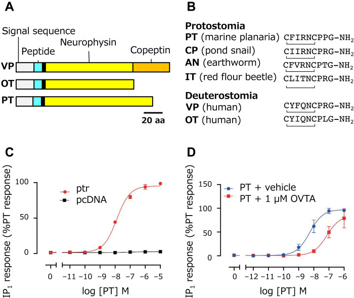
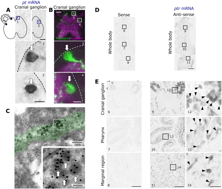
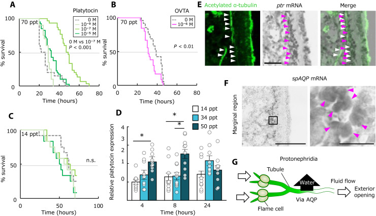
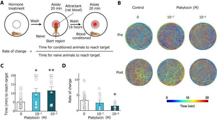
Vasopressin/oxytocin (VP/OT)-related peptides are essential for mammalian antidiuresis, sociosexual behavior, and reproduction. However, the evolutionary origin of this peptide system is still uncertain. Here, we identify orthologous genes to those for VP/OT in Platyhelminthes, intertidal planarians that have a simple bilaterian body structure but lack a coelom and body-fluid circulatory system. We report a comprehensive characterization of the neuropeptide derived from this VP/OT-type gene, identifying its functional receptor, and name it the "platytocin" system. Our experiments with these euryhaline planarians, living where environmental salinities fluctuate due to evaporation and rainfall, suggest that platytocin functions as an "antidiuretic hormone" and also organizes diverse actions including reproduction and chemosensory-associated behavior. We propose that bilaterians acquired physiological adaptations to amphibious lives by such regulation of the body fluids. This neuropeptide-secreting system clearly became indispensable for life even without the development of a vascular circulatory system or relevant synapses.
Figures
Similar articles
-
Comparative and Evolutionary Physiology of Vasopressin/ Oxytocin-Type Neuropeptide Signaling in Invertebrates.Front Endocrinol (Lausanne). 2020 Apr 17;11:225. doi: 10.3389/fendo.2020.00225. eCollection 2020. Front Endocrinol (Lausanne). 2020. PMID: 32362874 Free PMC article. Review.
-
A Putative Role of Vasopressin/Oxytocin-Type Neuropeptide in Osmoregulation and Feeding Inhibition of Apostichopus japonicus.Int J Mol Sci. 2023 Sep 20;24(18):14358. doi: 10.3390/ijms241814358. Int J Mol Sci. 2023. PMID: 37762661 Free PMC article.
-
Vasopressin and oxytocin receptor systems in the brain: Sex differences and sex-specific regulation of social behavior.Front Neuroendocrinol. 2016 Jan;40:1-23. doi: 10.1016/j.yfrne.2015.04.003. Epub 2015 May 4. Front Neuroendocrinol. 2016. PMID: 25951955 Free PMC article. Review.
-
Characterization of a novel vasopressin/oxytocin superfamily peptide and its receptor from an ascidian, Ciona intestinalis.Peptides. 2008 Oct;29(10):1672-8. doi: 10.1016/j.peptides.2008.05.030. Epub 2008 Jun 11. Peptides. 2008. PMID: 18586058
-
The Oxytocin-Vasopressin Pathway in the Context of Love and Fear.Front Endocrinol (Lausanne). 2017 Dec 22;8:356. doi: 10.3389/fendo.2017.00356. eCollection 2017. Front Endocrinol (Lausanne). 2017. PMID: 29312146 Free PMC article. Review.
Cited by
-
Recent advances in single-particle analysis with nanopore technology.Anal Sci. 2025 May;41(5):677-685. doi: 10.1007/s44211-025-00757-1. Epub 2025 Apr 5. Anal Sci. 2025. PMID: 40186842 Review.
-
Model systems for discovering evolutionary singularity of bilaterian physiological regulation: lessons from studies on simple/primitive flatworms.Biophys Physicobiol. 2024 Mar 2;21(Supplemental):e211012. doi: 10.2142/biophysico.bppb-v21.s012. eCollection 2024. Biophys Physicobiol. 2024. PMID: 39175867 Free PMC article. No abstract available.
-
Kidney collecting duct-derived vasopressin is not essential for appropriate concentration or dilution of urine.Am J Physiol Renal Physiol. 2024 Jun 1;326(6):F1091-F1100. doi: 10.1152/ajprenal.00057.2024. Epub 2024 May 2. Am J Physiol Renal Physiol. 2024. PMID: 38695074 Free PMC article.
-
Nanobiodevices and quantum life science for future healthcare.Anal Sci. 2025 May;41(5):601-607. doi: 10.1007/s44211-025-00742-8. Epub 2025 Mar 4. Anal Sci. 2025. PMID: 40035948 Review.
References
-
- Soloff M. S., Alexandrova M., Fernstrom M. J., Oxytocin receptors: Triggers for parturition and lactation? Science 204, 1313–1315 (1979). - PubMed
-
- Birnbaumer M., Seibold A., Gilbert S., Ishido M., Barberis C., Antaramian A., Brabet P., Rosenthal W., Molecular cloning of the receptor for human antidiuretic hormone. Nature 357, 333–335 (1992). - PubMed
-
- Schumacher M., Coirini H., Pfaff D. W., McEwen B. S., Behavioral effects of progesterone associated with rapid modulation of oxytocin receptors. Science 250, 691–694 (1990). - PubMed
-
- Donaldson Z. R., Young L. J., Oxytocin, vasopressin, and the neurogenetics of sociality. Science 322, 900–904 (2008). - PubMed
LinkOut - more resources
Full Text Sources
