. 2022 Mar 4;8(9):eabj5347.
doi: 10.1126/sciadv.abj5347. Epub 2022 Mar 4.

Structural insights into ligand recognition, activation, and signaling of the α2A adrenergic receptor

Affiliations

Structural insights into ligand recognition, activation, and signaling of the α2A adrenergic receptor

Jun Xu et al. Sci Adv. .

Abstract

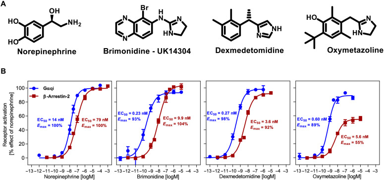
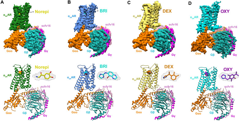
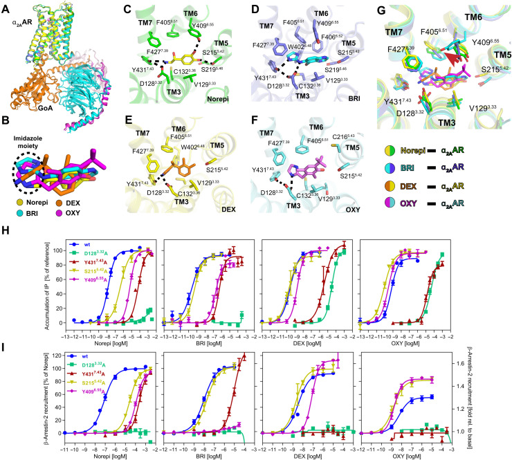
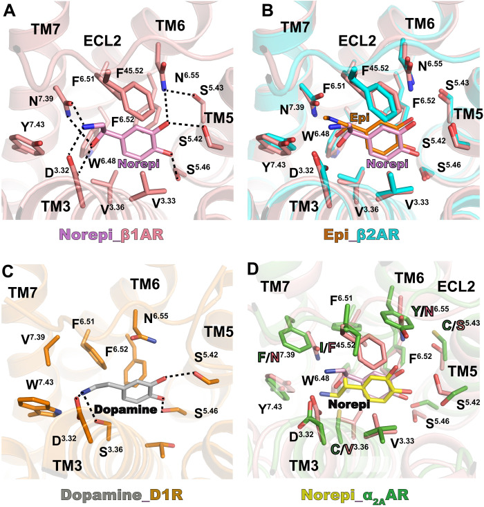
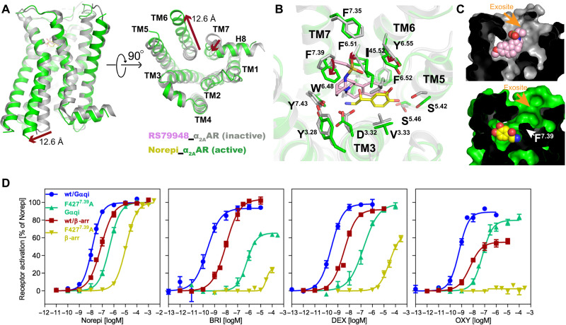
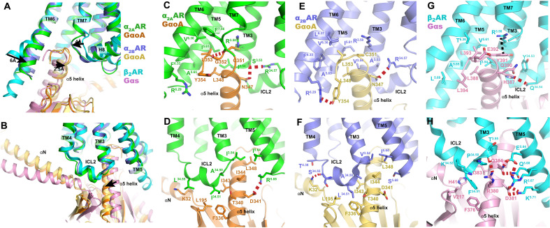
The α2A adrenergic receptor (α2AAR) is a G protein (heterotrimeric guanine nucleotide-binding protein)-coupled receptor that mediates important physiological functions in response to the endogenous neurotransmitters norepinephrine and epinephrine, as well as numerous chemically distinct drugs. However, the molecular mechanisms of drug actions remain poorly understood. Here, we report the cryo-electron microscopy structures of the human α2AAR-GoA complex bound to norepinephrine and three imidazoline derivatives (brimonidine, dexmedetomidine, and oxymetazoline). Together with mutagenesis and functional data, these structures provide important insights into the molecular basis of ligand recognition, activation, and signaling at the α2AAR. Further structural analyses uncover different molecular determinants between α2AAR and βARs for recognition of norepinephrine and key regions that determine the G protein coupling selectivity. Overall, our studies provide a framework for understanding the signal transduction of the adrenergic system at the atomic level, which will facilitate rational structure-based discovery of safer and more effective medications for α2AAR.


Figures

Fig. 1.
Fig. 1.. Structure and function of α2AAR agonists.
(A) Chemical structures of norepinephrine (Norepi), brimonidine (BRI) or UK14304, dexmedetomidine (DEX), and oxymetazoline (OXY). (B) Concentration-response curves of different agonists toward G protein activation (blue curve) and β-arrestin-2 recruitment (red curve) measured by a cell-based Gqi-inositol phosphate accumulation assay and the PathHunter assay respectively. Data are presented as means ± SEM of 4 to 10 independent experiments with repeats in duplicate.
Fig. 2.
Fig. 2.. Cryo-EM structures of Norepi, BRI-, DEX-, and OXY-bound α2AAR-GoA complexes.
Cryo-EM density maps and models of the α2AAR-GoA complex bound to Norepi (A), BRI (B), DEX (C), and OXY (D). The densities of the agonists (shown as sticks) are depicted as gray meshes. The maps are colored according to different subunits.
Fig. 3.
Fig. 3.. Orthosteric binding pocket of active α2AAR bound to different agonists.
(A) Alignment of four structures of α2AAR signaling complexes. (B) Superposition of Norepi, BRI, DEX, and OXY from cryo-EM structures. The imidazole moiety is highlighted by the dashed circle. (C to F) Detailed interactions of Norepi (C), BRI (D), DEX (E), and OXY (F) with α2AAR. Residues within 4 Å of agonist are shown in sticks. The polar interactions are indicated by black dashed lines. (G) Superposition of the α2AAR orthosteric binding pocket residues bound to four different agonists. (H and I) Concentration-response curves of different agonists for G protein activation (H) and β-arrestin-2 recruitment (I) for wild-type (wt) α2AAR and receptor mutants D1283.32A, Y4317.43A, S2155.42A, and Y4096.55A, respectively. Except for the activation of D1283.32A, which is normalized to DEX for G protein activation and relative to basal for arrestin recruitment, receptor activation is shown relative to the maximum effect of Norepi. Data are presented as means ± SEM of 3 to 11 independent experiments with repeats in duplicate.
Fig. 4.
Fig. 4.. Comparison of Norepi binding for α2A and β adrenergic receptors.
(A) Detailed interactions of Norepi with β1AR [Protein Data Bank (PDB) code: 7BU6]. Residues within 4 Å of agonist are shown in sticks. The polar interactions are indicated by black dashed lines. (B) Superposition of orthosteric pockets of β1AR-Norepi and β2AR-Epi (epinephrine) (PDB code: 4LDO). (C) Detailed interactions of dopamine with D1R (PDB code: 7CKZ). The polar interactions are indicated by black dashed lines. (D) Superposition of orthosteric pockets of β1AR-Norepi and α2AAR-Norepi. Residues within 4 Å of agonist are shown in sticks.
Fig. 5.
Fig. 5.. Comparison of inactive and active α2AAR.
(A) Structural comparison of inactive α2AAR bound to RS79948 and active α2AAR bound to Norepi, with changes highlighted as red arrows. The distance is calculated between positions of Cα of residue 6.29 in TM6. (B) Conformational changes within the orthosteric pocket are shown from the extracellular side, with changes highlighted as red arrows. (C) Cross sections of α2AAR bound to antagonist and agonist are shown, with the interior in black and the exosite highlighted. (D) Concentration-response curves of F4277.39 mutant for different agonists toward G protein activation and β-arrestin-2 recruitment. Data are presented as means ± SEM of 4 to 10 independent experiments with repeats in duplicate.
Fig. 6.
Fig. 6.. G protein binding interface.
(A and B) Superposition of the G protein coupling interfaces of α2AAR-GoA, α2BAR-GoA, and β2AR-Gs complexes, using receptor for alignment. (C to H) Detailed interactions of α2AAR with Goα (C and D), α2BAR with Goα (E and F), and β2AR with Gsα (G and H). The polar interactions are indicated by red dashed lines.

References

    1. Link R., Daunt D., Barsh G., Chruscinski A., Kobilka B., Cloning of two mouse genes encoding alpha 2-adrenergic receptor subtypes and identification of a single amino acid in the mouse alpha 2-C10 homolog responsible for an interspecies variation in antagonist binding. Mol. Pharmacol. 42, 16–27 (1992). - PubMed
    1. Ruffolo R. R. Jr., Nichols A. J., Stadel J. M., Hieble J. P., Pharmacologic and therapeutic applications of alpha 2-adrenoceptor subtypes. Annu. Rev. Pharmacol. Toxicol. 32, 243–279 (1993). - PubMed
    1. MacDonald E., Kobilka B. K., Scheinin M., Gene targeting - Homing in on α2-adrenoceptor-subtype function. Trends Pharmacol. Sci. 18, 211–219 (1997). - PubMed
    1. Wang C. D., Buck M. A., Fraser C. M., Site-directed mutagenesis of alpha 2A-adrenergic receptors: Identification of amino acids involved in ligand binding and receptor activation by agonists. Mol. Pharmacol. 40, 168–179 (1991). - PubMed
    1. Ramos B. P., Arnsten A. F. T., Adrenergic pharmacology and cognition: Focus on the prefrontal cortex. Pharmacol. Ther. 113, 523–536 (2007). - PMC - PubMed

Publication types