. 2022 Mar 1;149(5):dev200076.
doi: 10.1242/dev.200076. Epub 2022 Mar 4.

Selective requirement for polycomb repressor complex 2 in the generation of specific hypothalamic neuronal subtypes

Affiliations

Selective requirement for polycomb repressor complex 2 in the generation of specific hypothalamic neuronal subtypes

Behzad Yaghmaeian Salmani et al. Development. .

Abstract

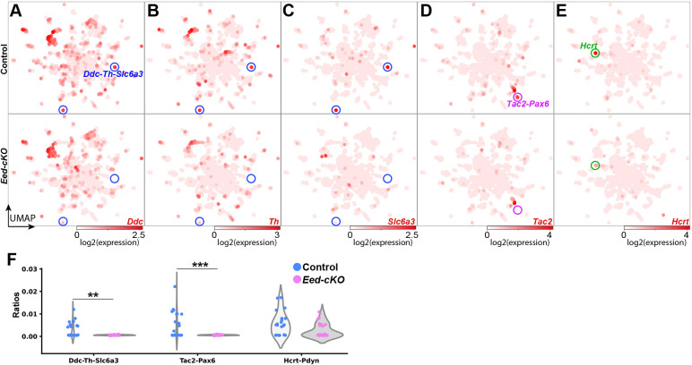
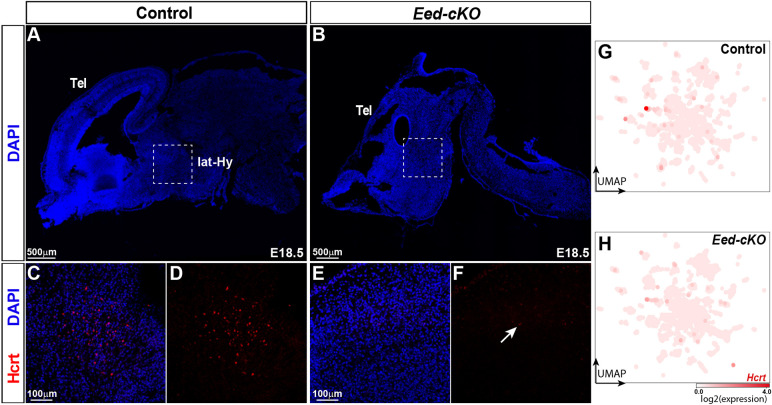
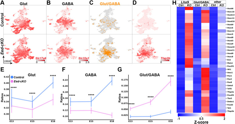
The hypothalamus displays staggering cellular diversity, chiefly established during embryogenesis by the interplay of several signalling pathways and a battery of transcription factors. However, the contribution of epigenetic cues to hypothalamus development remains unclear. We mutated the polycomb repressor complex 2 gene Eed in the developing mouse hypothalamus, which resulted in the loss of H3K27me3, a fundamental epigenetic repressor mark. This triggered ectopic expression of posteriorly expressed regulators (e.g. Hox homeotic genes), upregulation of cell cycle inhibitors and reduced proliferation. Surprisingly, despite these effects, single cell transcriptomic analysis revealed that most neuronal subtypes were still generated in Eed mutants. However, we observed an increase in glutamatergic/GABAergic double-positive cells, as well as loss/reduction of dopamine, hypocretin and Tac2-Pax6 neurons. These findings indicate that many aspects of the hypothalamic gene regulatory flow can proceed without the key H3K27me3 epigenetic repressor mark, but points to a unique sensitivity of particular neuronal subtypes to a disrupted epigenomic landscape.

Keywords: Cell specification; Epigenetics; H3K27me3; H3K4me1/3; Mouse; Neuropeptide neurons.


Conflict of interest statement

Competing interests The authors declare no competing or financial interests.

Figures

Fig. 1.
Fig. 1.
Single cell transcriptomic analysis of Eed-cKO mutants reveals upregulation of TFs and Hox genes in the hypothalamus. (A) Process for quality control (QC) and biological filtering of hypothalamic scRNA-seq cells. (B) UMAP of hypothalamic scRNA-seq cells, combined from control and Eed-cKO, at E18.5, based upon 300 genes (Table S1). (C) UMAP of All-Hypo cells in control and Eed-cKO. (D) Volcano plot showing DEGs in Eed-cKO. (E,F) UMAP of E18.5 hypothalamic scRNA-seq All-Hypo cells, showing expression of Cdkn2a (E) and Hoxd10 (F), in control and Eed-cKO. (G) Violin plots comparing the expression levels of Hox genes and the highest differentially expressed (DE) transcription factors between control and Eed-cKO in E18.5 hypothalamic scRNA-seq All-Hypo cells (adjusted P-value≤0.05).
Fig. 2.
Fig. 2.
Eed-cKO mutants display more Glut/GABA cells. (A-F) UMAP embedding, based upon 300 DEGs (Table S1), of E18.5 All-Hypo cells, showing astrocytes (Gfap; A), oligodendrocyte precursors (Olig1; B), ependymal cells (Foxj1; C), tanycytes (Rax; D), glutamatergic neurons (Slc17a6; E) and GABAergic neurons (Slc32a1; F). (G) Glut/GABA co-expression revealed by Slc32a1 and Slc17a6 expression. (H) Violin plots of the ratios of Glut, Glut/GABA and GABA cells detected per embryo stratified by control and Eed-cKO reveal an increase in Glut/GABA cells and a decrease in GABA and Glut cells in Eed-cKO (***adjusted P-value<1e-3; t-test on centred-log-ratios, see Table S10 for details). (I,J) Venn diagrams summarizing the percentage of Glut, Gluta/GABA and GABA cells in control (I) and Eed-cKO (J).
Fig. 3.
Fig. 3.
Eed-cKO mutants display ectopic Tfap2b and Pax2 expression in the hypothalamus. (A-H) Staining for DAPI, Tfap2b (A-D) and Pax2 (E-H) in sagittal sections of E18.5 brains in control and Eed-cKO. Dashed squares in A,B,E,F delineate regions of hypothalamic tissue magnified in C,D,G,H. In control, Tfap2b and Pax2 expression is observed in the mid- and hindbrain regions. In Eed-cKO mutants, Tfap2b and Pax2 expression expands into the anterior brain, including the hypothalamus. (I-L) UMAP embedding of E18.5 hypothalamic scRNA-seq All-Hypo cells revealing the ectopic expression of Tfap2b (I,J) and Pax2 (K,L) in control and Eed-cKO.
Fig. 4.
Fig. 4.
Eed-cKO is not necessary for most NP-DA cell types. (A,B) UMAP embedding of hypothalamic NP-DA cells, based on 122 genes (Table S1), in control (A) and Eed-cKO (B) at E18.5. Each cluster is labelled by the combination of NP-DA marker genes upregulated in that cluster. (C) Heatmap displaying average gene expression of the NP-DA marker genes in each UMAP cell cluster, in control (blue, top) and Eed-cKO (pink, bottom). Gene expression is measured in log2(CPT+1), scaled between 0 and 1 along each row (see Materials and Methods). The Ddc-Th-Slc6a3, Tac2-Pax6 and Hcrt clusters are absent in Eed-cKO, and hence a column of 0 expression was depicted. (D) Expression levels of NP-DA marker genes in control and Eed-cKO at E18.5 (see Table S2 for analysis).
Fig. 5.
Fig. 5.
Eed-cKO is necessary for DA, Hcrt and Tac2 cells. (A-E) UMAP embedding of E18.5 NP-DA cells, based upon 122 DE genes (Table S1), showing expression of Ddc (A), Th (B), Slc6a3 (C), Tac2 (D) and Hcrt (E). Two prominent clusters co-express Ddc-Th-Slc6a3, and these cluster are largely missing in Eed-cKO (A-C). Similarly, one specific Tac2 cluster and the Hcrt cluster are largely absent in Eed-cKO mutants (D,E). (F) Violin plots of the ratios of Ddc-Th-Slc6a3, Tac2-Pax6 and Hcrt-Pdyn cell types in control and Eed-cKO embryos (see Table S2 for analysis). **adjusted P-value<0.01, ***adjusted P-value<0.001; t-test on centred-log-ratios (see Table S5 for details).
Fig. 6.
Fig. 6.
Eed is crucial for Hcrt neurons. (A,B) Staining for DAPI in sagittal sections of E18.5 control and Eed-cKO mutant brains. Dashed boxed areas delineate the lateral hypothalamus (lat-Hy). (C-F) Magnifications of boxed areas in A and B showing staining for DAPI and Hcrt in the lateral hypothalamus in control (C,D) and Eed-cKO (E,F) embryos. A cluster of Hcrt cells is present in the control, whereas Eed-cKO mutants only show occasional weakly expressing cells. (G,H) UMAP embedding of E18.5 hypothalamic scRNA-seq NP-DA cells, with each cell coloured according to the expression level of Hcrt in control and Eed-cKO.
Fig. 7.
Fig. 7.
Developmental onset of Glut/GABA co-expression. (A-D) UMAP embedding of hypothalamic cells from E13.5, E15.5 and E18.5, depicting expression of Slc17a6 (A), Slc32a1 (B) and Tfap2b (D). Glut/GABA co-expression (C) and Tfap2b expression is limited in control but increases in Eed-cKO mutants. (E-G) Quantification of Glut-, GABA- and Glut/GABA-expressing cells at developmental time points (see Tables S9 and S10 for analysis). In Eed-cKO, Glut/GABA cells increase at the expense of both Glut and GABA cells. (H) Expression analysis of Lhx9 cells, Glut/GABA cells and ‘other’ cells, at E13.5-E18.5. In Eed-cKO, Lhx9 and Glut/GABA cells show ectopic expression of many developmental TFs, and display upregulation to a greater extent than all ‘other’ cells.

References

    1. Alpár, A., Benevento, M., Romanov, R. A., Hökfelt, T. and Harkany, T. (2019). Hypothalamic cell diversity: non-neuronal codes for long-distance volume transmission by neuropeptides. Curr. Opin. Neurobiol. 56, 16-23. 10.1016/j.conb.2018.10.012 - DOI - PubMed
    1. Alvarez-Bolado, G. (2019). Development of neuroendocrine neurons in the mammalian hypothalamus. Cell Tissue Res. 375, 23-39. 10.1007/s00441-018-2859-1 - DOI - PubMed
    1. Arcipowski, K. M., Martinez, C. A. and Ntziachristos, P. (2016). Histone demethylases in physiology and cancer: a tale of two enzymes, JMJD3 and UTX. Curr. Opin. Genet. Dev. 36, 59-67. 10.1016/j.gde.2016.03.010 - DOI - PMC - PubMed
    1. Ashburner, M., Ball, C. A., Blake, J. A., Botstein, D., Butler, H., Cherry, J. M., Davis, A. P., Dolinski, K., Dwight, S. S., Eppig, J. T.et al. (2000). Gene ontology: tool for the unification of biology. The Gene Ontology Consortium. Nat. Genet. 25, 25-29. 10.1038/75556 - DOI - PMC - PubMed
    1. Aylwin, C. F., Vigh-Conrad, K. and Lomniczi, A. (2019). The emerging role of chromatin remodeling factors in female pubertal development. Neuroendocrinology 109, 208-217. 10.1159/000497745 - DOI - PMC - PubMed

Publication types

Substances