Prenatal allergic inflammation in rats programs the developmental trajectory of dendritic spine patterning in brain regions associated with cognitive and social behavior
- PMID: 35245680
- PMCID: PMC9070022
- DOI: 10.1016/j.bbi.2022.02.026
Prenatal allergic inflammation in rats programs the developmental trajectory of dendritic spine patterning in brain regions associated with cognitive and social behavior
Abstract
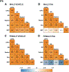
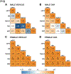
Allergic inflammation during pregnancy increases risk for a diagnosis of neurodevelopmental disorders such as Attention Deficit/Hyperactivity Disorder (ADHD) and Autism Spectrum Disorder (ASD) in the offspring. Previously, we found a model of such inflammation, allergy-induced maternal immune activation (MIA), produced symptoms analogous to those associated with neurodevelopmental disorders in rats, including reduced juvenile play behavior, hyperactivity, and cognitive inflexibility. These behaviors were preceded by perinatal changes in microglia colonization and phenotype in multiple relevant brain regions. Given the role that microglia play in synaptic patterning as well as evidence for altered synaptic architecture in neurodevelopmental disorders, we investigated whether allergic MIA altered the dynamics of dendritic spine patterning throughout key regions of the rat forebrain across neurodevelopment. Adult virgin female rats were sensitized to the allergen, ovalbumin, with alum adjuvant, bred, and allergically challenged on gestational day 15. Brain tissue was collected from male and female offspring on postnatal days (P) 5, 15, 30, and 100-120 and processed for Golgi-Cox staining. Mean dendritic spine density was calculated for neurons in brain regions associated with cognition and social behavior, including the medial prefrontal cortex (mPFC), basal ganglia, septum, nucleus accumbens (NAc), and amygdala. Allergic MIA reduced dendritic spine density in the neonatal (P5) and juvenile (P15) mPFC, but these mPFC spine deficits were normalized by P30. Allergic inflammation reduced spine density in the septum of juvenile (P30) rats, with an interaction suggesting increased density in males and reduced density in females. MIA-induced reductions in spine density were also found in the female basal ganglia at P15, as well as in the NAc at P30. Conversely, MIA-induced increases were found in the NAc in adulthood. While amygdala dendritic spine density was generally unaffected throughout development, MIA reduced density in both medial and basolateral subregions in adult offspring. Correlational analyses revealed disruption to amygdala-related networks in the neonatal animals and cortico-striatal related networks in juvenile and adult animals in a sex-specific manner. Collectively, these data suggest that communication within and between these cognitive and social brain regions may be altered dynamically throughout development after prenatal exposure to allergic inflammation. They also provide a basis for future intervention studies targeted at rescuing spine and behavior changes via immunomodulatory treatments.
Keywords: Allergy; Dendritic Spines; Development; Inflammation; Maternal Immune Activation; Microglia; Sex Differences.
Copyright © 2022 Elsevier Inc. All rights reserved.
Conflict of interest statement
Figures


Similar articles
-
Prenatal allergic inflammation in rats confers sex-specific alterations to oxytocin and vasopressin innervation in social brain regions.Horm Behav. 2024 Jan;157:105427. doi: 10.1016/j.yhbeh.2023.105427. Epub 2023 Sep 22. Horm Behav. 2024. PMID: 37743114 Free PMC article.
-
Prenatal Allergen Exposure Perturbs Sexual Differentiation and Programs Lifelong Changes in Adult Social and Sexual Behavior.Sci Rep. 2019 Mar 18;9(1):4837. doi: 10.1038/s41598-019-41258-2. Sci Rep. 2019. PMID: 30886382 Free PMC article.
-
Maternal allergic inflammation in rats impacts the offspring perinatal neuroimmune milieu and the development of social play, locomotor behavior, and cognitive flexibility.Brain Behav Immun. 2021 Jul;95:269-286. doi: 10.1016/j.bbi.2021.03.025. Epub 2021 Mar 30. Brain Behav Immun. 2021. PMID: 33798637 Free PMC article.
-
Impact of maternal immune activation on dendritic spine development.Dev Neurobiol. 2021 Jul;81(5):524-545. doi: 10.1002/dneu.22804. Epub 2021 Jan 15. Dev Neurobiol. 2021. PMID: 33382515 Review.
-
The Outcomes of Maternal Immune Activation Induced with the Viral Mimetic Poly I:C on Microglia in Exposed Rodent Offspring.Dev Neurosci. 2023;45(4):191-209. doi: 10.1159/000530185. Epub 2023 Mar 21. Dev Neurosci. 2023. PMID: 36944325 Review.
Cited by
-
Prenatal allergic inflammation in rats confers sex-specific alterations to oxytocin and vasopressin innervation in social brain regions.Horm Behav. 2024 Jan;157:105427. doi: 10.1016/j.yhbeh.2023.105427. Epub 2023 Sep 22. Horm Behav. 2024. PMID: 37743114 Free PMC article.
-
Urolithin A alleviates schizophrenic-like behaviors and cognitive impairment in rats through modulation of neuroinflammation, neurogenesis, and synaptic plasticity.Sci Rep. 2025 Mar 26;15(1):10477. doi: 10.1038/s41598-025-93554-9. Sci Rep. 2025. PMID: 40140679 Free PMC article.
-
Postnatal Allergic Inhalation Induces Glial Inflammation in the Olfactory Bulb and Leads to Autism-Like Traits in Mice.Int J Mol Sci. 2024 Sep 28;25(19):10464. doi: 10.3390/ijms251910464. Int J Mol Sci. 2024. PMID: 39408806 Free PMC article.
-
Offspring behavioral outcomes following maternal allergic asthma in the IL-4-deficient mouse.J Neuroimmunol. 2024 May 15;390:578341. doi: 10.1016/j.jneuroim.2024.578341. Epub 2024 Apr 8. J Neuroimmunol. 2024. PMID: 38613873 Free PMC article.
-
Deciduous tooth biomarkers reveal atypical fetal inflammatory regulation in autism spectrum disorder.iScience. 2023 Feb 21;26(3):106247. doi: 10.1016/j.isci.2023.106247. eCollection 2023 Mar 17. iScience. 2023. PMID: 36926653 Free PMC article.
References
-
- Diagnostic and Statistical Manual of Mental Disorders (5th ed.). 2013, Washington, DC: The American Psychiatric Association.
-
- Instanes JT, et al. Attention-Deficit/Hyperactivity Disorder in Offspring of Mothers With Inflammatory and Immune System Diseases. Biol Psychiatry, 2017. 81(5): p. 452–459. - PubMed
-
- Chen SW, et al. Maternal autoimmune diseases and the risk of autism spectrum disorders in offspring: A systematic review and meta-analysis. Behav Brain Res, 2016. 296: p. 61–69. - PubMed
-
- Croen LA, et al. Maternal Autoimmune Diseases, Asthma and Allergies, and Childhood Autism Spectrum Disorders. Archives of pediatrics & adolescent medicine, 2005. 159(2): p. 151–157. - PubMed
Publication types
MeSH terms
Grants and funding
LinkOut - more resources
Full Text Sources
Medical
Miscellaneous

