

Isolated IgG4-related cholecystitis with localized gallbladder wall thickening mimicking gallbladder cancer: a case report and literature review
- PMID: 35246051
- PMCID: PMC8895667
- DOI: 10.1186/s12876-022-02179-z
Isolated IgG4-related cholecystitis with localized gallbladder wall thickening mimicking gallbladder cancer: a case report and literature review
Abstract
Background: IgG4-related cholecystitis, which is a manifestation of IgG4-related disease in the gallbladder, is associated with autoimmune pancreatitis or IgG4-related sclerosing cholangitis in most cases; isolated gallbladder lesions without systemic manifestations are very rare. Gallbladder wall thickening is often diffuse, but sometimes localized, in which case, differentiation from gallbladder cancer becomes difficult. The characteristic features of IgG4-related cholecystitis on imaging that would enable differentiation from gallbladder cancer remain poorly described.
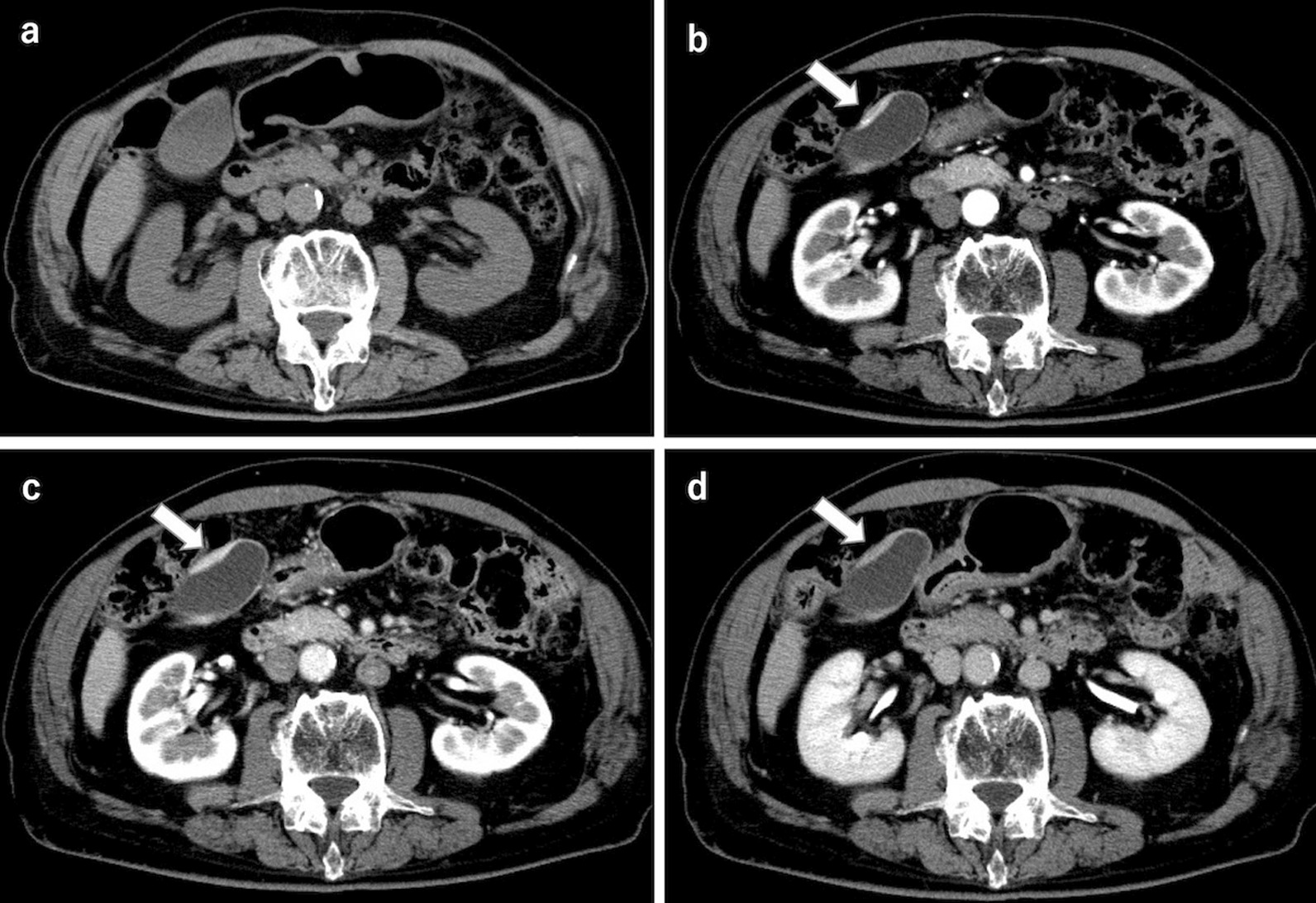
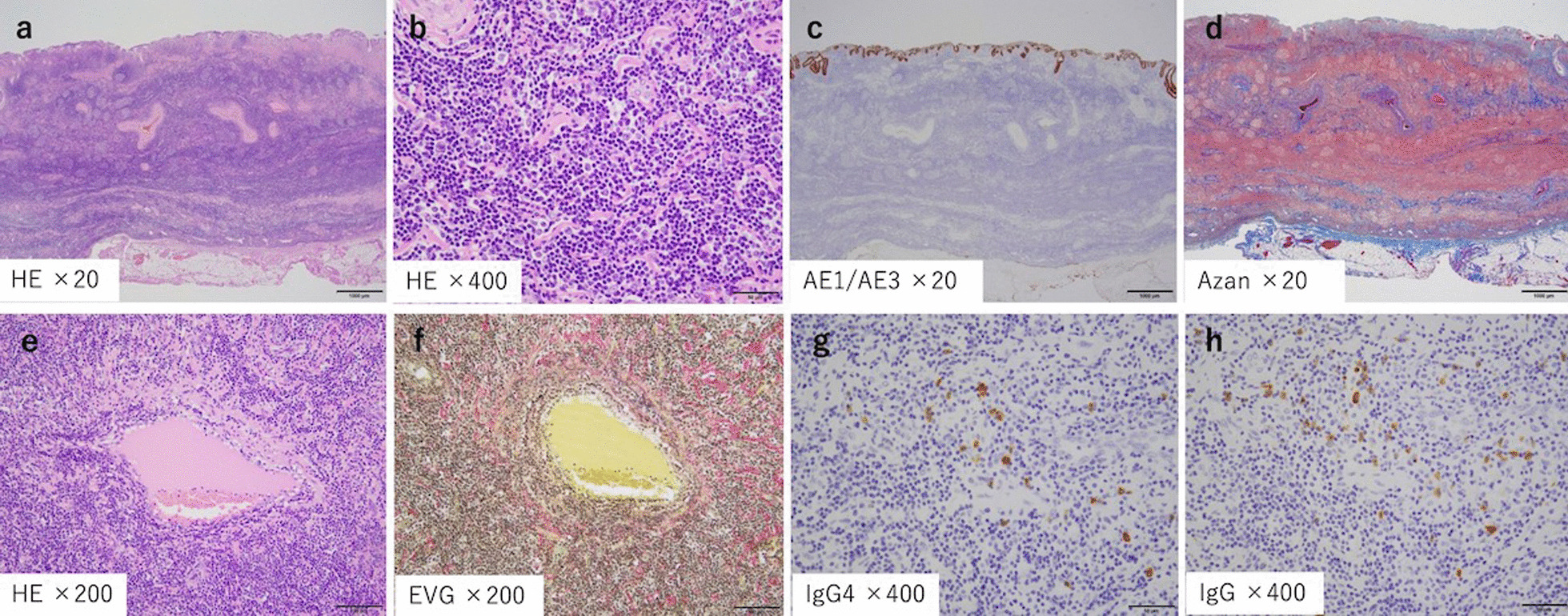
Case presentation: We present a rare case of isolated IgG4-related cholecystitis with localized gallbladder wall thickening that was clinically difficult to distinguish from malignancy before resection. An 82-year-old man was referred to our hospital because of gallbladder wall thickening on abdominal ultrasonography without any symptoms. Dynamic computed tomography of the abdomen showed localized wall thickening from the body to the fundus of the gallbladder that was enhanced from an early stage with a prolonged contrast effect. There were no other findings, such as pancreatic enlargement and bile duct dilatation. Magnetic resonance cholangiopancreatography revealed neither dilatation nor stenosis of the bile duct and pancreatic duct. Endoscopic ultrasonography (EUS) showed a smooth layered thickening of the gallbladder wall with a maximum thickness of 6 mm and a well-preserved outermost hyperechoic layer in the same area. Laparoscopic cholecystectomy was performed because malignancy could not be completely ruled out. Pathological examination of a resected specimen revealed IgG4-positive plasma cell infiltration, fibrosis, and phlebitis. Although the serum IgG4 level measured after resection was normal, the condition was ultimately diagnosed as probable IgG4-related cholecystitis according to the 2020 revised comprehensive diagnostic criteria for IgG4-related disease. The EUS images reflected the pathological findings, in which lymphocytic infiltration was distributed in a laminar fashion in the gallbladder wall.
Conclusions: Although rare, isolated IgG4-related cholecystitis with localized wall thickening mimicking gallbladder cancer remains a clinical problem. A smooth laminar thickening of the gallbladder wall on EUS imaging could be one of the most informative characteristics for differentiating IgG4-related cholecystitis from gallbladder cancer.
Keywords: Case report; Gallbladder cancer; Gallbladder wall thickening; IgG4-related cholecystitis.
© 2022. The Author(s).
Conflict of interest statement
The authors declare that they have no competing interests.
Figures


References
Publication types
MeSH terms
Substances
LinkOut - more resources
Full Text Sources
Medical
