

Retinal and choroidal thickness in pediatric patients with sickle cell disease: a cross-sectional cohort study
- PMID: 35246275
- PMCID: PMC8895628
- DOI: 10.1186/s40942-021-00351-3
Retinal and choroidal thickness in pediatric patients with sickle cell disease: a cross-sectional cohort study
Abstract
Background: To measure the retinal/choroidal thicknesses in the macular area of asymptomatic pediatric patients with sickle cell disease (SCD).
Methods: This cross-sectional cohort study included 40 children (79 eyes) with SCD and 19 control patients (36 eyes). All subjects underwent spectral-domain optical coherence tomography (SD-OCT) with enhanced-depth imaging OCT. Generalized Estimating Equations (GEE) were applied to compare the outcomes between groups. P ≤ 0.05 was considered significant.
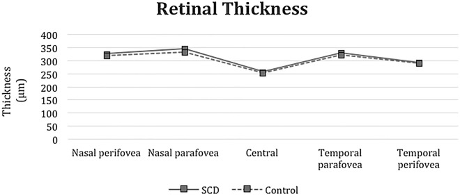
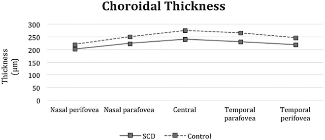
Results: The choroidal thickness in the macular area in the study subfields was significantly thinner in the SCD eyes compared with control eyes (subfoveal subfield and temporal parafoveal subfield, p < 0.0001; nasal parafoveal subfield, p < 0.0001 temporal perifoveal subfield, p < 0.0001; and nasal perifoveal subfield, p < 0.0001). The variations in the retinal thickness were not significant.
Conclusion: EDI-OCT showed that the macular choroidal thickness is thinner in asymptomatic pediatric patients with SCD.
Keywords: Choroidal thickness; Retinal thickness; Sickle cell disease.
© 2022. The Author(s).
Conflict of interest statement
The authors declare that they have no competing interests.
Figures

Similar articles
-
Choroidal thinning as a new finding in Alzheimer's disease: evidence from enhanced depth imaging spectral domain optical coherence tomography.J Alzheimers Dis. 2014;40(4):907-17. doi: 10.3233/JAD-132039. J Alzheimers Dis. 2014. PMID: 24577467
-
Comparison of macular choroidal thickness in patients with pseudoexfoliation syndrome to normal control subjects with enhanced depth SD-OCT imaging.J Curr Ophthalmol. 2017 Jul 12;29(4):258-263. doi: 10.1016/j.joco.2017.06.009. eCollection 2017 Dec. J Curr Ophthalmol. 2017. PMID: 29270471 Free PMC article.
-
Spectral domain optical coherence tomography in patients with sickle cell disease.Br J Ophthalmol. 2015 Jul;99(7):967-72. doi: 10.1136/bjophthalmol-2014-305532. Epub 2015 Jan 16. Br J Ophthalmol. 2015. PMID: 25595176
-
Clinical observation of macular choroidal thickness in primary chronic angle-closure glaucoma.Int Ophthalmol. 2021 Dec;41(12):4217-4223. doi: 10.1007/s10792-021-01988-7. Epub 2021 Jul 31. Int Ophthalmol. 2021. PMID: 34333686 Review.
-
[Pathophysiology of macular diseases--morphology and function].Nippon Ganka Gakkai Zasshi. 2011 Mar;115(3):238-74; discussion 275. Nippon Ganka Gakkai Zasshi. 2011. PMID: 21476310 Review. Japanese.
Cited by
-
Pitfalls of social media for aesthetic eye surgery patients: assessing YouTube's aesthetic canthoplasty content.Int Ophthalmol. 2024 Jun 25;44(1):279. doi: 10.1007/s10792-024-03197-4. Int Ophthalmol. 2024. PMID: 38918201
References
LinkOut - more resources
Full Text Sources
