. 2022 Mar 4;13(1):1065.
doi: 10.1038/s41467-022-28690-1.

The molecular basis of FimT-mediated DNA uptake during bacterial natural transformation

Affiliations

The molecular basis of FimT-mediated DNA uptake during bacterial natural transformation

Sebastian A G Braus et al. Nat Commun. .

Abstract

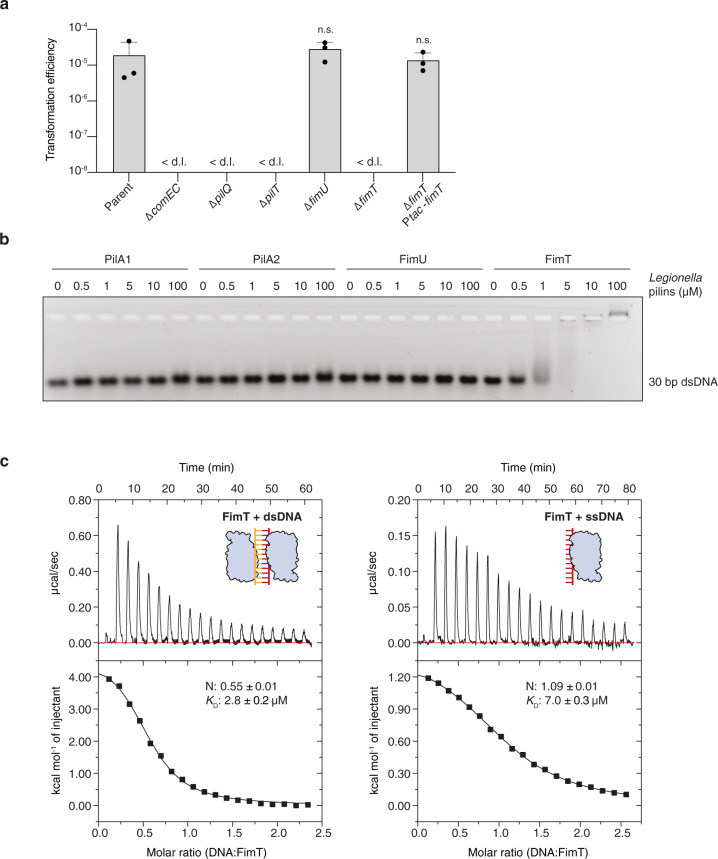
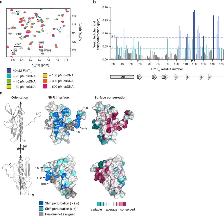
Naturally competent bacteria encode sophisticated protein machinery for the uptake and translocation of exogenous DNA into the cell. If this DNA is integrated into the bacterial genome, the bacterium is said to be naturally transformed. Most competent bacterial species utilise type IV pili for the initial DNA uptake step. These proteinaceous cell-surface structures are composed of thousands of pilus subunits (pilins), designated as major or minor according to their relative abundance in the pilus. Here, we show that the minor pilin FimT plays an important role in the natural transformation of Legionella pneumophila. We use NMR spectroscopy, in vitro DNA binding assays and in vivo transformation assays to understand the molecular basis of FimT's role in this process. FimT binds to DNA via an electropositive patch, rich in arginines, several of which are well-conserved and located in a conformationally flexible C-terminal tail. FimT orthologues from other Gammaproteobacteria share the ability to bind to DNA. Our results suggest that FimT plays an important role in DNA uptake in a wide range of competent species.


Conflict of interest statement

The authors declare no competing interests.

Figures

Fig. 1
Fig. 1. FimT is critical for the transformation of L. pneumophila and binds to DNA.
a Natural transformation efficiencies of the parental L. pneumophila Lp02 strain and Lp02 strains harbouring deletions of genes known to play a role in transformation compared to the fimU and fimT deletion strains. The ΔfimT strain was complemented by ectopic expression of wild-type FimT, under the control of an IPTG-inducible promoter. The mean transformation efficiencies of three independent biological replicates is shown (error bars represent standard deviation [SD]). < d.l., below detection limit (d.l.) (average d.l. = 2.0 × 10−8 ± 8.2 × 10−9). Statistical significances of transformation differences were determined on log-transformed data using an unpaired two-sided t-test with Welch’s correction. All strains were compared to the parental strain. n.s., not statistically significant, p > 0.05 (pΔfimU = 0.39; pΔfimT Ptac-fimT = 0.89). b In vitro DNA binding of purified L. pneumophila PilA1, PilA2, FimU and FimT assessed by an EMSA. A 30 bp dsDNA fragment (1 µM) was incubated with increasing concentrations of purified pilins (0–100 µM) and resolved by agarose gel electrophoresis. This experiment was independently performed three times with reproducible results. c ITC binding studies of wild-type FimT binding to 12meric dsDNA (left) and ssDNA (right). In both cases, DNA (syringe) was injected into FimT (cell). Data were fitted using the “one set” of sites model, assuming that both binding sites on the dsDNA are of equal affinity. Source data are provided as a Source Data file.
Fig. 2
Fig. 2. The structure of FimTLp.
a The solution structure of FimTLp 28–152 (state 18) in ribbon representation (left) and the corresponding topology diagram (right). Secondary structure elements are indicated: truncated N-terminal α-helix (α1C) (blue), β-sheet I formed by β1, β2, β3, and β5 (yellow), and β-sheet II formed by β4, β6, and β7 (magenta). A vertical arrow indicates the pilus axis from the cell surface towards the pilus tip. b Structure alignment of FimTLp (blue) and FimUPa (grey; PDB ID: 4IPV) (left) and the topology diagram of FimUPa (right). The disulphide bond of FimUPa is indicated in stick representation with sulphur atoms in yellow. c Superimposed 20 lowest energy structures calculated by NMR spectroscopy. An arrow indicates the conformational flexibility of the C-terminal tail (140–152). The pairwise backbone root-mean-square deviation (RMSD) for the structured region (residues 32–62, 70–139) is 1.13 Å. N- and C-termini are indicated in each panel. d Cα chemical shift values (top) and T2(1H) transverse relaxation data (bottom), encompassing the last 27 residues of FimTLp. Secondary structural elements are indicated and error bars represent the fitting errors of the respective exponential decay curves. Source data are provided as a Source Data file.
Fig. 3
Fig. 3. Identification of the DNA interaction surface of FimTLp.
a, Selected region of 1H, 15N-HSQC spectra showing 15N-labelled FimTLp alone and in the presence of increasing concentrations of 12 bp dsDNA. b, Weighted CSP map generated from a. Residues experiencing CSPs (Δppm > 1 σ), light blue; residues experiencing CSPs (Δppm > 2 σ), dark blue; P, prolines; *, residues not assigned. c, Left, FimTLp is shown in two orientations rotated by 120° in ribbon representation. Arrows indicate the pilus axis as in Fig. 2a. Middle, CSPs are mapped onto the surface of FimTLp and coloured as in b. Residues producing large shifts are labelled on the molecular surface. Right, surface residues of FimTLp are coloured according to conservation. This image was generated using the ConSurf server. Source data are provided as a Source Data file.
Fig. 4
Fig. 4. Characterisation of FimTLp binding to DNA in vitro and in vivo.
MST/TRIC binding assay of 12 bp FAM-labelled dsDNA with a, wild-type FimTLp performed at different NaCl concentrations (ionic strength) and b, wild-type FimTLp compared to FimT mutants predicted to disrupt DNA binding based on Fig. 3. n.d., not determined. The MST/TRIC data were fitted according to two binding sites with equal affinity. Error bars represent the mean ± SD (n = 3). c, d, Natural transformation efficiencies of parental Lp02, Lp02 ΔfimT, and the Lp02 ΔfimT strain complemented by ectopic expression of wild-type and FimTLp mutants that disrupt DNA binding (c) or do not contribute to DNA binding according to Fig. 3 (d). The mean transformation efficiencies of three independent biological replicates are plotted with error bars representing the SD. < d.l., below detection limit (d.l.) (average d.l. = 2.0 × 10⁻⁸ ± 8.2 × 10⁻⁹ (c) and 2.5 × 10⁻⁸ ± 9.5 ×10⁻⁹ (d)); #, below d.l. in at least one replicate (average d.l. used to calculate the mean transformation efficiency). The assay in panel c was performed in parallel to those displayed in Fig. 1a, and statistical differences were determined on log-transformed data using an unpaired two-sided t-test with Welch’s correction. The Lp02 ΔfimT strain complemented with mutants was compared to the Lp02 ΔfimT strain complemented with wild-type FimT, which was in turn compared to the parental strain. **, p < 0.01 (pR143Q = 0.003; pR146Q = 0.002; pR148Q = 0.004); n.s., not statistically significant, p > 0.05 (pWild-type(c) = 0.89; pWild-type(d) = 0.33; pS107Q = 0.38; pS122Q = 0.08; pG150Q = 0.89). Source data are provided as a Source Data file.
Fig. 5
Fig. 5. Bioinformatic and functional analysis of FimT orthologues.
a EMSA showing in vitro DNA binding of purified FimT and FimU orthologues from L. pneumophila, P. aeruginosa and X. campestris. A 30 bp dsDNA fragment (1 μM) was incubated with increasing concentrations of purified pilins (0–100 μM) and resolved by agarose gel electrophoresis. These experiments were independently performed three times with reproducible results. b A comparison of natural transformation efficiencies of the Lp02 ΔfimT strain complemented by ectopic expression of FimTLp, FimT orthologues from P. aeruginosa (FimTPa) and X. campestris (FimTXc), or chimeric FimT mutants (1–2). The corresponding composition of these FimT chimeras (1–2) is explained by a schematic drawing (top). The mean transformation efficiencies of three independent biological replicates are shown with error bars representing the SD. < d.l., below detection limit (d.l.) (average d.l. = 4.8 × 10−8 ± 2.1 × 10−8). An unpaired two-sided t-test with Welch’s correction, using log-transformed data, was used to analyse statistical significance. The Lp02 ΔfimT strain complemented with the chimeric construct was compared to the Lp02 ΔfimT strain complemented with wild-type FimT, which was in turn compared to the parental strain. n.s., not statistically significant, p > 0.05 (pfimTLp = 0.69; p1 = 0.1). c Phylogenetic tree of FimT homologues, comprising eight orders of γ-Proteobacteria illustrated by the coloured circumferential ring. Branches coloured in orange represent FimTs encoded as orphan genes, whereas those coloured red represent FimTs encoded within minor pilin operons. The positions of the four functionally characterised FimT orthologues in the tree are indicated (Lp, L. pneumophila; Ab, A. baylyi; Pa, P. aeruginosa; and Xc, X. campestris). The scale bar indicates the average number of substitutions per site. d Top, multisequence alignment of representative FimT orthologues across six orders (indicated by a coloured line as in c) focusing on their C-terminal region (Lc, Legionella cherrii; La, Legionella anisa; Fd, Fluoribacter dumoffii; Vg, Ventosimonas gracilis; Pc, Pseudomonas chloritidismutans; Ml, Marinicella litoralis; He, Halomonas endophytica; Xt, Xylella taiwanensis; Sp, Shewanella polaris; Si, Shewanella indica; Eh, Ectothiorhodospira haloalkaliphile). Residues are coloured according to sequence identity. Bottom, sequence logo generated from the full multisequence alignment of 196 high-confidence FimTs. Source data are provided as a Source Data file.

References

    1. Johnsborg O, Eldholm V, Håvarstein LS. Natural genetic transformation: prevalence, mechanisms and function. Res. Microbiol. 2007;158:767–778. - PubMed
    1. Johnston C, Martin B, Fichant G, Polard P, Claverys J-P. Bacterial transformation: distribution, shared mechanisms and divergent control. Nat. Rev. Microbiol. 2014;12:181–196. - PubMed
    1. Stone BJ, Kwaik YA. Natural competence for DNA transformation by Legionella pneumophila and its association with expression of type IV Pili. J. Bacteriol. 1999;181:1395–1402. - PMC - PubMed
    1. Gomez-Valero L, et al. Extensive recombination events and horizontal gene transfer shaped the Legionella pneumophila genomes. BMC Genomics. 2011;12:536. - PMC - PubMed
    1. Sánchez-Busó L, Comas I, Jorques G, González-Candelas F. Recombination drives genome evolution in outbreak-related Legionella pneumophila isolates. Nat. Genet. 2014;46:1205–1211. - PubMed

Publication types

MeSH terms

LinkOut - more resources