. 2022 Mar 4;12(1):3592.
doi: 10.1038/s41598-022-07324-y.

Modular polymer platform as a novel approach to head and neck cancer therapy

Affiliations

Modular polymer platform as a novel approach to head and neck cancer therapy

Yazeed Alhiyari et al. Sci Rep. .

Abstract

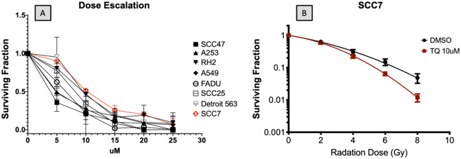
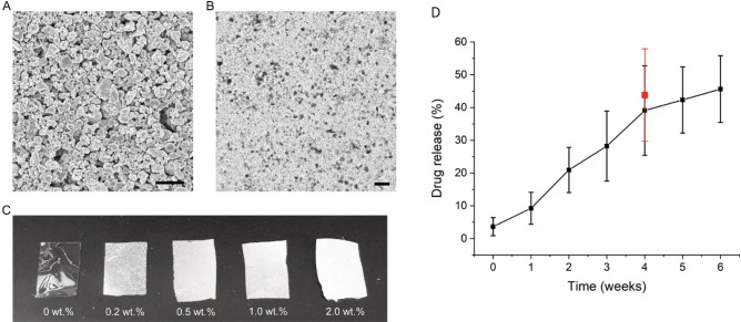
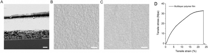
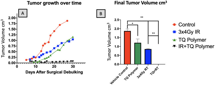
Head and neck cancer is the sixth most common cancer in the world, with more than 300,000 deaths attributed to the disease annually. Aggressive surgical resection often with adjuvant chemoradiation is the cornerstone of treatment. However, the necessary chemoradiation treatment can result in collateral damage to adjacent vital structures causing a profound impact on quality of life. Here, we present a novel polymer of poly(lactic-co-glycolic) acid and polyvinyl alcohol that can serve as a versatile multidrug delivery platform as well as for detection on cross-sectional imaging while functioning as a fiduciary marker for postoperative radiotherapy and radiotherapeutic dosing. In a mouse xenograft model, the dual-layered polymer composed of calcium carbonate/thymoquinone was used for both polymer localization and narrow-field infusion of a natural therapeutic compound. A similar approach can be applied in the treatment of head and neck cancer patients, where immunotherapy and traditional chemotherapy can be delivered simultaneously with independent release kinetics.


Conflict of interest statement

The authors declare no competing interests.

Figures

Figure 1
Figure 1
TQ treatment decreases cancer cell survival. (A) Dose escalation studies using a panel of human head and neck cell lines and SCCVIISF mouse derived line, demonstrates that 15 μM of TQ is sufficient for reducing colony formation by 50% or more (P < 0.001). (B) Linear quadratic survival curve depicting TQ-treated SCCVIISF vs control. TQ synergizes with radiation causing a significant decrease in cancer cell survival at 8 Gy with p < 0.05 and with 4.86% (SD 2.7%) for RT only and 1.2% (SD 0.6%) for RT in combination with 10 μM TQ.
Figure 2
Figure 2
Characterization of CaCO3 micron particles and CaCO3/PVA films and release kinetics. (A) SEM image of CaCO3 micron particles (scale bar: 50 μm). (B) Microscopic image of CaCO3/PVA film (scale bar: 50 μm). (C) CaCO3/PVA films with varying concentrations of CaCO3 (0, 0.2, 0.5, 1.0, and 2.0 wt%). (D) Drug release characteristics measured in vitro (black line) while red point represents drug release upon collection from mouse terminal surgery.
Figure 3
Figure 3
Characterizations of multilayer polymer film. (A) SEM image of the cross-section of multilayer polymer film (scale bar: 50 μm). (B) and (C) SEM images of the surface of CaCO3/PVA layer and TQ/PLGA layer from multilayer polymer film (scale bar: 5 μm). (Dual-layer polymer blend with treatment layer loaded with TQ and imaging layer composed of CaCO3). In (D) mechanical performance of the multiplayer polymer film is depicted as tensile strain vs tensile stress. The tensile strength of the multilayer polymer film was measured using dynamic mechanical analysis; the film has a very high tensile strength (32.9 Mpa), which is useful for clinical applications.
Figure 4
Figure 4
CT imaging demonstrating polymer localization in the tumor bed for precision radiotherapy localization.
Figure 5
Figure 5
Localization of polymer via Gross and FDG-PET imaging. (A1) Tumor exposed with TQ polymer removed. (B1D1) demonstrate transverse, coronal, and sagittal sections of tumor in (A1). FDG-PET signal is decreased around the TQ-embedded polymer treated areas with no tumor regrowth. Arrows mark location of polymer. The polymer imaging layer is visible in a standard FDG-PET scan. (A2) Depicts exposed tumor upon which plain control polymer (lacking TQ drug layer) was placed. (B2D2) are the corresponding PET images of (A2). (A1) mouse tumor had 0.024%ID/g FDG-PET uptake, whereas (A2) has 0.055%ID/g.
Figure 6
Figure 6
Histological assessment of the tumor at the site of polymer removal. (AD) demonstrates tumors collected from four different mouse treatment groups: vehicle control, TQ-embedded polymer, 3 × 4 Gy RT, and 3 × 4 Gy + TQ polymer from left to right with their corresponding insets below. Arrows signify the location of the polymer upon tumor removal. (A) vehicle control: Poorly differentiated carcinoma with viable tumor persists. Along the polymer boundary, a thin layer of dark pink fibrotic and inflammatory tissue can be observed. (B) TQ-embedded polymer: Tumor necrosis is seen with no viable tumor in the treatment area; The pink areas correspond to necrosis seen extending below the area of the drug treatment layer (dotted line). (C) 3 × 4 Gy RT : Narrow band of necrosis is noted along the polymer site with viable pockets of tumor tissue below. (D) 3 × 4 Gy + TQ polymer: No viable tumor present.
Figure 7
Figure 7
Final volume measurements. Tumor volume growth after 50% surgical debulking per each treatment group. N >  = 4; validated three times. (A) Shows tumor measurements taken by calipers over the course of 30 days post-surgical debulking. (B) Tumor volume upon resection. *p < 0.05, **p < 0.001, error bars are plotted as SEM. TQ + fractionated RT reflects no physically measurable tumor by caliper assessment; this was confirmed by histological analysis, where no residual cancer was seen (Fig. 6).

Similar articles

Cited by

References

    1. Cancer.Net, Vol. 2021 (2021).
    1. Gleich LL, et al. Recurrent advanced (T3 or T4) head and neck squamous cell carcinoma: is salvage possible? Arch. Otolaryngol. Head Neck Surg. 2004;130:35–38. doi: 10.1001/archotol.130.1.35. - DOI - PubMed
    1. Ross DA, et al. Microsurgical free flap reconstruction outcomes in head and neck cancer patients after surgical extirpation and intraoperative brachytherapy. Laryngoscope. 2004;114:1170–1176. doi: 10.1097/00005537-200407000-00007. - DOI - PubMed
    1. Lo, S. & Reimann, M., Vol. 2022 (Cancer.gov; 2021).
    1. Smith RV, Krevitt L, Yi SM, Beitler JJ. Early wound complications in advanced head and neck cancer treated with surgery and Ir 192 brachytherapy. Laryngoscope. 2000;110:8–12. doi: 10.1097/00005537-200001000-00002. - DOI - PubMed