

Neural stem cell therapy in conjunction with curcumin loaded in niosomal nanoparticles enhanced recovery from traumatic brain injury
- PMID: 35246564
- PMCID: PMC8897489
- DOI: 10.1038/s41598-022-07367-1
Neural stem cell therapy in conjunction with curcumin loaded in niosomal nanoparticles enhanced recovery from traumatic brain injury
Abstract
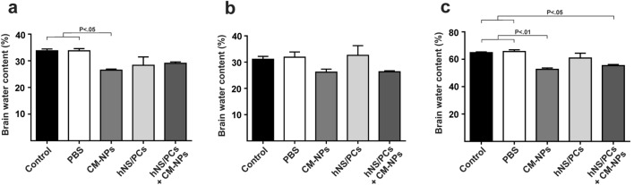
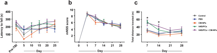
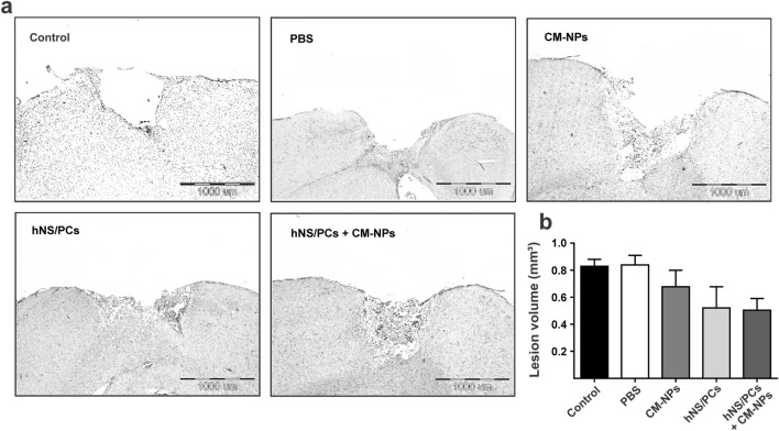
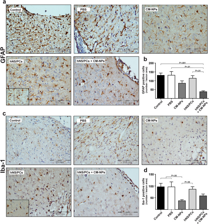
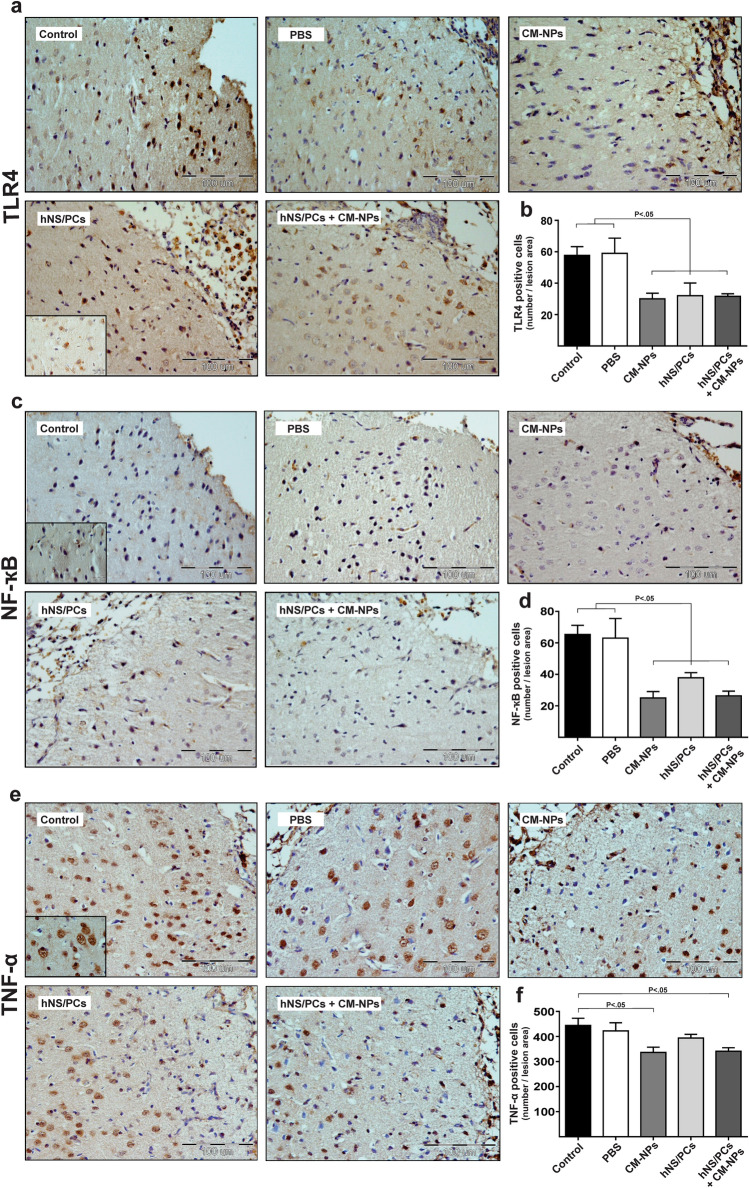
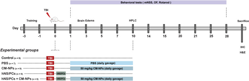
Despite a great amount of effort, there is still a need for reliable treatments of traumatic brain injury (TBI). Recently, stem cell therapy has emerged as a new avenue to address neuronal regeneration after TBI. However, the environment of TBI lesions exerts negative effects on the stem cells efficacy. Therefore, to maximize the beneficial effects of stem cells in the course of TBI, we evaluated the effect of human neural stem/progenitor cells (hNS/PCs) and curcumin-loaded niosome nanoparticles (CM-NPs) on behavioral changes, brain edema, gliosis, and inflammatory responses in a rat model of TBI. After TBI, hNS/PCs were transplanted within the injury site and CM-NPs were orally administered for 10 days. Finally, the effect of combination therapy was compared to several control groups. Our results indicated a significant improvement of general locomotor activity in the hNS/PCs + CM-NPs treatment group compared to the control groups. We also observed a significant improvement in brain edema in the hNS/PCs + CM-NPs treatment group compared to the other groups. Furthermore, a significant decrease in astrogliosis was seen in the combined treatment group. Moreover, TLR4-, NF-κB-, and TNF-α- positive cells were significantly decreased in hNS/PCs + CM-NPs group compared to the control groups. Taken together, this study indicated that combination therapy of stem cells with CM-NPs can be an effective therapy for TBI.
© 2022. The Author(s).
Conflict of interest statement
The authors declare no competing interests.
Figures
Similar articles
-
Human Neural Stem/Progenitor Cells Derived From Epileptic Human Brain in a Self-Assembling Peptide Nanoscaffold Improve Traumatic Brain Injury in Rats.Mol Neurobiol. 2018 Dec;55(12):9122-9138. doi: 10.1007/s12035-018-1050-8. Epub 2018 Apr 12. Mol Neurobiol. 2018. PMID: 29651746
-
Combination of curcumin with autologous transplantation of adult neural stem/progenitor cells leads to more efficient repair of damaged cerebral tissue of rat.Exp Physiol. 2020 Sep;105(9):1610-1622. doi: 10.1113/EP088697. Epub 2020 Jul 31. Exp Physiol. 2020. PMID: 32627273
-
Transplantation of human meningioma stem cells loaded on a self-assembling peptide nanoscaffold containing IKVAV improves traumatic brain injury in rats.Acta Biomater. 2019 Jul 1;92:132-144. doi: 10.1016/j.actbio.2019.05.010. Epub 2019 May 7. Acta Biomater. 2019. PMID: 31075516
-
Combination of drug and stem cells neurotherapy: Potential interventions in neurotrauma and traumatic brain injury.Neuropharmacology. 2019 Feb;145(Pt B):177-198. doi: 10.1016/j.neuropharm.2018.09.032. Epub 2018 Sep 26. Neuropharmacology. 2019. PMID: 30267729 Review.
-
The Potential of Stem Cells in Treatment of Traumatic Brain Injury.Curr Neurol Neurosci Rep. 2018 Jan 25;18(1):1. doi: 10.1007/s11910-018-0812-z. Curr Neurol Neurosci Rep. 2018. PMID: 29372464 Free PMC article. Review.
Cited by
-
Therapeutic Application of Stem Cells in the Repair of Traumatic Brain Injury.Stem Cells Cloning. 2022 Jul 13;15:53-61. doi: 10.2147/SCCAA.S369577. eCollection 2022. Stem Cells Cloning. 2022. PMID: 35859889 Free PMC article. Review.
-
Multi-Mechanistic Approaches to the Treatment of Traumatic Brain Injury: A Review.J Clin Med. 2023 Mar 11;12(6):2179. doi: 10.3390/jcm12062179. J Clin Med. 2023. PMID: 36983181 Free PMC article. Review.
-
Natural Antioxidant Compounds as Potential Pharmaceutical Tools against Neurodegenerative Diseases.ACS Omega. 2022 Jul 19;7(30):25974-25990. doi: 10.1021/acsomega.2c03291. eCollection 2022 Aug 2. ACS Omega. 2022. PMID: 35936442 Free PMC article. Review.
-
Curcumin, inflammation, and neurological disorders: How are they linked?Integr Med Res. 2023 Sep;12(3):100968. doi: 10.1016/j.imr.2023.100968. Epub 2023 Jun 25. Integr Med Res. 2023. PMID: 37664456 Free PMC article. Review.
-
Traumatic brain injury: Bridging pathophysiological insights and precision treatment strategies.Neural Regen Res. 2026 Mar 1;21(3):887-907. doi: 10.4103/NRR.NRR-D-24-01398. Epub 2025 Mar 25. Neural Regen Res. 2026. PMID: 40145994 Free PMC article.
References
-
- Feng Y, et al. Protective role of apocynin via suppression of neuronal autophagy and TLR4/NF-κB signaling pathway in a rat model of traumatic brain injury. Neurochem. Res. 2017;42:3296–3309. - PubMed
-
- Evran S, et al. The effect of high mobility group box-1 protein on cerebral edema, blood-brain barrier, oxidative stress and apoptosis in an experimental traumatic brain injury model. Brain Res. Bull. 2020;154:68–80. - PubMed
-
- Shi H, Hua X, Kong D, Stein D, Hua F. Role of Toll-like receptor mediated signaling in traumatic brain injury. Neuropharmacology. 2019;145:259–267. - PubMed
Publication types
MeSH terms
Substances
LinkOut - more resources
Full Text Sources
Medical
