. 2022 Mar 5;12(1):21.
doi: 10.1186/s13613-022-00982-1.

Recombinant human interleukin-7 reverses T cell exhaustion ex vivo in critically ill COVID-19 patients

Collaborators, Affiliations

Recombinant human interleukin-7 reverses T cell exhaustion ex vivo in critically ill COVID-19 patients

Frank Bidar et al. Ann Intensive Care. .

Erratum in

Abstract

Background: Lymphopenia is a hallmark of severe coronavirus disease 19 (COVID-19). Similar alterations have been described in bacterial sepsis and therapeutic strategies targeting T cell function such as recombinant human interleukin 7 (rhIL-7) have been proposed in this clinical context. As COVID-19 is a viral sepsis, the objectives of this study were to characterize T lymphocyte response over time in severe COVID-19 patients and to assess the effect of ex vivo administration of rhIL-7.

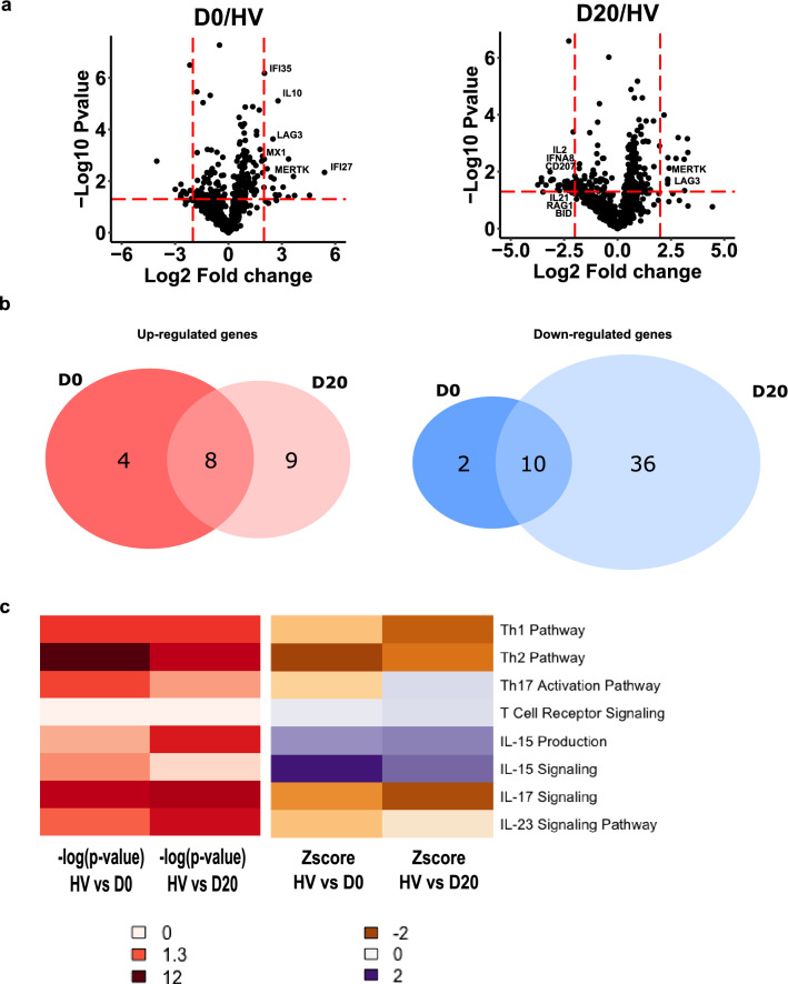
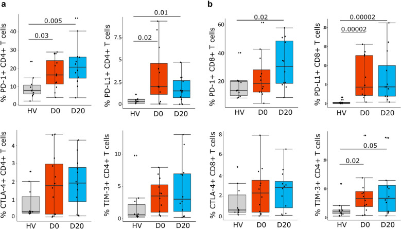
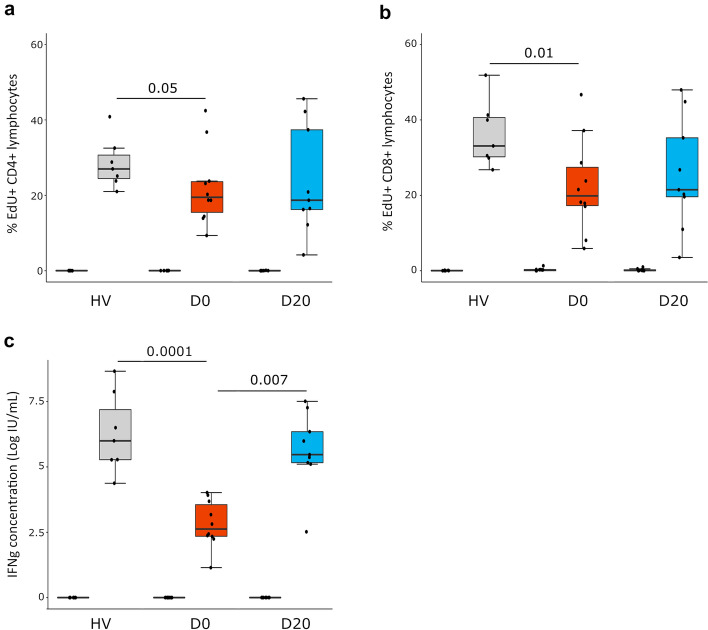
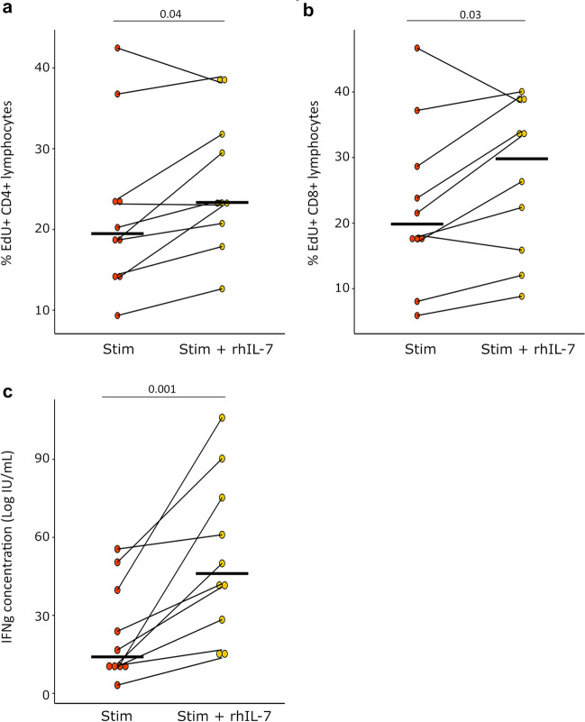
Results: Peripheral blood mononuclear cells from COVID-19 patients hospitalized in intensive care unit (ICU) were collected at admission and after 20 days. Transcriptomic profile was evaluated through NanoString technology. Inhibitory immune checkpoints expressions were determined by flow cytometry. T lymphocyte proliferation and IFN-γ production were evaluated after ex vivo stimulation in the presence or not of rhIL-7. COVID-19 ICU patients were markedly lymphopenic at admission. Mononuclear cells presented with inhibited transcriptomic profile prevalently with impaired T cell activation pathways. CD4 + and CD8 + T cells presented with over-expression of co-inhibitory molecules PD-1, PD-L1, CTLA-4 and TIM-3. CD4 + and CD8 + T cell proliferation and IFN-γ production were markedly altered in samples collected at ICU admission. These alterations, characteristic of a T cell exhaustion state, were more pronounced at ICU admission and alleviated over time. Treatment with rhIL-7 ex vivo significantly improved both T cell proliferation and IFN-γ production in cells from COVID-19 patients.

Conclusions: Severe COVID-19 patients present with features of profound T cell exhaustion upon ICU admission which can be reversed ex vivo by rhIL-7. These results reinforce our understanding of severe COVID-19 pathophysiology and opens novel therapeutic avenues to treat such critically ill patients based of immunomodulation approaches. Defining the appropriate timing for initiating such immune-adjuvant therapy in clinical setting and the pertinent markers for a careful selection of patients are now warranted to confirm the ex vivo results described so far. Trial registration ClinicalTrials.gov identifier: NCT04392401 Registered 18 May 2020, http:// clinicaltrials.gov/ct2/show/NCT04392401.

Keywords: Critically ill; Exhaustion; Immunostimulation; Interleukin-7; SARS-CoV-2; T lymphocytes.


Conflict of interest statement

The authors declare no competing financial interests in relation to the work. MAC, KBP and MM are bioMérieux’s employees. This private company had no role in the study design, result analysis and decision to publish this study.

Figures

Fig. 1
Fig. 1
Transcriptomic profile of mononuclear cells in severe COVID-19 patients. RNA extracted from PBMCs of COVID-19 patients at day 0 and day 20 (n = 10) and healthy volunteers (HV, n = 10) was analyzed through NanoString technology. a Volcano plots of differentially expressed genes between patients sampled at D0 or at D20 and HV are showed. Limits of significance are illustrated by red dotted lines (i.e. Log2 Fold Change = −2 or + 2 and −Log 10 P value = 1.3). Selected genes are mentioned. b Venn diagrams of significantly up-regulated (left diagram, n = 21) or down-regulated (right diagram, n = 48) genes between patients and HV are showed. c Ingenuity Pathway Analysis was applied on the list of differentially expressed genes at D0 and D20. Heatmaps of Log 10 P-value (from white indicating the absence of significance to dark red indicating a strong significance) and Z-score (from orange indicating a down-regulation to purple indicating an up-regulation) for pathways related to T cell activation at D0 and D20 are presented
Fig. 2
Fig. 2
Immune-inhibitory receptors expressions on CD4 + and CD8 + T cells in severe COVID-19 patients. PD-1, PD-L1, CTLA-4 and TIM-3 expressions were assessed by flow cytometry in PBMCs from COVID-19 patients (n = 12) at day 0 (D0) and day 20 (D20) and healthy volunteers (HV, n = 10). Results are presented as individual values and boxplots. a Expressions on CD4 + T cells are presented. b Expressions on CD8 + T cells are presented. Nonparametric Mann–Whitney U test was used to compare results between HV and COVID-19 patients. Only statistically significant differences are shown
Fig. 3
Fig. 3
Lymphocyte proliferation and IFN-γ production after TCR stimulation in severe COVID-19 patients. PBMCs were collected from healthy volunteers (HV, n = 7) and COVID-19 patients at D0 and D20 (n = 10). Cells were cultured for 3 days in the presence (TCR stimulation) or not (Control) of anti-CD2/CD3/CD28 Ab-coated beads (ratio of beads/cells = 2:1). Cellular proliferation was assessed by monitoring EdU AF488 incorporation into cells and expressed as percentages of cells incorporating EdU. IFN-γ was measured in cell culture supernatants after stimulation. Results are presented as individual values and boxplots. a CD4 + T cell proliferation is presented. b CD8 + T cell proliferation is presented. c Natural log transformed IFN-γ production in culture supernatants is presented. Nonparametric Mann–Whitney U test was used to compare results between HV and patients. Wilcoxon signed-rank test was used for paired comparisons between D0 and D20. Only statistically significant differences are shown
Fig. 4
Fig. 4
Lymphocyte proliferation and IFN-γ production after rhIL-7 and TCR stimulation in severe COVID-19 patients. PBMCs were collected in COVID-19 patients at D0 (n = 10). Cells were cultured for 3 days in the presence or not of anti-CD2/CD3/CD28 Ab-coated beads (TCR Stimulation, ratio of beads/cells = 2:1) and rhIL-7 (TCR Stimulatio n + rhIL-7,100 ng/ml). Cellular proliferation was assessed by monitoring EdU AF488 incorporation into cells and expressed as percentages of cells incorporating EdU. IFN-γ was measured in cell supernatants after proliferation. Results are presented as individual values and medians. a CD4 + T cell proliferation is presented. b CD8 + T cell proliferation is presented. c Natural log transformed IFN-γ production in culture supernatants is shown. Wilcoxon signed-rank test was used to compare results between groups. Only statistically significant differences are shown

References

    1. Network C-IGobotR, the C-ICUI. Clinical characteristics and day-90 outcomes of 4244 critically ill adults with COVID-19: a prospective cohort study. Intensive Care Med. 2021;47(1):60–73. - PMC - PubMed
    1. Carenzo L, Costantini E, Greco M, Barra FL, Rendiniello V, Mainetti M, et al. Hospital surge capacity in a tertiary emergency referral centre during the COVID-19 outbreak in Italy. Anaesthesia. 2020;75(7):928–934. - PubMed
    1. Hadjadj J, Yatim N, Barnabei L, Corneau A, Boussier J, Smith N, et al. Impaired type I interferon activity and inflammatory responses in severe COVID-19 patients. Science. 2020;369(6504):718–724. - PMC - PubMed
    1. Qin C, Zhou L, Hu Z, Zhang S, Yang S, Tao Y, et al. Dysregulation of immune response in patients with COVID-19 in Wuhan, China. Clin Infect Dis. 2020. - PMC - PubMed
    1. Yang X, Yu Y, Xu J, Shu H, Xia J, Liu H, et al. Clinical course and outcomes of critically ill patients with SARS-CoV-2 pneumonia in Wuhan, China: a single-centered, retrospective, observational study. Lancet Respir Med. 2020;8(5):475–481. - PMC - PubMed

Associated data