Review
. 2023 Apr;290(8):1973-1985.
doi: 10.1111/febs.16419. Epub 2022 Mar 11.

Impact of structural dynamics on biological functions of flaviviruses

Affiliations
Review

Impact of structural dynamics on biological functions of flaviviruses

Karin Stiasny et al. FEBS J. 2023 Apr.

Abstract

Flaviviruses comprise a number of mosquito- or tick-transmitted human pathogens of global public health importance. Advances in structural biology techniques have contributed substantially to our current understanding of the life cycle of these small enveloped RNA viruses and led to deep insights into details of virus assembly, maturation and cell entry. In addition to large-scale conformational changes and oligomeric rearrangements of envelope proteins during these processes, there is increasing evidence that smaller-scale protein dynamics (referred to as virus "breathing") can confer extra flexibility to these viruses for the fine-tuning of their interactions with the immune system and possibly with cellular factors they encounter in their complex ecological cycles in arthropod and vertebrate hosts. In this review, we discuss how work with tick-borne encephalitis virus has extended our view on flavivirus breathing, leading to the identification of a novel mechanism of antibody-mediated infection enhancement and demonstrating breathing intermediates of the envelope protein in the process of membrane fusion. These data are discussed in the context of other flaviviruses and the perspective of a potential role of virus breathing to cope with the requirements of adaptation and replication in evolutionarily very different hosts.

Keywords: antibody-dependent enhancement; envelope protein; flavivirus; membrane fusion; particle dynamics; particle heterogeneity; tick-borne encephalitis virus; virus breathing.


Conflict of interest statement

None to declare with this review. The Center for Virology received a research grant from Pfizer on the epidemiology of TBE in Austria, with KS as principal investigator (2018‐2021). FXH and KS are inventors on a patent of the Medical University of Vienna on flavivirus IgM serodiagnosis.

Figures

Fig. 1
Fig. 1
Structural organization of mature flaviviruses. (A) Flavivirus RNA genome with the single open reading frame. The three structural proteins are coloured yellow, the nonstructural proteins grey. (B) Schematic representation of a flavivirus particle in its fully mature state. (C) Cryo‐EM structure of the mature TBEV particle (PDB: 5O6A, [19]). One E dimer is encircled in white. (D) Top view of the TBEV E dimer ectodomain (without the stem‐anchor region) and (E) side view of the full‐length TBEV E‐M complex (PDB: 5O6A, [19]). The flexible linkers/hinges between the domains are encircled in black in one E monomer. The E protein is coloured according to its domains: domain I (DI), red; domain II (DII), yellow; domain III (DIII), blue; fusion loop (FL), orange; stem, green and transmembrane anchor (TM), grey. M is displayed in pink; the viral membrane in light blue. Molecular structures were generated with UCSF Chimera (panel C) (https://www.cgl.ucsf.edu/chimera, [93] and PyMol (panels D and E) (https://pymol.org/2).
Fig. 2
Fig. 2
Flavivirus membrane fusion. (A) E dimers on the surface of the mature particle. (B) Low‐pH‐induced dissociation of the E dimers in endosomes, fusion‐loop exposure and insertion into the endosomal membrane. (C) Formation of trimers in an extended conformation bridging the two membranes, blue arrows indicate the subsequent relocation of domain III from the end to the side of the molecule (see also panel E). (D) Formation of the hairpin‐like post‐fusion trimer with the fusion loops and membrane anchors juxtaposed and opening of the fusion pore. (E) Ribbon representations of E monomers in their pre‐ and post‐fusion conformations, showing the relocation of domain III (DIII), indicated by a blue arrow in the pre‐fusion monomer. Colour code E: domain I, red; domain II, yellow; domain III, blue; fusion loop (FL), orange; stem, green and membrane anchor, grey. Molecular structures in panel E were generated with PyMol (https://pymol.org/2) using coordinates of TBEV (PDBs: 1SVB, 1URZ [20, 27]).
Fig. 3
Fig. 3
Rearrangements of viral surface proteins during maturation. Schematic representations in upper panels and cryo EM reconstructions in lower panels. (A) Immature virions assembled in the endoplasmic reticulum (ER) at neutral pH with trimeric prM/E spikes on the particle surface. (B) Formation of smooth‐surfaced particles with the herringbone‐like arrangement of E dimers induced by the acidic pH in the trans‐Golgi network (TGN) and cleavage of prM into pr and M. The cleavage product pr remains associated with the particles at acidic pH. (C) Mature infectious virion with the E dimers in a metastable conformation, primed for fusion. The cleaved pr parts have been released from the particles at neutral pH. Colour code of E and prM/M in the schematics as in Figs. 1 and 2. Molecular structures were generated with UCSF Chimera (https://www.cgl.ucsf.edu/chimera, [93]) using coordinates of dengue viruses (PDBs: 4B03, 3C6R, 4CCT; [34, 37]). E is shown in grey, prM and M in pink.
Fig. 4
Fig. 4
Schematic illustrations of structural heterogeneity of flavivirus particles. (A) E breathing (indicated by curved lines) of E on the surface of a mature virion, leading to the transient exposure of the fusion loop (FL). (B) Partially mature particles with a prM/E spike and E dimer representing the immature and mature patches on the virus surface, respectively. The border zone between the immature and mature regions (symbolized as a breathing E monomer) is irregular and therefore structurally not resolved. (C) Binding of a fusion‐loop (FL) specific antibody to E upon exposure of the fusion loop by breathing. Colour code of E and prM/M in the schematics as in Figs. 1 and 2.
Fig. 5
Fig. 5
Promotion of TBEV cell binding and enhancement of infection induced by a monoclonal antibody (mab A5). (A) Surface representation of the TBEV E dimer (upper panel) and its dissociation into the two subunits by antibody A5. Amino acids contributing to the epitope of mab A5 (black and grey circles) are shown in cyan. (B) Schematic representation of antibody‐induced exposure of the fusion loop (FL), its attachment to the plasma membrane (upper panel) and inhibition of binding by a fusion‐loop‐specific antibody (lower panel). Colour code of E and pr/M as in Figs. 1 and 2. Molecular structures were generated with UCSF Chimera (https://www.cgl.ucsf.edu/chimera, [93]) using coordinates of the TBEV E dimer (PDB: 1SVB, [20]).
Fig. 6
Fig. 6
Dynamic trimeric fusion intermediates in flavivirus membrane fusion. (A) Side view of the E protein trimer (left), and open book view of two subunits and the missing “open” subunit. The dynamically exposed regions in fusion intermediates are shown in grey. (B) Schematic of the pre‐fusion organization of the E stem based on cryo‐EM of TBEV [19]. Pre‐F‐H1, stem helix 1 in its pre‐fusion conformation; Pre‐F‐H2, stem helix 2 in its pre‐fusion conformation; (Pre‐F‐) H3, stem helix 3 in pre‐ and post‐fusion form; CS, conserved sequence. (C) Dynamic intermediate after relocation of domain III with incompletely zippered stem (“breathing trimers”). (D) Final post‐fusion trimer with the fully zippered stem and formation of the fusion pore. Only one of the three stem‐anchor regions in the trimer is shown in panels (C) and (D). Colour code E as in Figs. 1 and 2. Molecular structures shown in panel A were generated with PyMol (https://pymol.org/2) using coordinates of TBEV (PDBs: 1URZ, 6S8C [27, 31]).
Fig. 7
Fig. 7
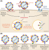
Schematic representation of different modes of cell attachment. (A) Known cell‐binding modes of flaviviruses. Left panel: after binding to a specific receptor. Middle panel: antibody‐induced exposure of the FL and attachment to the plasma membrane. Right panel: FcR‐mediated uptake of an antibody‐virus complex. (B) Hypothetical cell‐binding modes of dynamic and/or heterogeneous flavivirus particles. Colour code E: DI, red; DII, yellow; DIII, blue; fusion loop (FL), orange; stem, green and membrane anchor, grey. The (pr)M protein is shown in pink.

References

    1. Pierson TC, Diamond MS. The continued threat of emerging flaviviruses. Nat Microbiol. 2020;5:796–812. - PMC - PubMed
    1. Bhatt S, Gething PW, Brady OJ, Messina JP, Farlow AW, Moyes CL, et al. The global distribution and burden of dengue. Nature. 2013;496:504–7. - PMC - PubMed
    1. Guzman MG, Harris E. Dengue. Lancet. 2015;385:453–65. - PubMed
    1. WHO . Vaccines against tick‐borne encephalitis: WHO position paper. Wkly Epidemiol Rec. 2011;86:241–56. - PubMed
    1. Caminade C, McIntyre KM, Jones AE. Impact of recent and future climate change on vector‐borne diseases. Ann N Y Acad Sci. 2019;1436:157–73. - PMC - PubMed

Publication types