

Comparison of lung ultrasound, chest radiographs, C-reactive protein, and clinical findings in dogs treated for aspiration pneumonia
- PMID: 35247005
- PMCID: PMC8965265
- DOI: 10.1111/jvim.16379
Comparison of lung ultrasound, chest radiographs, C-reactive protein, and clinical findings in dogs treated for aspiration pneumonia
Abstract
Background: Comparison of clinical findings, chest radiographs (CXR), lung ultrasound (LUS) findings, and C-reactive protein (CRP) concentrations at admission and serial follow-up in dogs with aspiration pneumonia (AP) is lacking.
Hypothesis: Lung ultrasound lesions in dogs with AP are similar to those described in humans with community-acquired pneumonia (comAP); the severity of CXR and LUS lesions are similar; normalization of CRP concentration precedes resolution of imaging abnormalities and more closely reflects the clinical improvement of dogs.
Animals: Seventeen dogs with AP.
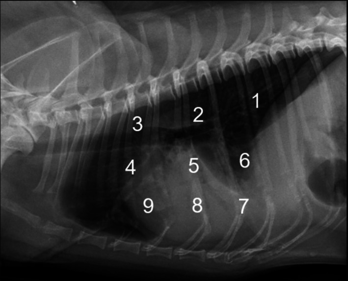
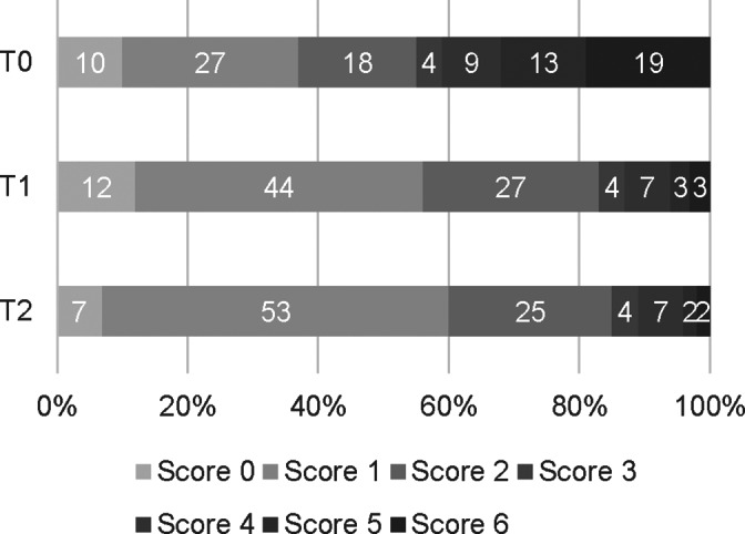
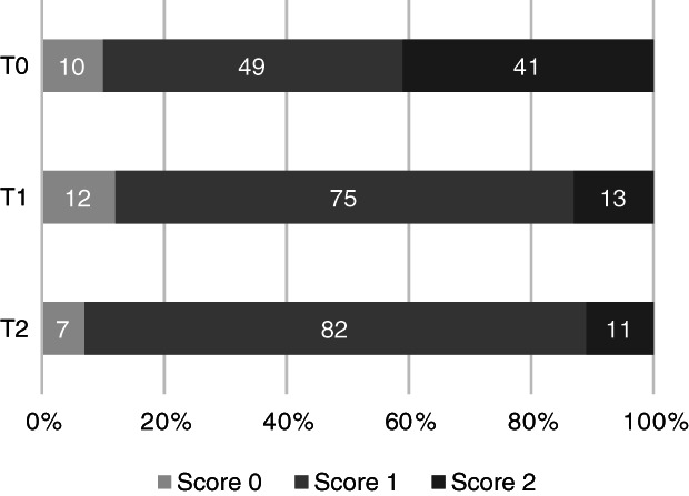
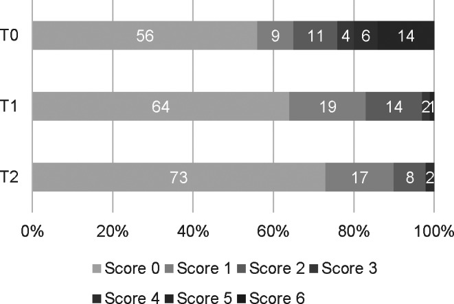
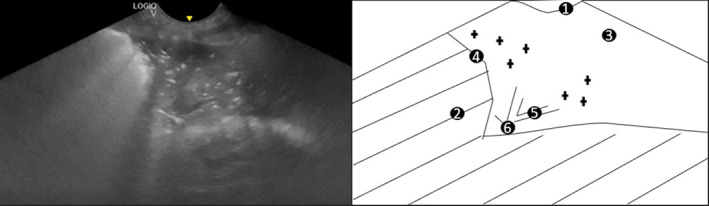
Methods: Prospective observational study. Clinical examination, CXR, LUS, and CRP measurements performed at admission (n = 17), 2 weeks (n = 13), and 1 month after diagnosis (n = 6). All dogs received antimicrobial therapy. Lung ultrasound and CXR canine aspiration scoring systems used to compare abnormalities.
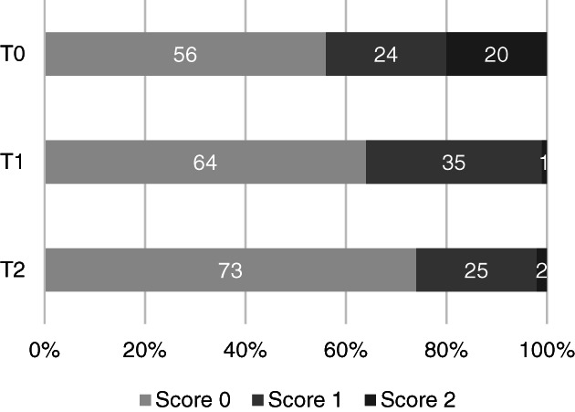
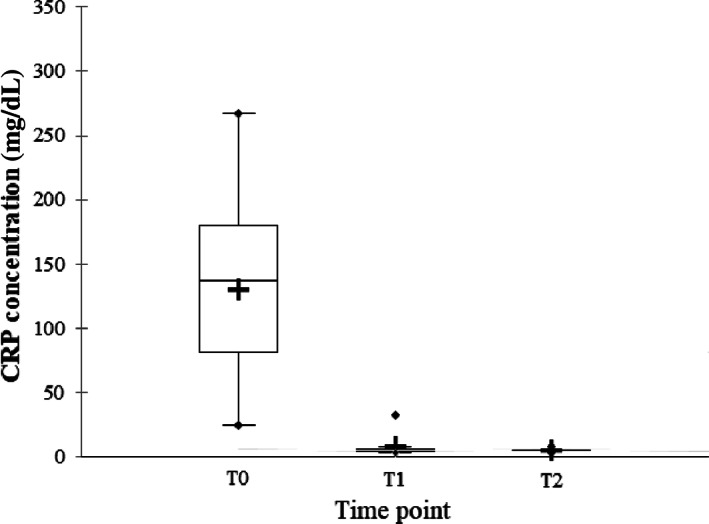
Results: B-lines and shred signs with or without bronchograms were identified on LUS in 14 of 17 and 16 of 17, at admission. Chest radiographs and LUS scores differed significantly using both canine AP scoring systems at each time point (18 regions per dog, P < .001). Clinical and CRP normalization occurred in all dogs during follow up. Shred signs disappeared on LUS in all but 1 of 6 dogs at 1 month follow-up, while B-lines and CXR abnormalities persisted in 4 of 6 and all dogs, respectively.
Conclusion and clinical importance: Lung ultrasound findings resemble those of humans with comAP and differ from CXR findings. Shred signs and high CRP concentrations better reflect clinical findings during serial evaluation of dogs.
Keywords: acute phase proteins; canine aspiration pneumonia score; community-acquired pneumonia; point of care ultrasound; simplified canine aspiration pneumonia score.
© 2022 The Authors. Journal of Veterinary Internal Medicine published by Wiley Periodicals LLC on behalf of American College of Veterinary Internal Medicine.
Conflict of interest statement
Authors declare no conflict of interest.
Figures
Comment in
-
Letter regarding "Comparison of lung ultrasound, chest radiographs, C-reactive protein, and clinical findings in dogs treated for aspiration pneumonia".J Vet Intern Med. 2022 Nov;36(6):1855. doi: 10.1111/jvim.16557. Epub 2022 Sep 30. J Vet Intern Med. 2022. PMID: 36178173 Free PMC article. No abstract available.
Similar articles
-
Antimicrobial discontinuation in dogs with acute aspiration pneumonia based on clinical improvement and normalization of C-reactive protein concentration.J Vet Intern Med. 2022 May;36(3):1082-1088. doi: 10.1111/jvim.16405. Epub 2022 Mar 29. J Vet Intern Med. 2022. PMID: 35348224 Free PMC article.
-
Response to letter regarding "Comparison of lung ultrasound, chest radiographs, C-reactive protein, and clinical findings in dogs treated for aspiration pneumonia".J Vet Intern Med. 2022 Nov;36(6):1856-1857. doi: 10.1111/jvim.16558. Epub 2022 Oct 3. J Vet Intern Med. 2022. PMID: 36189851 Free PMC article. No abstract available.
-
Letter regarding "Comparison of lung ultrasound, chest radiographs, C-reactive protein, and clinical findings in dogs treated for aspiration pneumonia".J Vet Intern Med. 2022 Nov;36(6):1855. doi: 10.1111/jvim.16557. Epub 2022 Sep 30. J Vet Intern Med. 2022. PMID: 36178173 Free PMC article. No abstract available.
-
The utility of chest x-ray and lung ultrasound in the management of infants and children presenting with severe pneumonia in low-and middle-income countries: A pragmatic scoping review.J Glob Health. 2022 Dec 23;12:10013. doi: 10.7189/jogh.12.10013. J Glob Health. 2022. PMID: 36560909 Free PMC article.
-
Point-of-care lung ultrasound for the assessment of pneumonia: a narrative review in the COVID-19 era.J Med Ultrason (2001). 2021 Jan;48(1):31-43. doi: 10.1007/s10396-020-01074-y. Epub 2021 Jan 13. J Med Ultrason (2001). 2021. PMID: 33438132 Free PMC article. Review.
Cited by
-
Confidence level of Australian veterinarians with point-of-care ultrasound before and after a training course.Can Vet J. 2024 Sep;65(9):910-919. Can Vet J. 2024. PMID: 39219614 Free PMC article.
-
Retrospective Evaluation of Subpleural Consolidations Using Lung Ultrasound in 634 Dogs and 347 Cats.Animals (Basel). 2025 Feb 13;15(4):549. doi: 10.3390/ani15040549. Animals (Basel). 2025. PMID: 40003031 Free PMC article.
-
Utility of serum amyloid A in monitoring clinical response to antimicrobial treatment in horses with bacterial pneumonia.J Vet Intern Med. 2023 Sep-Oct;37(5):1917-1922. doi: 10.1111/jvim.16818. Epub 2023 Jul 31. J Vet Intern Med. 2023. PMID: 37522636 Free PMC article.
-
Emergency Removal of a Proximal Tracheal Foreign Body by Tracheotomy in a Dog and a Cat.Case Rep Vet Med. 2023 Sep 16;2023:6478643. doi: 10.1155/2023/6478643. eCollection 2023. Case Rep Vet Med. 2023. PMID: 37745017 Free PMC article.
-
Recognition and Diagnosis of Underlying Disease Processes in Bacterial Pneumonia.Animals (Basel). 2024 May 29;14(11):1601. doi: 10.3390/ani14111601. Animals (Basel). 2024. PMID: 38891647 Free PMC article. Review.
References
-
- Sherman R, Karagiannis M. Aspiration pneumonia in the dog: a review. Topics in Compan an Med. 2017;32:1‐7. - PubMed
-
- Kogan DA, Johnson LR, Jandrey KE, Pollard RE. Clinical, clinicopathologic, and radiographic findings in dogs with aspiration pneumonia: 88 cases (2004–2006). J Am Vet Med Assoc. 2008;233:1742‐1747. - PubMed
-
- Kogan DA, Lynelle RJ, Sturges BK, et al. Etiology and clinical outcome in dogs with aspiration pneumonia: 88 cases (2004–2006). J Am Vet Med Assoc. 2008;233:1748‐1755. - PubMed
-
- Tart KM, Babski DM, Lee JA. Potential risks, prognostic indicators, and diagnostic and treatment modalities affecting survival in dogs with presumptive aspiration pneumonia: 125 cases (2005‐2008). J Vet Emerg Crit Care. 2010;20:319‐329. - PubMed
Publication types
MeSH terms
Substances
LinkOut - more resources
Full Text Sources
Medical
Research Materials
Miscellaneous
