. 2022 Mar 5;22(1):107.
doi: 10.1186/s12935-022-02487-0.

DFNA5 regulates immune cells infiltration and exhaustion

Affiliations

DFNA5 regulates immune cells infiltration and exhaustion

Jian Hu et al. Cancer Cell Int. .

Abstract

Background: DFNA5 (GSDME) belongs to Gasdermin familily that is involved in a variety of cancers and triggers cell pyroptosis after chemical treatment. However, the relationship in DFNA5 between prognosis and immune cells in diverse cancers has been receiving little attention. Tumor immune cells infiltration and exhaustion may associate with patients prognosis. The roles of DFNA5 in tumor immune cells infiltration and exhaustion have not been clarified.

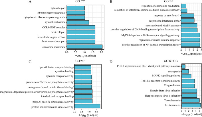
Methods: The expression level of DFNA5 was determined by the Tumour Immune Estimation Resource and the Oncomine database. Then the impacts of DFNA5 in prognosis were assessed by Kaplan-Meier plotter and ULACAN. The correlations between DFNA5 and tumour-infiltrating lymphocytes were explored by TIMER. In addition, the relationships in the expression levels of DFNA5 and typical genes combination of tumour-infiltrating lymphocytes were analysed by GEPIA and TIMER. In this study, we screened the chemokine and immune related proteins interacted with DFNA5 using TurboID system to explore the instantaneous or weak interactions.

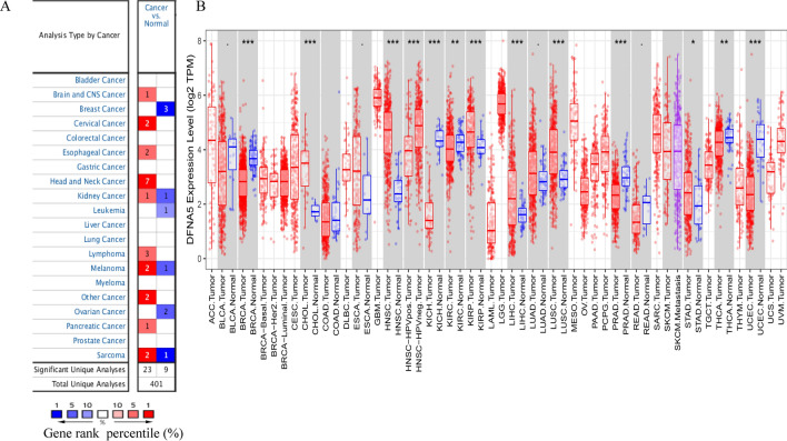
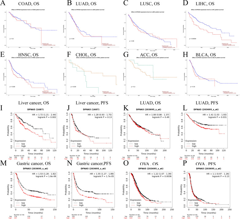
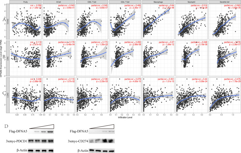
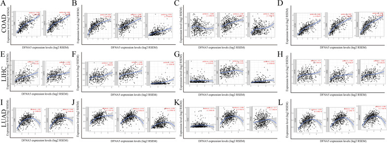
Results: DFNA5 significantly influences the prognosis in different cancers according to The Cancer Genome Atlas (TCGA). The expression levels of DFNA5 showed positive correlations to the infiltration of macrophages, CD8 + T cells, CD4 + T cells in liver hepatocellular carcinoma (LIHC), colon adenocarcinoma (COAD), and lung adenocarcinoma (LUAD). DFNA5 expression displayed obvious correlations with multiple lymphocytes gene makers in COAD, LIHC and LUAD. DFNA5 expression has effects on the prognosis of liver hepatocellular carcinoma and LUAD. DFNA5 upregulated the expression levels of PDCD1 and CD274 in a dose-dependent manner. Chemokine and immune related proteins interact with DFNA5.

Conclusions: These results indicate that DFNA5 is related to patient prognosis and immune cells, consisting of macrophages, CD4 + T cells, and CD8 + T cells, in diverse cancers. In addition, DFNA5 expression might contribute to the regulation of T cell exhaustion, tumour-associated macrophages (TAMs), and Tregs in COAD, LIHC and LUAD. DFNA5 may regulate immune infiltration via EIF2AK2. IFNGR1 was related to the functions of PD-L1 expression and PD-1 checkpoint pathway. These results indicate that DFNA5 levels may be act as a prognostic factor and predict the degrees of immune cells infiltration in LIHC and LUAD.

Keywords: CD274; Cancer; DFNA5; Prognosis; Tumour-infiltrating lymphocytes.


Conflict of interest statement

The authors declare no competing financial interest.

Figures

Fig. 1
Fig. 1
DFNA5 expression levels in diverse types of human cancers. A DFNA5 expression levels in diverse cancers relative to normal tissues in the Oncomine database. B Human DFNA5 expression levels in adjacent normal tissues and related tumour types in the TCGA database were obtained by TIMER (*P < 0.05, **P < 0.01, ***P < 0.001)
Fig. 2
Fig. 2
Kaplan–Meier survival curves comparing with high and low expression of DFNA5 in different types of cancer in the UALCAN (A–H) and Kaplan–Meier plotter databases (I–P). AH Survival curves of OS in COAD, LUAD, LUSC, LIHC, HNSC, CHOL, ACC, BLCA. I, J Survival curves of OS and DFS in the liver cancer cohort. K, L High LUAD expression was correlated with poor PFS in the LUAD and survival curves of OS in the LUAD. M, N OS and PFS survival curves of gastric cancer. O, P OS and PFS of OVA. OS overall survival, PFS Progression Free Survival
Fig. 3
Fig. 3
Correlation of DFNA5 expression with tumour-infiltrating lymphocytes in COAD (colon adenocarcinoma), LIHC (hepatocellular carcinoma), and LUAD (lung adenocarcinoma). A DFNA5 expression is significantly negatively related to tumour purity and has significant positive correlations with infiltrating levels of CD8 + T cells, CD4 + T cells, macrophages, neutrophils, and dendritic cells in COAD but not B cells. B DFNA5 expression has significant correlations with tumour purity and infiltrating levels of B cells, CD8 + T cells, CD4 + T cells, macrophages, neutrophils, and dendritic cells in LIHC. C DFNA5 expression is significantly negatively related to tumour purity and has significant positive correlations with infiltrating levels of B cells, CD8 + T cells, CD4 + T cells, macrophages, neutrophils, and dendritic cells in LUAD. D The increased content of Flag-DFNA5 plasmids co-transfected with 3x-myc-PDCD1 or 3x-myc-CD274 plasmid, the expression levels of FLAG, 3x-myc and β-actin were detected by western blot. The band grey value of the rightmost well of 3x-myc-PDCD1 or 3x-myc-CD274 relative to the corresponding beta-actin were set at 1.0. The other treatments were compared with them
Fig. 4
Fig. 4
DFNA5 expression was correlated with macrophage polarization in COAD, LIHC and LUAD. The markers consist of CD86 and CSF1R of monocytes; CCL2, CD68, and IL10 of TAMs (tumour-associated macrophages); NOS2, IRF5, and PTGS2 of M1 macrophages; and CD163, VSIG4, and MS4A4A of M2 macrophages. AD Scatter diagram of relationships between DFNA5 expression and gene markers of monocytes (A), TAMs (B), and M1 (C) and M2 macrophages (D) in COAD. EH Scatter diagram of relationships between DFNA5 expression and gene markers of monocytes (E), TAMs (F), and M1 (G) and M2 macrophages (H) in LIHC. IL Scatter diagram of relavances between DFNA5 expression and gene markers of monocytes (I), TAMs (J), and M1 (K) and M2 macrophages (L) in LUAD
Fig. 5
Fig. 5
GO and KEGG enrichment analysis of chemokine and immune related Genes interacted with DFNA5 (DAVID). A BP, B CC, C MF, D KEGG

Similar articles

Cited by

References

    1. Zhang C, et al. Multiomics analysis reveals a distinct response mechanism in multiple primary lung adenocarcinoma after neoadjuvant immunotherapy. J Immunother Cancer. 2021;9:e002312. doi: 10.1136/jitc-2020-002312. - DOI - PMC - PubMed
    1. Wang X, et al. Hypoxia-induced myeloid derived growth factor promotes hepatocellular carcinoma progression through remodeling tumor microenvironment. Theranostics. 2021;11:209–221. doi: 10.7150/thno.49327. - DOI - PMC - PubMed
    1. Shigeta K, et al. Regorafenib combined with PD1 blockade increases CD8 T-cell infiltration by inducing CXCL10 expression in hepatocellular carcinoma. J Immunother Cancer. 2020;8:e001435. doi: 10.1136/jitc-2020-001435. - DOI - PMC - PubMed
    1. Peng QH, et al. CMTM6 and PD-L1 coexpression is associated with an active immune microenvironment and a favorable prognosis in colorectal cancer. J Immunother Cancer. 2021;9:e001638. doi: 10.1136/jitc-2020-001638. - DOI - PMC - PubMed
    1. Doroshow DB, et al. PD-L1 as a biomarker of response to immune-checkpoint inhibitors. Nat Rev Clin Oncol. 2021;18:345–362. doi: 10.1038/s41571-021-00473-5. - DOI - PubMed