

Ki-67 assessment of pancreatic neuroendocrine neoplasms: Systematic review and meta-analysis of manual vs. digital pathology scoring
- PMID: 35249100
- PMCID: PMC9174054
- DOI: 10.1038/s41379-022-01055-1
Ki-67 assessment of pancreatic neuroendocrine neoplasms: Systematic review and meta-analysis of manual vs. digital pathology scoring
Abstract
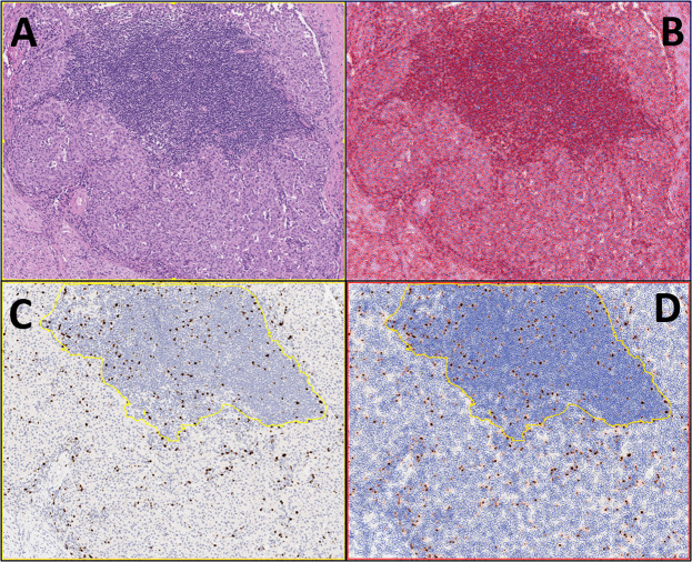
Ki-67 assessment is a key step in the diagnosis of neuroendocrine neoplasms (NENs) from all anatomic locations. Several challenges exist related to quantifying the Ki-67 proliferation index due to lack of method standardization and inter-reader variability. The application of digital pathology coupled with machine learning has been shown to be highly accurate and reproducible for the evaluation of Ki-67 in NENs. We systematically reviewed all published studies on the subject of Ki-67 assessment in pancreatic NENs (PanNENs) employing digital image analysis (DIA). The most common advantages of DIA were improvement in the standardization and reliability of Ki-67 evaluation, as well as its speed and practicality, compared to the current gold standard approach of manual counts from captured images, which is cumbersome and time consuming. The main limitations were attributed to higher costs, lack of widespread availability (as of yet), operator qualification and training issues (if it is not done by pathologists), and most importantly, the drawback of image algorithms counting contaminating non-neoplastic cells and other signals like hemosiderin. However, solutions are rapidly developing for all of these challenging issues. A comparative meta-analysis for DIA versus manual counting shows very high concordance (global coefficient of concordance: 0.94, 95% CI: 0.83-0.98) between these two modalities. These findings support the widespread adoption of validated DIA methods for Ki-67 assessment in PanNENs, provided that measures are in place to ensure counting of only tumor cells either by software modifications or education of non-pathologist operators, as well as selection of standard regions of interest for analysis. NENs, being cellular and monotonous neoplasms, are naturally more amenable to Ki-67 assessment. However, lessons of this review may be applicable to other neoplasms where proliferation activity has become an integral part of theranostic evaluation including breast, brain, and hematolymphoid neoplasms.
© 2022. The Author(s).
Conflict of interest statement
The authors declare no competing interests.
Figures
References
Publication types
MeSH terms
Substances
Grants and funding
LinkOut - more resources
Full Text Sources
Medical
