. 2022 Apr;44(2):1157-1168.
doi: 10.1007/s11357-022-00531-5. Epub 2022 Mar 6.

Brain cellular senescence in mouse models of Alzheimer's disease

Affiliations

Brain cellular senescence in mouse models of Alzheimer's disease

Angela O Dorigatti et al. Geroscience. 2022 Apr.

Abstract

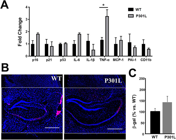
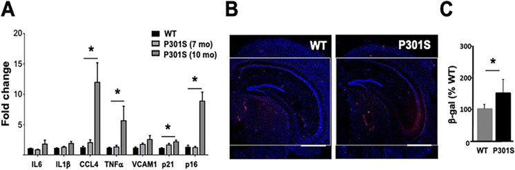
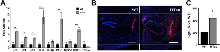
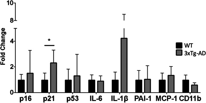
The accumulation of senescent cells contributes to aging pathologies, including neurodegenerative diseases, and its selective removal improves physiological and cognitive function in wild-type mice as well as in Alzheimer's disease (AD) models. AD models recapitulate some, but not all components of disease and do so at different rates. Whether brain cellular senescence is recapitulated in some or all AD models and whether the emergence of cellular senescence in AD mouse models occurs before or after the expected onset of AD-like cognitive deficits in these models are not yet known. The goal of this study was to identify mouse models of AD and AD-related dementias that develop measurable markers of cellular senescence in brain and thus may be useful to study the role of cellular senescence in these conditions. We measured the levels of cellular senescence markers in the brains of P301S(PS19), P301L, hTau, and 3xTg-AD mice that model amyloidopathy and/or tauopathy in AD and related dementias and in wild-type, age-matched control mice for each strain. Expression of cellular senescence markers in brains of transgenic P301L and 3xTg-AD mice was largely indistinguishable from that in WT control age-matched mice. In contrast, markers of cellular senescence were differentially increased in brains of transgenic hTau and P301S(PS19) mice as compared to WT control mice before the onset of AD-like cognitive deficits. Taken together, our data suggest that P301S(PS19) and hTau mice may be useful models for the study of brain cellular senescence in tauopathies including, but not limited to, AD.

Keywords: Aging; Amyloidopathy; Inflammation; Tauopathies.


Figures

Fig. 1
Fig. 1
Increased leves of TNFα but not of markers of senescence, in brains of P301L mice modeling tauopathy. A Levels of markers of cell damage/cell cycle arrest p16, p21, and p52 and of components of the senescence-associated secretory phenotype (SASP; IL-6, IL-1α, PAI-1, MCP-1, and CD11-b) in cortices, including hippocampus, of 6-month-old P301L mice and WT controls measured using qRT-PCR. Transcript levels were normalized to those of GAPDH and are shown as fold changes compared to the WT group. n = 5, *P < 0.05, unpaired Student’s t test. BC Quantitative analyses of SA-βgal activity, as percent red fluorescent area (indicating SA-βgal activity, panel B, representative images) normalized (panel C, quantitative analyses of data in B) to blue fluorescent area as a measure of total cell number in hippocampi of WT and P301S(PS19) mice. Scale bar, 500 µM. *P < 0.05, unpaired Student’s t test. WT, n = 4; P301L, n = 3 mice per group at 6 months of age. Data are means ± SEM
Fig. 2
Fig. 2
Specific markers of cellular senescence are increased in brains of P301S mice modeling tauopathy. A Levels of cell damage/cell cycle arrest markers p16 and p21 and of components of the senescence-associated secretory phenotype (SASP; IL-6, IL-1α, CCL-4, VCAM1, and TNFα) in hippocampus of 7- and 10-month-old P301S(PS19) mice and non-transgenic littermates (WT) measured using qRT-PCR. Transcript levels were normalized to those of β-actin and are shown as fold changes compared to the WT group. n = 12–13 for 7-month-old WT and P301S(PS19) and n = 3 for 10-month-old P301S(PS19) groups. Scale bar, 500 µM. *P < 0.01 as a result of pairwise comparisons between groups indicated, unpaired Student’s t test. BC Quantitative analyses of SA-βgal activity, as percent red fluorescent area (indicating SA-βgal activity, panel B, representative images) normalized (panel C, quantitative analyses of data in B) to blue fluorescent area as a measure of total cell number in hippocampi of WT and hTau mice. Scale bar, 500 µM. *P < 0.05, unpaired Student’s t test. NTg, n = 12, P301S(PS19), n = 13 at 7 months of age and 3 at 10 months of age. Data are means ± SEM
Fig. 3
Fig. 3
Markers of cellular senescence are increased in brains of hTau mice modeling tauopathy of AD. A Levels of cell damage/cell cycle arrest markers p16, p21, and p53 and of components of the senescence-associated secretory phenotype (SASP; IL-6, IL-1α, PAI-1, MCP-1, CD11-b, and TNFα) in cortical tissues, including hippocampus, of 7–9-month-old hTau and non-transgenic littermate mice (WT) measured with qRT-PCR. Transcript levels were normalized to those of GAPDH and are shown as fold changes compared to the WT group. WT, n = 4; hTau, n = 7; *P < 0.05; **P < 0.01; ***P < 0.001, and ****P < 0.0001 as a result of unpaired Student’s t test between the groups indicated. WT animals were 9–19-month-old; hTau were 7–9-month-old. BC Quantitative analyses of SA-βgal activity, measured as percent red fluorescent area (indicating SA-βgal activity, panel B, representative images) normalized (panel C, quantitative analyses of data in B) to blue fluorescent area as a measure of total cell number in hippocampi of WT and P301S(PS19) mice. Scale bar, 500 µM. *P < 0.05, unpaired Student’s t test. WT, n = 4 aged 14–19 months; hTau, n = 7 aged 7–9 months. Data are means ± SEM
Fig. 4
Fig. 4
p21 but not other markers of cell damage/cell cycle arrest or SASP are increased in the brains of 3xTg-AD mice modeling amyloidopathy and non-AD tauopathy. Levels of cell damage/cell cycle arrest markers p16, p21, and p53 and of components of the senescence-associated secretory phenotype (SASP; IL-6, IL-1α, PAI-1, MCP-1 and CD11-b) in cortical tissues of 12-month-old 3xTg-AD and non-transgenic mice (WT) measured with qRT-PCR. Transcript levels were normalized to those of GAPDH and are shown as fold changes compared to the WT group. Scale bar, 500 µM. *P < 0.05 as a result of unpaired Student’s t test between the groups indicated. WT, n = 5, 3xTg-AD, n = 5 at 12 months of age. Data are means ± SEM
Fig. 5
Fig. 5
Increased inflammatory cytokines, but no change in markers of cell damage/cell cycle arrest in brains of Tg2576 mice. A Levels of cell damage/cell cycle arrest markers p16, p21, and p53 and of components of the senescence-associated secretory phenotype (SASP; IL-6, IL-1α, TNFα PAI-1, MCP-1, PAI-1, and CD11-b) in mRNA fractions isolated from vasculature-depleted brain homogenates of 19-month-old Tg2576 and non-transgenic (WT) mice measured with qRT-PCR. Transcript levels were normalized to those of GAPDH and are shown as fold changes compared to the WT group. WT, n = 6; Tg2576, n = 6; *P < 0.05 and ****P < 0.0001 as a result of unpaired Student’s t test between the groups indicated. B Quantitative analyses of SA-βgal activity, measured as percent red fluorescent area (indicating SA-βgal activity, panel B, representative images) normalized (panel C, quantitative analyses of data in B) to blue fluorescent area as a measure of total cell number in hippocampi of WT and Tg2576 mice. *P < 0.05, unpaired Student’s t test. WT, n = 6; Tg2576, n = 6, at 19 months of age. Data are means ± SEM

References

    1. Childs BG, et al. Cellular senescence in aging and age-related disease: from mechanisms to therapy. Nat Med. 2015;21(12):1424–1435. doi: 10.1038/nm.4000. - DOI - PMC - PubMed
    1. Ferrucci L, Fabbri E. Inflammageing: chronic inflammation in ageing, cardiovascular disease, and frailty. Nat Rev Cardiol. 2018;15(9):505–522. doi: 10.1038/s41569-018-0064-2. - DOI - PMC - PubMed
    1. Chen WW, Zhang X, Huang WJ. Role of neuroinflammation in neurodegenerative diseases (review) Mol Med Rep. 2016;13(4):3391–3396. doi: 10.3892/mmr.2016.4948. - DOI - PMC - PubMed
    1. Song, P., J. An, and M.H. Zou, Immune clearance of senescent cells to combat ageing and chronic diseases. Cells, 2020. 9(3). - PMC - PubMed
    1. Chitnis T, Weiner HL. CNS inflammation and neurodegeneration. J Clin Invest. 2017;127(10):3577–3587. doi: 10.1172/JCI90609. - DOI - PMC - PubMed

Publication types