. 2022 Aug;241(2):297-336.
doi: 10.1111/joa.13646. Epub 2022 Mar 6.

A path to gigantism: Three-dimensional study of the sauropodomorph limb long bone shape variation in the context of the emergence of the sauropod bauplan

Affiliations

A path to gigantism: Three-dimensional study of the sauropodomorph limb long bone shape variation in the context of the emergence of the sauropod bauplan

Rémi Lefebvre et al. J Anat. 2022 Aug.

Abstract

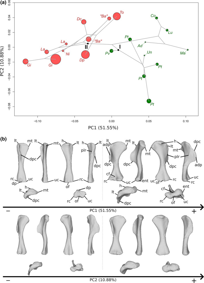
Sauropodomorph dinosaurs include the largest terrestrial animals that ever lived on Earth. The early representatives of this clade were, however, relatively small and partially to totally bipedal, conversely to the gigantic and quadrupedal sauropods. Although the sauropod bauplan is well defined, notably by the acquisition of columnar limbs, the evolutionary sequence leading to its emergence remains debated. Here, we aim to tackle this evolutionary episode by investigating shape variation in the six limb long bones for the first time using three-dimensional geometric morphometrics. The morphological features of the forelimb zeugopod bones related to the sauropod bauplan tend to appear abruptly, whereas the pattern is more gradual for the hindlimb zeugopod bones. The stylopod bones tend to show the same pattern as their respective zeugopods. The abrupt emergence of the sauropod forelimb questions the locomotor abilities of non-sauropodan sauropodomorphs inferred as quadrupeds. Features characterizing sauropods tend to corroborate a view of their locomotion mainly based on stylopod retraction. An allometric investigation of the shape variation in accordance with size highlight differences in hindlimb bone allometries between the sauropods and the non-sauropodan sauropodomorphs. These differences notably correspond to an unexpected robustness decrease trend in the sauropod hindlimb zeugopod. In addition to forelimb bones that appear to be proportionally more gracile than in non-sauropodan sauropodomorphs, sauropods may have relied on limb architecture and features related to the size increase, rather than general robustness, to deal with the role of weight-bearing.

Keywords: Dinosauria; functional morphology; gigantism; graviportality; phylomorphospace.


Figures

FIGURE 1
FIGURE 1
Phylogeny of the sauropodomorph dinosaurs used in this study. On the right, the Venn diagram in grey gives an overview of the taxonomy of the group, with clades in capital letters. The taxa belonging to the clade Sauropoda (in red) are examined in this study compared to taxa belonging to non‐sauropodiform sauropodomorphs (in green) and non‐sauropodan sauropodiforms (in yellow) grades. Based on phylogenies of Holwerda and Pol (2018) and Peyre de Fabrègues and Allain (2019). Taxa marked with an asterisk were added informally to the consensus of these two phylogenies (see Section 2). Silhouettes by Scott Hartman
FIGURE 2
FIGURE 2
(a) Phylomorphospace of the humerus analysis along with the first two PCs. Green dots represent non‐sauropodiform sauropodomorphs, red dots represent sauropods. The diameter of the dots represents the centroid size of the specimen. Node I corresponds to the root of the tree and Node II to the estimation of the node Sauropoda. Taxonomic abbreviations: Ad: Adeopapposaurus; Ba: Barosaurus africanus; Co: Coloradisaurus; Dc: Dicraeosaurus; Dp: Diplodocus; Gi: Giraffatitan; La: Lapparentosaurus; Lu: Lufengosaurus; Ma: Massospondylus; Ni: Nigersaurus; Pt: Plateosaurus; Pv: Plateosauravus; To: Tornieria; Un: Unaysaurus. (b) Thin‐plate splines visualization of aligned theoretical shapes of the humerus analysis. The shape changes along with PC1 negative and positive shapes, and PC2 negative and positive shapes are observed. Each shape is represented, from left to right and top to down, in anterior, medial, posterior, lateral, proximal and distal views. In the proximal view, the top corresponds to the posterior side; in the distal view, the top corresponds to the anterior side. Anatomical abbreviations: Adp: Apex of deltopectoral crest; cf: cuboid fossa; dp: distal processes; dpc: deltopectoral crest; ent: entepycondyle; h: humeral head; lt: lateral tubercle; mt: medial tuberosity; of: olecranon fossa; plr: posterolateral ridge; rc: radial condyle; uc: ulnar condyle. PC, principal components
FIGURE 3
FIGURE 3
(a) Phylomorphospace of the radius analysis along with the first two PCs. Green dots represent the non‐sauropodiform sauropodomorphs, yellow dots represent non‐sauropodan sauropodiforms and red dots represent sauropods. The diameter of the dots represents the centroid size of the specimen. Node I corresponds to the root of the tree, Node II to the estimated node Sauropodiformes and Node III to the estimated node Sauropoda. Taxonomic abbreviations: Aa: Aardonyx; Ad: Adeopapposaurus; An: Antetonitrus; Dc: Dicraeosaurus; Dp: Diplodocus; ind: Indeterminate sauropod from Tendaguru; L4: MNHN.F.LES.400; La: Lapparentosaurus; Ma: Massospondylus; Me: Melanorosaurus; Pa: Patagosaurus; Pt: Plateosaurus; Pv: Plateosauravus; Se: Sefapanosaurus. (b) Thin‐plate splines visualization of aligned theoretical shapes of the radius analysis. The shape changes along with PC1 negative and positive shapes, and PC2 negative and positive shapes are observed. Each shape is represented, from left to right and top to down, in anterior, medial, posterior, lateral, proximal, and distal views. In proximal and distal views, the top corresponds to the anterior side. Anatomical abbreviations: Hc: Humeral cotyle; Dp: distal process; pp: proximal process; ua: ulnar articulation. PC, principal components
FIGURE 4
FIGURE 4
(a) Phylomorphospace of the ulna analysis along with the first two PCs. Green dots represent the non‐sauropodiform sauropodomorphs, yellow dots represent non‐sauropodan sauropodiforms and red dots represent sauropods. The diameter of the dots represents the centroid size of the specimen. Node I corresponds to the root of the tree, Node II to the estimated node Sauropodiformes and Node III to the estimated node Sauropoda. Taxonomic abbreviations: Ad: Adeopapposaurus; An: Antetonitrus; Ba: Barosaurus africanus; Dc: Dicraeosaurus; Dp: Diplodocus; Kh: Kholumolumo; La: Lapparentosaurus; Ls: Lessemsaurus; Ma: Massospondylus; Me: Melanorosaurus; Mu: Mussaurus; Ni: Nigersaurus; Ri: Riojasaurus; Ru: Ruehleia; Pa: Patagosaurus; Pt: Plateosaurus; Pv: Plateosauravus; Se: Sefapanosaurus; To: Tornieria. (b) Thin‐plate splines visualization of aligned theoretical shapes of the ulna analysis. The shape changes along with PC1 negative and positive shapes, and PC2 negative and positive shapes are observed. Each shape is represented, from left to right and top to down, in anterior, medial, posterior, lateral, proximal and distal views. In proximal and distal views, the top corresponds to the anterior side. Anatomical abbreviations: Amp: Anteromedial process; lc: lateral concavity; ldc: laterodistal concavity; lp: lateral process; mc: medial concavity; mdc: mediodistal concavity; olp: olecranon process; rf: radial fossa. PC, principal components
FIGURE 5
FIGURE 5
(a) Phylomorphospace of the femur analysis along with the first two PCs. Green dots represent the non‐sauropodiform sauropodomorphs, yellow dots represent non‐sauropodan sauropodiforms and red dots represent sauropods. Diameter of the dots represents the centroid size of the specimen. Node I corresponds to the root of the tree, Node II to the estimated node Sauropodiformes and Node III to the estimated node Sauropoda. Taxonomic abbreviations: Ad: Adeopapposaurus; Ba: Barosaurus africanus; Co: Coloradisaurus; Gi: Giraffatitan; Gy: Gyposaurus sinensis; Kh: Kholumolumo; La: Lapparentosaurus; Ls: Lessemsaurus; Ma: Massospondylus; Mr: Meroktenos; Ni: Nigersaurus; Ri: Riojasaurus; Ru: Ruehleia; Pt: Plateosaurus. (b) Thin‐plate splines visualization of aligned theoretical shapes of the femur analysis. The shape changes along with PC1 negative and positive shapes, and PC2 negative and positive shapes are observed. Each shape is represented, from left to right and top to down, in anterior, medial, posterior, lateral, proximal and distal views. In proximal and distal views, the top corresponds to the posterior side. Anatomical abbreviations: 4t: fourth trochanter; aif: anterior intercondylar fossa; an: anterior notch; ar: anterior ridge; gt: greater trochanter; h: Femoral head; lc: lateral condyle; lt: lesser trochanter; mc: medial condyle; pif: posterior intercondylar fossa. PC, principal components
FIGURE 6
FIGURE 6
(a) Phylomorphospace of the tibia analysis along with the first two PCs. Green dots represent the non‐sauropodiform sauropodomorphs, yellow dots represent non‐sauropodan sauropodiforms and red dots represent sauropods. The diameter of the dots represents the centroid size of the specimen. The diameter of the dots represents the centroid size of the specimen. Node I corresponds to the root of the tree, Node II to the estimated node Sauropodiformes and Node III to the estimated node Sauropoda. Taxonomic abbreviations: Ad: Adeopapposaurus; An: Antetonitrus; Ba: Barosaurus africanus; Co: Coloradisaurus; Dp: Diplodocus; Gi: Giraffatitan; La: Lapparentosaurus; Ls: Lessemsaurus; Ma: Massospondylus; Me: Melanorosaurus; Mu: Mussaurus; Ni: Nigersaurus; Ri: Riojasaurus; Pa: Panphagia; Pt: Plateosaurus; Pv: Plateosauravus; To: Tornieria; Vo: Volkheimeria. (b) Thin‐plate splines visualization of aligned theoretical shapes of the tibia analysis. The shape changes along with PC1 negative and positive shapes, and PC2 negative and positive shapes are observed. Each shape is represented, from left to right and top to down, in anterior, medial, posterior, lateral, proximal and distal views. In proximal and distal views, the top corresponds to the lateral side. Anatomical abbreviations: Afap: Ascending process articular facet; cc: cnemial crest; dp: descending process; fc: fibular condyle; ic: internal condyle; mc: medial corner of the distal end; PC: posterior corner of the distal end. PC, principal components
FIGURE 7
FIGURE 7
(a) Phylomorphospace of the fibula analysis along with the first two PCs. Green dots represent the non‐sauropodiform sauropodomorphs, yellow dots represent non‐sauropodan sauropodiforms and red dots represent sauropods. The diameter of the dots represents the centroid size of the specimen. Node I corresponds to the root of the tree, Node II to the estimated node Sauropodiformes and Node III to the estimated node Sauropoda. Taxonomic abbreviations: Ad: Adeopapposaurus; An: Antetonitrus; Ba: Barosaurus africanus; Dc: Dicraeosaurus; Dp: Diplodocus; Gi: Giraffatitan; ind: indeterminate sauropod from Tendaguru; La: Lapparentosaurus; Ma: Massospondylus; Ni: Nigersaurus; Pt: Plateosaurus; Ri: Riojasaurus; Se: Sefapanosaurus Ta: Tazoudasaurus; To; Tornieria. (b) Thin‐plate splines visualization of aligned theoretical shapes of the fibula analysis. The shape changes along with PC1 negative and positive shapes, and PC2 negative and positive shapes are observed. Each shape is represented, from left to right and top to down, in anterior, medial, posterior, lateral, proximal and distal views. In proximal and distal views, the top corresponds to the lateral side. Anatomical abbreviations: amc: anteromedial concavity; anteromedial depression; amp: amteromedial process; lb: lateral bulge; or: oblique ridge. PC, principal components
FIGURE 8
FIGURE 8
Allometric pattern of the humerus: Top: bivariate plot showing the CAC scores against the natural logarithm of the centroid size. Open circles represent the non‐columnar sauropodomorphs, filled circles the columnar sauropods. Bottom: Thin‐plate splines visualizations of shape changes along with the CAC. Shape changes at minimal size are displayed in cyan, whereas shape changes at maximal size are displayed in orange. Shape variation is represented, from left to right and top to down, in anterior, medial, posterior, lateral, proximal and distal views. In the proximal view, the top corresponds to the posterior side; in the distal view, the top corresponds to the anterior side. Taxonomic abbreviations: Ad: Adeopapposaurus; Ba: Barosaurus africanus; Co: Coloradisaurus; Dc: Dicraeosaurus; Dp: Diplodocus; Gi: Giraffatitan; La: Lapparentosaurus; Lu: Lufengosaurus; Ma: Massospondylus; Ni: Nigersaurus; Pt: Plateosaurus; Pv: Plateosauravus; To: Tornieria; Un: Unaysaurus. CAC, common allometric component
FIGURE 9
FIGURE 9
Allometric pattern of the radius: Top: bivariate plot showing the CAC scores against the natural logarithm of the centroid size. Open circles represent the non‐columnar sauropodomorphs and filled circles the columnar sauropods. Bottom: Thin‐plate splines visualizations of shape changes along with the CAC. Shape changes at minimal size are displayed in cyan, whereas shape changes at maximal size are displayed in orange. Shape variation is represented, from left to right and top to down, in anterior, medial, posterior, lateral, proximal and distal views. In proximal and distal views, the top corresponds to the anterior side. Taxonomic abbreviations: Aa: Aardonyx; Ad: Adeopapposaurus; An: Antetonitrus; Dc: Dicraeosaurus; Dp: Diplodocus; ind: indeterminate sauropod from Tendaguru; L4: MNHN.F.LES.400; La: Lapparentosaurus; Ma: Massospondylus; Me: Melanorosaurus; Pa: Patagosaurus; Pt: Plateosaurus; Pv: Plateosauravus; Se: Sefapanosaurus. CAC, common allometric component
FIGURE 10
FIGURE 10
Allometric pattern of the ulna: Top: bivariate plot showing the CAC scores against the natural logarithm of the centroid size. Open circles represent the non‐columnar sauropodomorphs and filled circles the columnar sauropods. Bottom: Thin‐plate splines visualizations of shape changes along with the CAC. Shape changes at minimal size are displayed in cyan, whereas shape changes at maximal size are displayed in orange. Shape variation is represented, from left to right and top to down, in anterior, medial, posterior, lateral, proximal and distal views. In proximal and distal views, the top corresponds to the anterior side. Taxonomic abbreviations: Ad: Adeopapposaurus; An: Antetonitrus; Ba: Barosaurus africanus; Dc: Dicraeosaurus; Dp: Diplodocus; Kh: Kholumolumo; La: Lapparentosaurus; Ls: Lessemsaurus; Ma: Massospondylus; Me: Melanorosaurus; Mu: Mussaurus; Ni: Nigersaurus; Ri: Riojasaurus; Ru: Ruehleia; Pa: Patagosaurus; Pt: Plateosaurus; Pv: Plateosauravus; Se: Sefapanosaurus; To: Tornieria. CAC, common allometric component
FIGURE 11
FIGURE 11
Allometric pattern of the femur: Top: bivariate plot showing the CAC scores against the natural logarithm of the centroid size. Open circles represent the non‐columnar sauropodomorphs and filled circles the columnar sauropods. Bottom: Thin‐plate splines visualizations of shape changes along with the CAC. Shape changes at minimal size are displayed in cyan, whereas shape changes at maximal size are displayed in orange. Shape variation is represented, from left to right and top to down, in anterior, medial, posterior, lateral, proximal and distal views. In proximal and distal views, the top corresponds to the posterior side. Taxonomic abbreviations: Ad: Adeopapposaurus; Ba: Barosaurus africanus; Co: Coloradisaurus; Gi: Giraffatitan; Gy: Gyposaurus sinensis; Kh: Kholumolumo; La: Lapparentosaurus; Ls: Lessemsaurus; Ma: Massospondylus; Mr: Meroktenos; Ni: Nigersaurus; Ri: Riojasaurus; Ru: Ruehleia; Pt: Plateosaurus. CAC, common allometric component
FIGURE 12
FIGURE 12
Allometric pattern of the tibia for non‐columnar non‐sauropodan sauropodomorphs: Top: bivariate plot showing the CAC scores against the natural logarithm of the centroid size. Open circles represent the non‐columnar sauropodomorphs and filled circles the columnar sauropods. Bottom: Thin‐plate splines visualizations of shape changes along with the CAC. Shape changes at minimal size are displayed in cyan, whereas shape changes at maximal size are displayed in orange. Shape variation is represented, from left to right and top to down, in anterior, medial, posterior, lateral, proximal and distal views. In proximal and distal views, the top corresponds to the lateral side. Taxonomic abbreviations: Ad: Adeopapposaurus; An: Antetonitrus; Co: Coloradisaurus; Ls: Lessemsaurus; Ma: Massospondylus; Me: Melanorosaurus; Mu: Mussaurus; Ri: Riojasaurus; Pa: Panphagia; Pt: Plateosaurus; Pv: Plateosauravus. CAC, common allometric component
FIGURE 13
FIGURE 13
Allometric pattern of the tibia for columnar sauropods: Top: bivariate plot showing the CAC scores against the natural logarithm of the centroid size. Open circles represent the non‐columnar sauropodomorphs and filled circles the columnar sauropods. Bottom: Thin‐plate splines visualizations of shape changes along with the CAC. Shape changes at minimal size are displayed in cyan, whereas shape changes at maximal size are displayed in orange. Shape variation is represented, from left to right and top to down, in anterior, medial, posterior, lateral, proximal and distal views. In proximal and distal views, the top corresponds to the lateral side. Taxonomic abbreviations: Ba: Barosaurus africanus; Dp: Diplodocus; Gi: Giraffatitan; La: Lapparentosaurus; Ni: Nigersaurus; To; Tornieria; Vo: Volkheimeria. CAC, common allometric component
FIGURE 14
FIGURE 14
Allometric pattern of the fibula for non‐columnar non‐sauropodan sauropodomorphs: Top: bivariate plot showing the CAC scores against the natural logarithm of the centroid size. Open circles represent the non‐columnar sauropodomorphs and filled circles the columnar sauropods. Bottom: Thin‐plate splines visualizations of shape changes along with the CAC. Shape changes at minimal size are displayed in cyan, whereas shape changes at maximal size are displayed in orange. Shape variation is represented, from left to right and top to down, in anterior, medial, posterior, lateral, proximal and distal views. In proximal and distal views, the top corresponds to the lateral side. Taxonomic abbreviations: Ad: Adeopapposaurus; An: Antetonitrus; Ma: Massospondylus; Ri: Riojasaurus; Pt: Plateosaurus; Se: Sefapanosaurus. CAC, common allometric component. Note that the associated test of the effect of size on shape failed to reject the null hypothesis (cf. Table S3)
FIGURE 15
FIGURE 15
Allometric pattern of the fibula for columnar sauropods: Top: bivariate plot showing the CAC scores against the natural logarithm of the centroid size. Open circles represent the non‐columnar sauropodomorphs and filled circles the columnar sauropods. Bottom: Thin‐plate splinesvisualizations of shape changes along with the CAC. Shape changes at minimal size are displayed in cyan, whereas shape changes at maximal size are displayed in orange. Shape variation is represented, from left to right and top to down, in anterior, medial, posterior, lateral, proximal and distal views. In proximal and distal views, the top corresponds to the lateral side. Taxonomic abbreviations: Ba: Barosaurus africanus; Dc: Dicraeosaurus; Dp: Diplodocus; Gi: Giraffatitan; ind: indeterminate sauropod from Tendaguru; La: Lapparentosaurus; Ni: Nigersaurus; Ta: Tazoudasaurus; To; Tornieria. CAC, common allometric component

References

    1. 3D Systems . (2017) Geomagic wrap. Rock Hill, SC: 3D Systems.
    1. Adams D.C., Collyer M., Kaliontzopoulou A. (2020) Geomorph: software for geometric morphometric analyses. R Package Version 3.3.1.
    1. Adams, D.C. , Rohlf, F.J. & Slice, D.E. (2013) A field comes of age: geometric morphometrics in the 21st century. Hystrix, 24, 7.
    1. Agisoft LLC . (2018) Photoscan professional edition. St. Petersburg: Agisoft.
    1. Alexander, R. (1998) All‐time giants: the largest animals and their problems. Palaeontology, 41, 1231–1246.

Publication types