Review
. 2022 Apr;29(3):159-170.
doi: 10.1016/j.arcped.2022.01.016. Epub 2022 Mar 3.

Neonatal gastrointestinal emergencies: a radiological review

Affiliations
Review

Neonatal gastrointestinal emergencies: a radiological review

Luis Octavio Tierradentro-Garcia et al. Arch Pediatr. 2022 Apr.

Abstract

Background: Abdominal emergencies in neonates require surgical management in almost all cases and complications may include bowel perforation, sepsis, shock, and even death. Radiological imaging has become a very important aid in the clinical setting as it shortens time to diagnosis.

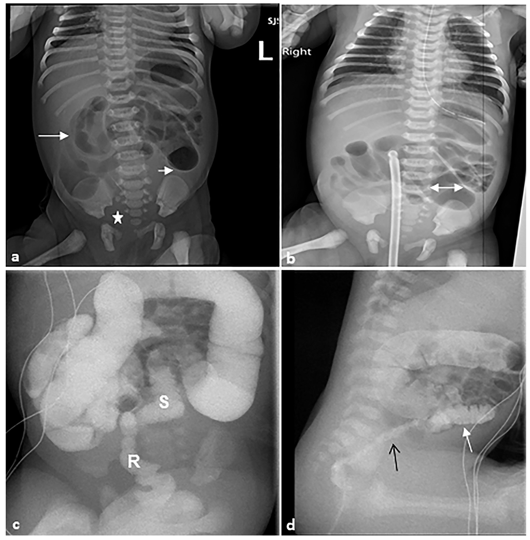
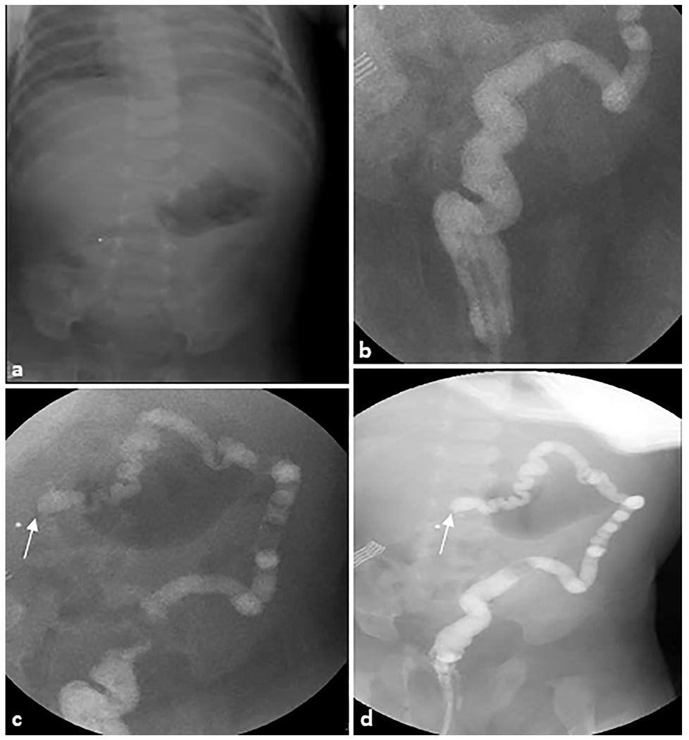
Objective: The objective of this review is to discuss the more prevalent neonatal gastrointestinal emergencies, review appropriate imaging options, and illustrate common radiological presentations of these entities.

Conclusion: Despite advancements in imaging techniques, it is important to keep in mind that neonates have a higher susceptibility to the adverse effects of ionizing radiation, and therefore radiography and ultrasonography remain the main diagnostic modalities for ruling out the diseases with the worst prognosis. Other modalities (fluoroscopy, computed tomography, and magnetic resonance imaging) may have limited use in very specific conditions. All providers in an emergency department should be familiar with the basic radiological findings that may indicate a gastrointestinal emergency, especially in health institutions that do not have 24-h radiologist coverage.

Keywords: Abdominal emergencies; Gastrointestinal emergencies; Neonatal imaging; Pediatric radiology; Radiology.


Conflict of interest statement

Conflict of interest statement None declared.

Figures

Fig. 1.
Fig. 1.. Hypertrophic pyloric stenosis.
A 28-day-old female patient presented with non-bilious emesis and abdominal distension. (a) Anteroposterior abdominal radiograph demonstrating a markedly distended gas-filled stomach (S) without signs of perforation. (b) Sagittal grayscale ultrasound image reveals a thickening of the pyloric muscle wall (W) that measures 4 mm (double-headed arrow). The pyloric channel is elongated and measures 19 mm (line) and the pylorus does not open on dynamic imaging. (c, d) Sagittal and anteroposterior view contrast upper GI series show contrast crosses the lower esophageal sphincter into the air-distended stomach. There is no contrast passage across the pylorus (P), which is consistent with the previous findings.
Fig. 2.
Fig. 2.. Duodenal atresia.
A 1-day-old female patient presented with abdominal distension. (a) Anteroposterior abdominal radiograph demonstrating gas-distended stomach and proximal duodenum (arrow) with no bowel gas seen distally. (b) Transverse real-time ultrasound examination revealed apparent dilation of the stomach and duodenal bulb (arrow) consistent with duodenal atresia. Superior mesenteric artery (SMA) and superior mesenteric vein (SMV) are shown (arrows). SMV is slightly to the left than expected of its normal location likely due to the mass effect from dilated duodenum.
Fig. 3.
Fig. 3.. Midgut volvulus.
A 4-day-old male, full-term neonate presenting with bilious emesis. (a) Anteroposterior chest and abdomen radiograph (“babygram”) shows high-grade proximal bowel obstruction. The stomach and proximal duodenum are markedly distended (double-headed arrow); there is a paucity of bowel gas in the remainder of the abdomen. (b, c) Anteroposterior and lateral upper GI series show a distended stomach with contrast passing unobstructed across the pylorus (dotted arrow). Enteric contrast is seen freely progressing into the proximal duodenum with a discrete cut-off and blunted, rounded appearance of the opacified duodenum (arrows).
Fig. 4.
Fig. 4.. Midgut volvulus.
A 3-year-old female patient presented to the emergency department with intermittent abdominal pain. Transverse grayscale ultrasound without (a) and with (b) color Doppler was performed, revealing the “whirlpool” sign of midgut volvulus (arrows).
Fig. 5.
Fig. 5.. Midgut volvulus.
A 4-month-old female patient with a history of Beckwith–Wiedemann syndrome presented with a distended abdomen. (a) Axial T2-weighted magnetic resonance image of the abdomen demonstrates a distended loop of bowel in the left abdomen (arrow) with an apparent twist in the mesentery. (b) Anteroposterior abdominal radiograph demonstrating multiple dilated loops of bowel with an enteric tube terminating in the pelvis. (c) Coronal upper GI series with contrast passed through the enteric feeding tube and revealed a “bird’s beak” sign (arrow), indicating twist around mesenteric axis. Subsequent exploratory laparotomy revealed jejunal volvulus.
Fig. 6.
Fig. 6.. Jejuno-ileal atresia.
Ex-premature infant. (a) Anteroposterior chest and abdomen radiograph (“babygram”) of a 1-day-old ex-premature infant born to parents with cystic fibrosis presenting with bilious emesis. The radiograph demonstrates dilated bowel loops (arrows). (b) Sagittal grayscale ultrasound image of the midline abdomen shows a dilated (L) loop of bowel with thickened walls (W). Echogenic foci are intraluminal gas (G). (c) Axial grayscale ultrasound of the right upper quadrant with color Doppler demonstrates the superior mesenteric vein and artery in normal position arguing against malrotation with midgut volvulus. (d) Water-soluble contrast enema demonstrates microcolon (arrows). (e) Upper gastrointestinal series demonstrates a normal duodeno-jejunal junction in the left upper quadrant (arrows) followed by abrupt cutoffs of the jejunum (arrows) in (f) sagittal and (g) anteroposterior views. At surgery, this was determined to be jejunal atresia, type IIIB.
Fig. 7.
Fig. 7.. Necrotizing enterocolitis.
Anteroposterior chest and abdomen radiograph (“babygram”) of a 2-day-old ex-30-week premature infant admitted to the intensive care unit with profound metabolic acidosis and tense abdomen. The radiograph revealed apparent decreased density of the liver (“lucent liver sign”) with a crescentic lucency outlining the liver edge (arrows). Subtle small lucencies were seen overlying multiple bowel loops, most notable in the right lower quadrant. This patient had necrotizing enterocolitis with segmental volvulus and bowel perforation.
Fig. 8.
Fig. 8.. Necrotizing enterocolitis.
(a) Anteroposterior chest and abdomen radiograph (“babygram”) of a 2-month-old ex-premature infant who presented to the intensive care unit with a tense and distended abdomen. Multiple distended loops of bowel (arrows) are seen in the left abdomen with a paucity of bowel in the right lower quadrant. (b) Transverse grayscale ultrasound image with color Doppler overlay of the right lower quadrant demonstrates multiple loops of distended small bowel containing complex fluid with thickened walls (arrows) and echogenic contents. No color Doppler flow is seen in the bowel wall concerning for ischemia. At surgery, this patient had 22 cm of ischemic bowel.
Fig. 9.
Fig. 9.. Necrotizing enterocolitis.
An 8-day-old premature girl was born at 31 weeks. (a) Sagittal and (b) transverse grayscale ultrasound images show hypoperistaltic bowel loops with areas of wall thickening and mucosal hyperechogenicity consistent with “zebra pattern” (arrows). No discrete pneumatosis or portal venous air is present.
Fig. 10.
Fig. 10.. Necrotizing enterocolitis.
A 1-day-old formerly premature girl was born at 29 weeks with gaseous distention on abdominal radiography. (a) Grayscale ultrasound in the left upper quadrant shows multiple dilated loops of bowel with wall thickening and hypoperistalsis to aperistalsis in real time. (b) Corresponding color Doppler image shows apparent flow in the mesentery but no appreciable flow in the bowel. However, the interpretation was limited by pulsatile motion from the patient’s high-frequency oscillator. (c, d) Dual-screen contrast-enhanced ultrasound displays loops of bowel (arrowheads) in the right upper quadrant that do not enhance (arrowheads) regardless of the high-frequency oscillator. (Images reprinted from Benjamin J et al. [31] with permission).
Fig. 11.
Fig. 11.. Inguinal hernia.
A 14-day-old male patient presented with vomiting and right scrotal mass. (a) Cross-table lateral abdominal radiograph showing mildly dilated loops of small bowel with multiple air–fluid levels concerning for mechanical obstruction. (b, c) Transverse grayscale ultrasound examination of the right inguinal region shows a right inguinal hernia containing multiple loops of small bowel (arrow). The herniated bowel is hyperperistaltic with mild wall thickening. Motility and inflammation indicate viable bowel but urgent intervention is required to prevent incarceration and ischemia.
Fig. 12.
Fig. 12.. Intussusception.
A 1-month-old female infant presented with bloody stools and abdominal distension. (a) Transverse grayscale ultrasound showed an elongated mass containing concentric layers of different echogenicity in the mid upper abdomen, consistent with ileocolic intussusception (arrow). The mass is medial to the liver and pylorus. Therapeutic air enema was performed. (b) A preliminary spot radiograph of the abdomen shows a soft tissue density (arrow) in the mid upper abdomen thought to represent the intussusceptum/intussuscepiens complex. At the first attempt the intussusception was easily reduced to the level of the ileocecal valve. During the second attempt (c), the intussusception was completely reduced past the ileocecal valve, with air entering the terminal ileum and distal small bowel. At the end of the evaluation (d), there was no radiographic evidence of intussusception.
Fig. 13.
Fig. 13.. Hirschsprung disease.
A 1-day-old female patient who presented with a distended abdomen and emesis. Rectal biopsy confirmed Hirschsprung disease. (a) Anteroposterior abdominal radiograph demonstrating multiple loops of air distended bowel (arrows) with paucity of bowel gas within the pelvis (star). (b) There are dilated proximal bowel loops with the maximal diameter of 2.7 cm (double-headed arrow) with featureless appearance of bowel loops in the right upper quadrant. (c) Fluoroscopic contrast enema demonstrating the sigmoid colon and rectum are small (S) in caliber up to the descending colon-sigmoid colon junction. A rectal catheter (R) is present with tip to the right of L2–L3. (d) Sagittal fluoroscopic image demonstrating an abnormal rectosigmoid ratio (<1) with the rectum (black arrow) of much smaller caliber than the sigmoid colon (white arrow) and upstream colon.
Fig. 14.
Fig. 14.. Colonic atresia.
A 30-day-old female patient was evaluated for status post failed ileocolonic anastomosis due to intestinal atresia. (a) Scout radiograph demonstrates an unremarkable bowel gas pattern. (b–d) Fluoroscopic contrast enema demonstrates contrast refluxing into the rectum, sigmoid, descending, and distal transverse colon, which is small in caliber. There is abrupt termination of the contrast column in the mid transverse colon (arrows), highly suggestive of an area of colonic atresia.

References

    1. Louie JP. Essential diagnosis of abdominal emergencies in the first year of life. Emerg Med Clin North Am 2007;25:1009–40 vi. - PubMed
    1. Ezomike UO, Ekenze SO, Amah CC, et al. Infantile hypertrophic pyloric stenosis - our experience and challenges in a developing country. Afr J Paediatr Surg 2018;15:26–30. - PMC - PubMed
    1. Costa Dias S, Swinson S, Torrão H, et al. Hypertrophic pyloric stenosis: tips and tricks for ultrasound diagnosis. Insights Imaging 2012;3:247–50. - PMC - PubMed
    1. Argyropoulou MI, Hadjigeorgi CG, Kiortsis DN. Antro-pyloric canal values from early prematurity to full-term gestational age: an ultrasound study. Pediatr Radiol 1998;28:933–6. - PubMed
    1. Hiorns MP. Gastrointestinal tract imaging in children: current techniques. Pediatr Radiol 2011;41:42–54. - PubMed