

[27-P-CAUA induces apoptosis and mitochondrial autophagy in breast cancer cells by inhibiting HER2/PI3K/AKT signaling pathway]
- PMID: 35249871
- PMCID: PMC8901400
- DOI: 10.12122/j.issn.1673-4254.2022.01.07
[27-P-CAUA induces apoptosis and mitochondrial autophagy in breast cancer cells by inhibiting HER2/PI3K/AKT signaling pathway]
Abstract
Objective: To investigate the inhibitory effect of 27-P-coumayl-ursolic acid (27-P-CAUA), the active ingredient in triterpenoids from the leaves of Ilex latifolia Thunb, against breast cancer cells and explore the underlying mechanism.
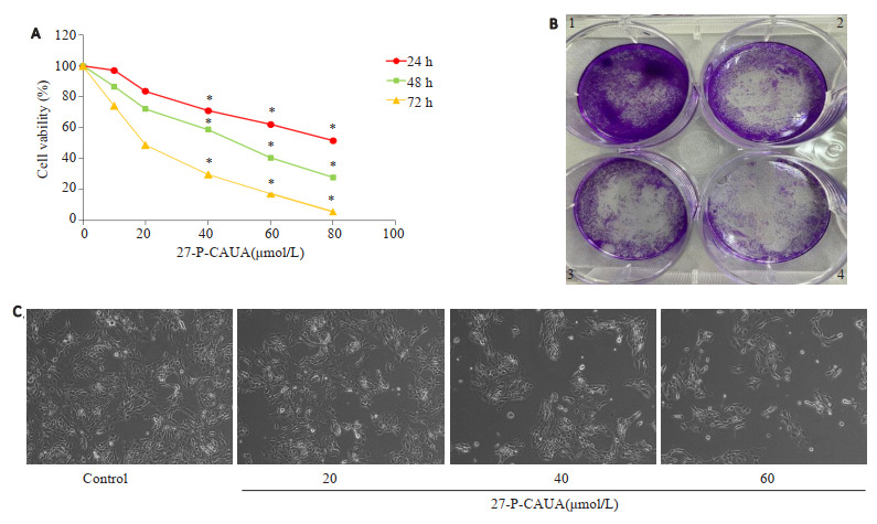
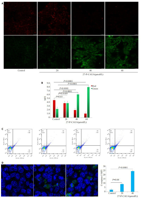
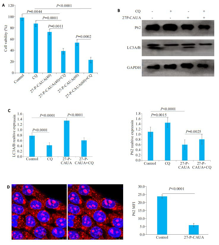
Methods: CCK-8 assay was used to assess the changes in viability of breast cancer HCC-1806 cells after 27-P-CAUA treatment for 24, 48, or 72 h. The inhibitory effect of 27-P-CAUA on proliferation of the cells was determined by clonogenic assay. JC-1 was used to detect the changes in mitochondrial membrane potential and flow cytometry was performed for analyzing cell apoptosis following 27-P-CAUA treatment. Immunofluorescence assay was used to observe the expression of cl-caspase-3 and P62 in the treated cells. Western blotting was performed to observe the effect of 27-P-CAUA and chloroquine pretreatment on the expressions of LC3I/II, P62 and HER2 signaling pathway proteins in the cells.
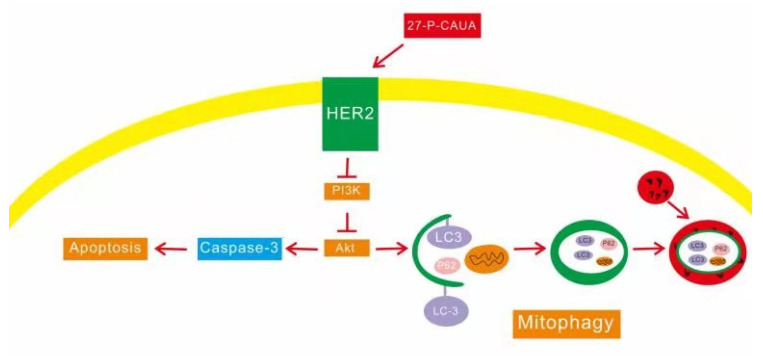
Results: The results of CCK-8 and clonogenic assays showed that 27-P-CAUA treatment significantly inhibited the proliferation of HCC-1806 cells (P < 0.01) with IC50 values of 81.473, 48.392 and 18.467 μmol/L at 24, 48, and 72 h, respectively. 27-P-CAUA treatment also caused obvious changes in mitochondrial membrane potential (P < 0.01) and induced cell apoptosis in HCC-1806 cells with a 3.34% increase of the early apoptosis rate. Immunofluorescence assay revealed a significant increase of cl-caspase3 expression in 27-P-CAUA-treated HCC-1806 cells, and treatment with 40 μmol/L 27-P-CAUA resulted in significant cell apoptosis (P < 0.01). 27-P-CAUA obviously reduced the expression of LC3II, caused P62 degradation and induced autophagy in HCC-1806 cells. Chloroquine pretreatment obviously blocked the autophagy-inducing effect of 27-P-CAUA. 27-P-CAUA treatment also inhibited the phosphorylation of HER2 and AKT proteins and progressively lowered the expressions of HER2 and phosphorylated AKT protein in HCC-1806 cells (P < 0.01).
Conclusion: 27-P-CAUA can inhibit the proliferation and induce mitochondrial autophagy and apoptosis of HCC-1806 cells by inhibiting the HER2/PI3K/AKT signaling pathway.
目的: 探讨大叶冬青叶中三萜类活性成分27-P-香豆酰基-乌索酸(27-P-CAUA)对乳腺癌细胞增殖抑制的作用机制。
方法: 27-P-CAUA处理HCC-1806细胞24、48、72 h后,CCK-8法检测HCC-1806细胞存活率;集落克隆法检测27-P-CAUA对细胞的增殖抑制作用;JC-1检测线粒体膜电位改变;流式细胞术检测细胞凋亡情况;细胞免疫荧光观察C-Caspase-3、P62表达;氯喹预处理后,Western blot观察27-P-CAUA对LC3I/II、P62以及HER2信号通路蛋白的影响。
结果: CCK-8和集落克隆结果显示,随着药物浓度增加和时间延长,27-P-CAUA对HCC-1806乳腺癌细胞的抑制作用逐渐增强(P < 0.01),24、48、72 h的IC50值分别为83.671、49.479、19.578 μmol/L;JC-1检测结果显示,线粒体膜电位改变明显(P < 0.01);流式细胞术结果显示,27-P-CAUA可诱导细胞发生凋亡,细胞早期调亡比率增加3.34%;免疫荧光结果显示,C-Caspase-3表达增强,促进细胞凋亡,40 μmol /L 27-PCAUA能显著诱导细胞凋亡(P < 0.01)。Western blot和细胞免疫荧光结果显示,27-P-CAUA降低LC3II表达,导致P62降解,诱导自噬发生。经氯喹预处理后,27-P-CAUA诱导自噬可被逆转。此外,27-P-CAUA可抑制细胞内Her2及AKT蛋白的磷酸化,细胞内的Her2以及磷酸化AKT蛋白表达逐渐减少(P < 0.01)。
结论: 27-P-CAUA可通过抑制HER2/PI3K/AKT信号通路,抑制HCC-1806细胞增殖,诱导细胞发生线粒体自噬和凋亡。
Keywords: 27-P-coumayl-ursolic acid; HCC-1806 cells; HER2; apoptosis; mitochondrial autophagy.
Figures

References
-
- 郭 怡, 姜 成燕, 焦 瑞华, et al. Antimycin类天然产物抗三阴性乳腺癌细胞MDA-MB-231作用机制初步研究. https://www.cnki.com.cn/Article/CJFDTOTAL-NJDZ202102019.htm. 南京大学学报: 自然科学. 2021;57(2):334–43. [郭怡, 姜成燕, 焦瑞华, 等. Antimycin类天然产物抗三阴性乳腺癌细胞MDA-MB-231作用机制初步研究[J]. 南京大学学报: 自然科学, 2021, 57(2): 334-43.] - PubMed
-
- 袁 永贵, 张 夏炎, 朱 晓俊, et al. 白花丹素对乳腺癌细胞凋亡和自噬的影响. 中国中医药信息杂志. 2022;29(1):90–5. [袁永贵, 张夏炎, 朱晓俊, 等. 白花丹素对乳腺癌细胞凋亡和自噬的影响[J]. 中国中医药信息杂志, 2022, 29(1): 90-5.]
-
- 李 佳鑫, 石 金凤, 吴 亿晗, et al. 雷公藤甲素抗乳腺癌的机制及应用进展. https://www.cnki.com.cn/Article/CJFDTOTAL-ZGZY202113009.htm. 中国中药杂志. 2021;46(13):3249–56. [李佳鑫, 石金凤, 吴亿晗, 等. 雷公藤甲素抗乳腺癌的机制及应用进展[J]. 中国中药杂志, 2021, 46(13): 3249-56.] - PubMed
-
- 林 艳, 闫 庆梓, 李 亚梅, et al. 夏枯草抗乳腺癌最佳组分筛选及其作用机制研究. 中草药. 2019;50(21):5298–306. doi: 10.7501/j.issn.0253-2670.2019.21.024. [林艳, 闫庆梓, 李亚梅, 等. 夏枯草抗乳腺癌最佳组分筛选及其作用机制研究[J]. 中草药, 2019, 50(21): 5298-306.] - DOI
MeSH terms
Substances
LinkOut - more resources
Full Text Sources
Medical
Research Materials
Miscellaneous