Case Reports
. 2022 Oct 1;61(19):2891-2894.
doi: 10.2169/internalmedicine.9170-21. Epub 2022 Mar 5.

Fatal Cardiac Tamponade Due to a Pericardial Inflammatory Myofibroblastic Tumor

Affiliations
Case Reports

Fatal Cardiac Tamponade Due to a Pericardial Inflammatory Myofibroblastic Tumor

Hiromichi Ohsaka et al. Intern Med. .

Abstract

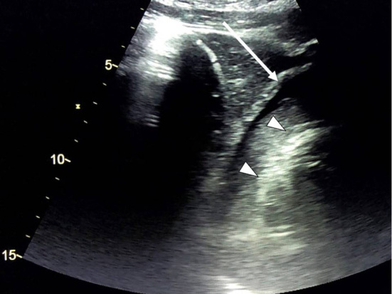
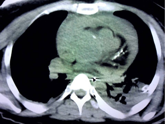
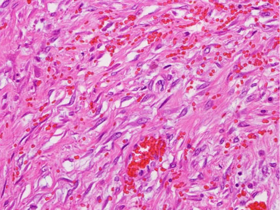
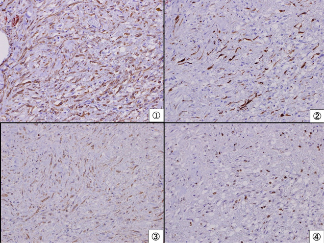
The patient was a 34-year-old woman who suddenly collapsed. On arrival, she was in cardiac arrest. Cardiac ultrasound revealed cardiac tamponade; thus, urgent thoracotomy with pericardiotomy was performed. Spontaneous circulation was temporarily obtained; however, her circulation was not stabilized, and she ultimately died. An autopsy revealed a pericardial inflammatory myofibroblastic tumor (IMT) causing bloody cardiac tamponade. There were no signs of cardiac rupture, myocardial infarction or aortic dissection. We reported the first case of fatal bloody cardiac tamponade due to pericardial IMT in an adult. An autopsy is important for clarifying the etiology in cases of fatal cardiac tamponade of unknown cause.

Keywords: cardiac tamponade; inflammatory myofibroblastic tumor; pericardium.


Conflict of interest statement

The authors state that they have no Conflict of Interest (COI).

Figures

Figure 1.
Figure 1.
Cardiac ultrasound on arrival. Cardiac ultrasound revealed cardiac tamponade (arrow). Triangles indicate the outline of the cardiac wall. The tamponade consisted of two layers: an outer low-echoic layer and an inner high-echoic layer.
Figure 2.
Figure 2.
Computed tomography (CT) after obtaining spontaneous circulation. CT demonstrated residual cardiac tamponade.
Figure 3.
Figure 3.
A histopathological examination of the pericardium (Hematoxylin and Eosin staining ×400). Infiltration with spindle cells and lymphocytes with hemorrhaging was observed from the pericardium to the origin of the aorta.
Figure 4.
Figure 4.
Results of an immunohistochemical examination of the pericardium. Spindle cells were positive for smooth muscle actin (①), desmin (②), anaplastic lymphoma kinase (③), and Ki67 (④) and negative for CD31/CD34. These results did not contradict a diagnosis of an inflammatory myofibroblastic tumor.

References

    1. Appleton C, Gillam L, Koulogiannis K. Cardiac tamponade. Cardiol Clin 35: 525-537, 2017. - PubMed
    1. Hoit BD. Pericardial effusion and cardiac tamponade in the new millennium. Curr Cardiol Rep 19: 57, 2017. - PubMed
    1. Asad ZUA, Ijaz SH, Chaudhary AMD, Khan SU, Pakala A. Hemorrhagic cardiac tamponade associated with apixaban: a case report and systematic review of literature. Cardiovasc Revasc Med 20: 15-20, 2019. - PMC - PubMed
    1. Čanádyová J, Zmeko D, Mokráček A. Re-exploration for bleeding or tamponade after cardiac operation. Interact Cardiovasc Thorac Surg 14: 704-707, 2012. - PMC - PubMed
    1. Yanagawa Y, Jitsuiki K, Ota S, et al. . Significance of medical intervention for non-traumatic hemorrhagic cardiac tamponade. Am J Emerg Med 50: 636-639, 2021. - PubMed

Publication types