

Vestibulo-Ocular Reflex Is Modulated by Noisy Galvanic Vestibular Stimulation
- PMID: 35250830
- PMCID: PMC8893018
- DOI: 10.3389/fneur.2022.826739
Vestibulo-Ocular Reflex Is Modulated by Noisy Galvanic Vestibular Stimulation
Abstract
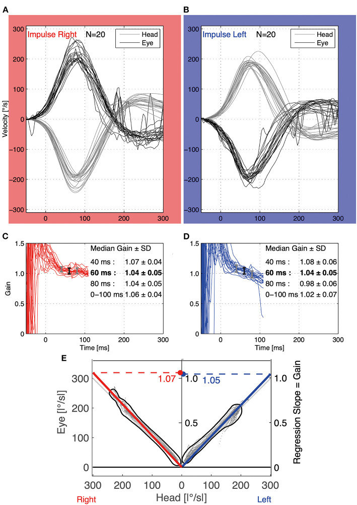
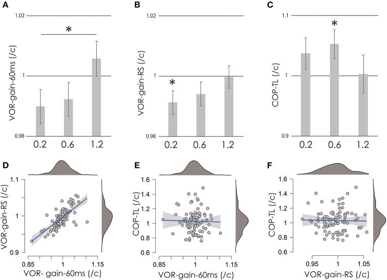
We investigated whether noisy galvanic vestibular stimulation (nGVS) modulates the vestibulo-ocular reflex (VOR) and whether this effect is correlated with the effect of nGVS on body sway. Thirty healthy young adults participated. The video head impulse test (vHIT) was used to estimate the ratio of eye motion velocity/head motion velocity to VOR-gain. The gain 60 ms after the start of head motion (VOR-gain-60 ms) and regression slope (RS) (i.e., gain in eye and head motion; VOR-gain-RS) were calculated. The total path length of the foot center of pressure (COP-TL) during upright standing was calculated to estimate body sway. Noisy Galvanic Vestibular Stimulation at 0.2, 0.6, 1.2 mA, or sham stimulation (direct current: 0 mA) was delivered to the bilateral mastoid process in random order during vHIT and COP measurements. Application of nGVS at 0.2 mA significantly reduced VOR-gain-RS, while application of nGVS at 0.6 mA significantly increased COP-TL. Vestibulo-ocular reflex-gain-60 ms differed significantly between 0.2 and 1.2 mA. There was no significant correlation between COP-TL and VOR-related parameters. These findings suggest that nGVS at 0.2 mA inhibits the VOR, while nGVS at 0.6 mA increases body sway during upright standing, although there may be no relationship between the respective effects in healthy individuals.
Keywords: galvanic vestibular stimulation; noise stimulation; postural control; vestibulo-ocular reflex; video head impulse test.
Copyright © 2022 Matsugi, Shiozaki and Tanaka.
Conflict of interest statement
The authors declare that the research was conducted in the absence of any commercial or financial relationships that could be construed as a potential conflict of interest.
Figures
Similar articles
-
Changes in vestibular-related responses to combined noisy galvanic vestibular stimulation and cerebellar transcranial direct current stimulation.Exp Brain Res. 2024 Jan;242(1):99-108. doi: 10.1007/s00221-023-06731-5. Epub 2023 Nov 15. Exp Brain Res. 2024. PMID: 37966504
-
Noisy galvanic vestibular stimulation effect on center of pressure sway during one-legged standing.J Clin Neurosci. 2020 Dec;82(Pt A):173-178. doi: 10.1016/j.jocn.2020.10.050. Epub 2020 Nov 12. J Clin Neurosci. 2020. PMID: 33317728
-
Effect of noisy galvanic vestibular stimulation on center of pressure sway of static standing posture.Brain Stimul. 2018 Jan-Feb;11(1):85-93. doi: 10.1016/j.brs.2017.10.007. Epub 2017 Oct 17. Brain Stimul. 2018. PMID: 29079459
-
Factors affecting variability in vestibulo-ocular reflex gain in the Video Head Impulse Test in individuals without vestibulopathy: A systematic review of literature.Front Neurol. 2023 Mar 9;14:1125951. doi: 10.3389/fneur.2023.1125951. eCollection 2023. Front Neurol. 2023. PMID: 36970532 Free PMC article.
-
Video head impulse test: a review of the literature.Eur Arch Otorhinolaryngol. 2017 Mar;274(3):1215-1222. doi: 10.1007/s00405-016-4157-4. Epub 2016 Jun 21. Eur Arch Otorhinolaryngol. 2017. PMID: 27328962 Review.
Cited by
-
Exploring the Potentials of Wearable Technologies in Managing Vestibular Hypofunction.Bioengineering (Basel). 2024 Jun 24;11(7):641. doi: 10.3390/bioengineering11070641. Bioengineering (Basel). 2024. PMID: 39061723 Free PMC article. Review.
-
Changes in vestibular-related responses to combined noisy galvanic vestibular stimulation and cerebellar transcranial direct current stimulation.Exp Brain Res. 2024 Jan;242(1):99-108. doi: 10.1007/s00221-023-06731-5. Epub 2023 Nov 15. Exp Brain Res. 2024. PMID: 37966504
-
Scoping out noisy galvanic vestibular stimulation: a review of the parameters used to improve postural control.Front Neurosci. 2023 May 2;17:1156796. doi: 10.3389/fnins.2023.1156796. eCollection 2023. Front Neurosci. 2023. PMID: 37205050 Free PMC article. Review.
-
Properties of standing balance control under noisy galvanic vestibular stimulation.Front Neurol. 2025 Apr 15;16:1500308. doi: 10.3389/fneur.2025.1500308. eCollection 2025. Front Neurol. 2025. PMID: 40303888 Free PMC article.
References
LinkOut - more resources
Full Text Sources