. 2022 Feb 11:12:788040.
doi: 10.3389/fmicb.2021.788040. eCollection 2021.

Effects of Lactobacillus acidophilus KLDS1.0901 on Proliferation and Apoptosis of Colon Cancer Cells

Affiliations

Effects of Lactobacillus acidophilus KLDS1.0901 on Proliferation and Apoptosis of Colon Cancer Cells

Yingxue Yue et al. Front Microbiol. .

Abstract

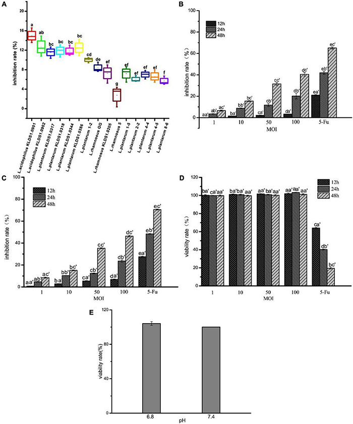
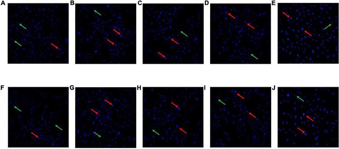
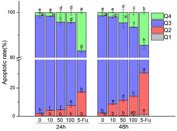
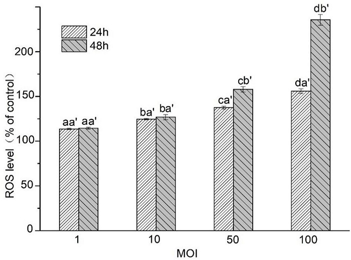
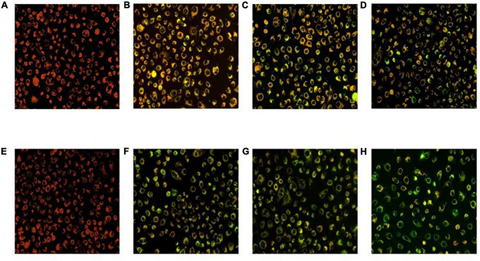
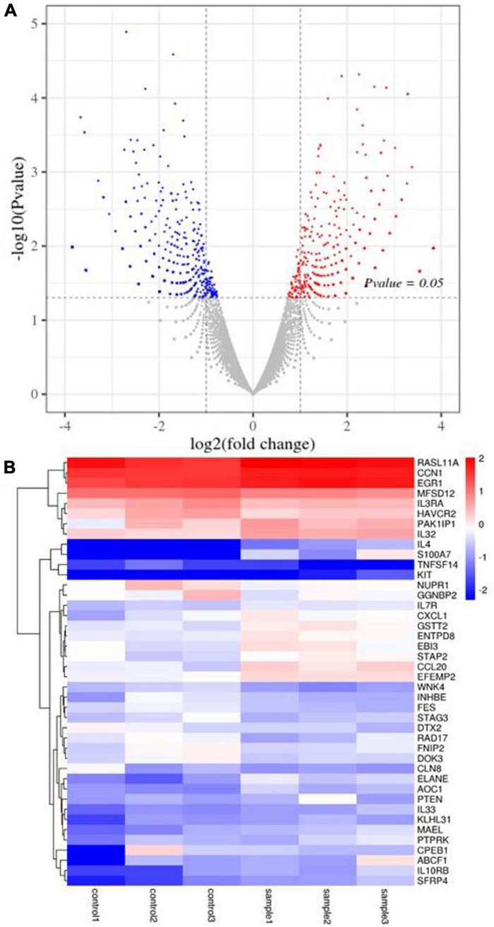
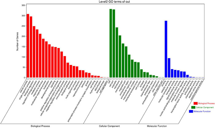
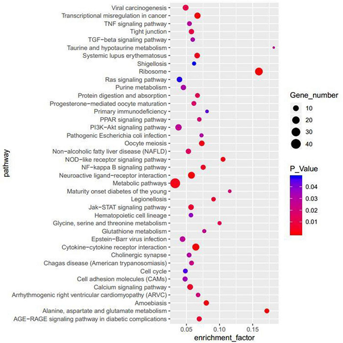
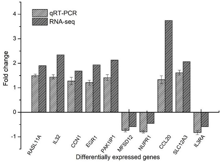
Colon cancer is the most common type of malignant tumor. The cytotoxicity effect of lactic acid bacteria may be active by inhibiting cancer cell proliferation, producing anticancer compounds, and inducing apoptosis in cancer cells, but the mechanism is unclear. Our previous study revealed that Lactobacillus acidophilus KLDS1.0901 has good probiotic properties. In this study, We screened out the highest inhibition rate of L. acidophilus KLDS1.0901 and assessed the effects on the proliferation of HT-29, Caco-2, and IEC-6 cells. Then, the apoptosis mechanism of HT-29 cells was studied when treated with L. acidophilus KLDS1.0901. Results showed that L. acidophilus KLDS1.0901 inhibited the proliferation of HT-29 and Caco-2 cells in a dose-dependent manner and reached the maximum under the condition of multiplicity of infection (MOI) = 100 (rate of Lactobacillus to cells) at 48 h. With the increase in time and MOI, reactive oxygen species in HT-29 cells, the apoptosis rates of HT-29 cells were increased, and the amount of blue fluorescence of the cells was also increased after Hoechst 33258 staining. Furthermore, L. acidophilus KLDS1.0901 reduced the mitochondrial membrane potential of HT-29 cells. Notably, 1,133 differentially expressed genes were screened by transcriptomics research, including 531 up-regulated genes and 602 down-regulated genes. These genes were involved in the nuclear factor κB and PI3K-AKT signaling pathways related to the apoptosis of HT-29 cells. These findings suggested that L. acidophilus KLDS1.0901 has the potential to be used in the development of a new type of functional foods for adjuvant treatment of colon cancer.

Keywords: HT-29 cell; Lactobacillus acidophilus; apoptosis; colon cancer; proliferation.


Conflict of interest statement

QX was employed by Heilongjiang Feihe Dairy Co., Ltd. The remaining authors declare that the research was conducted in the absence of any commercial or financial relationships that could be construed as a potential conflict of interest.

Figures

FIGURE 1
FIGURE 1
Effect of 15 strains on the inhibition rate of HT-29 cells (A). Effect of L. acidophilus KLDS1.0901 on the inhibition rate of HT-29 cells (B), Caco-2 cells (C), and the viability rate of IEC-6 cells (D). The viability rate of HT-29 cells in different pH of DMEM cell culture medium (E). Treatment of HT-29 cells with 15 strains MOI = 10 for 48 h. Treatment of HT-29 cells, Caco-2 cells and IEC-6 cells with L. acidophilus KLDS1.0901 of different MOI (1, 10, 50, 100) and 5-FU (100 μg/mL) of treatment in 12, 24, and 48 h. Treatment of HT-29 cells with pH 6.8 and 7.4 of DMEM cell culture medium for 48 h. All data are presented as means ± SD (n = 3). Different lowercase letters (a–f) above the columns indicate significant data differences between different groups (p < 0.05), (a’, b’, and c’) indicate significant data differences between the same group.
FIGURE 2
FIGURE 2
Morphological features of the HT-29 cells treated with L. acidophilus KLDS1.0901 of different MOI (0, 10, 50, and 100) and 5-FU (100 μmol/L) for 24 h (A–E) and 48 h (F–J). A fluorescence microscope was used to photograph images (20×). The red and green arrows indicate the corresponding apoptosis and intact cells.
FIGURE 3
FIGURE 3
Effect of L. acidophilus KLDS1.0901 on apoptosis of HT-29 cells. L. acidophilus KLDS1.0901 were incubated in HT-29 cells of different MOI (0, 10, 50, and 100) and 5-FU (100 μmol/L) for 24 and 48 h. Q1–Q4 represents necrotic cells, late apoptotic cells, nonapoptotic cells, and early apoptotic cells.
FIGURE 4
FIGURE 4
Effects of L. acidophilus KLDS1.0901 on intracellular ROS production of HT-29 cells. HT-29 cells were treated with L. acidophilus KLDS1.0901 of different MOI (1, 10, 50, and 100) for 24 and 48 h. All data are presented as means ± SD (n = 3). Different lowercase letters (a, b, c, and d) above the columns indicate significant data differences between different groups (p < 0.05), a’ and b’ indicate significant data differences between the same group.
FIGURE 5
FIGURE 5
Mitochondrial membrane potential of the HT-29 cells treated with L. acidophilus KLDS1.0901. L. acidophilus KLDS1.0901 treated with HT-29 cells of different MOI (10, 50, and 100) for 24 h (A–D) and 48 h (E–H). A fluorescence microscope was used to photograph images (20×).
FIGURE 6
FIGURE 6
DEG analysis of HT-29 cells treated with L. acidophilus (n = 3). (A) The volcano plot of a differential gene. (B) Clustering heatmap of differentially expressed gene expression. Controls 1, 2, and 3 were nontreated with L. acidophilus in HT-29 cells. Samples 1, 2, and 3 were treated with L. acidophilus of MOI 100 for 48 h in HT-29 cells. The red dots in the differentially expressed gene volcano map represent up-regulated, differentially expressed genes; blue dots represent down-regulated, differentially expressed genes, and gray dots represent non-differentially expressed genes. The heatmap expresses cancer-related genes, where red represents genes with high log2 (FPKM/average FPKM) expression, and blue represents low expression.
FIGURE 7
FIGURE 7
GO classification of differentially expressed genes.
FIGURE 8
FIGURE 8
The top 40 terms enriched in the KEGG of the DEGs.
FIGURE 9
FIGURE 9
Diagram of protein–protein interactions predicted by string.
FIGURE 10
FIGURE 10
RT-qPCR to verify differentially expressed genes.

References

    1. An Y., Cai H., Yang Y., Zhang Y., Liu S., Wu X., et al. (2018). Identification of ENTPD8 and cytidine in pancreatic cancer by metabolomic and transcriptomic conjoint analysis. Cancer Sci. 109 2811–2821. 10.1111/cas.13733 - DOI - PMC - PubMed
    1. Balakrishnan M., Floch M. H. (2012). Prebiotics, probiotics and digestive health. Curr. Opin. Clin. Nutr. Metab. Care 15 580–585. - PubMed
    1. Baldwin C., Millette M., Oth D., Ruiz M. T., Luquet F.-M., Lacroix M. (2010). Probiotic Lactobacillus acidophilus and L. casei mix sensitize colorectal tumoral cells to 5-fluorouracil-induced apoptosis. Nutr. Cancer 62 371–378. 10.1080/01635580903407197 - DOI - PubMed
    1. Bian Q., Xu L., Wang S., Xia Y., Tan L., Chen J., et al. (2004). Study on the relation between occupational fenvalerate exposure and spermatozoa DNA damage of pesticide factory workers. Occup. Environ. Med. 61 999–1005. 10.1136/oem.2004.014597 - DOI - PMC - PubMed
    1. Celebioglu H. U., Erden Y., Ozel H. B. (2021). In vitro cytotoxic effects of lactobacilli grown with lime honey on human breast and colon cancer cells. Food Biosci. 41:101020. 10.1016/j.fbio.2021.101020 - DOI

LinkOut - more resources